"So, naturalists observe, a flea has smaller fleas that on him prey; and these have smaller still to bite ’em; and so proceed ad infinitum."
- Jonathan Swift

December 14, 2022

Stenurus globicephalae

Whales are big animals, even the "smaller" cetaceans such as dolphins and pilot whales are large animals in the range of body sizes across the animal kingdom. The thing about big animals is that they provide a lot of room for parasites. The myriad array of spacious organs found in an animal like a whale provide ample opportunities for parasites to become very specialised not just on a particular species of host, but on a very specific niche within the host's anatomy. Unsurprisingly, there are a wide range of parasites that make their home inside whales. With so many different species inhabiting different parts of the whale anatomy, it is perhaps not surprising that there are worms that specialise in living within the voluminous lungs and sinuses of whales. One of them is a genus named Stenurus.

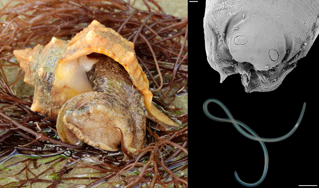
Numerous Stenurus worms in the pterygoid sinuses of a short-finned pilot whale (Globicephala macrorhynchus).
Photo from Fig. 1 of the paper. 

There are nine known species of Stenurus, all of them live in the respiratory system and sinuses of whales. Each of those worms differ in the particular cetacean species that they infect, as well as the part of the host's respiratory system they inhabit. The study being covered in today's blog post were based on samples collected from fourteen toothed whales that had been stranded along the Galicia coast between 2009 and 2019. There were a mix of six different whale species in total, and considering the population of parasites that each whale could support, it provided researchers with plenty of material to work with. Unlike most other parasitological studies where the dissection takes place in the controlled environment of a laboratory, you can't exactly bring a dead whale back to your lab. So instead, the parasites were collected via on-site necropsies.

The researchers found many different species of lung parasites from the different whales, including three species of Stenurus. But out of them, the most abundant was Stenurus globicephalae, which was found in three host species including Risso's Dolphin, Short-finned pilot whales, and Long-finned pilot whales. Each whale harboured between 18 to over 1700 of those worms, which were mostly found in the pterygoid sinus, located deep within the nasal passage near the back of the whale's throat. Previous studies have also recorded S. globicephalae dwelling in the lungs as well as other canals and cavities in a whale's head including the middle ear cavities and cranial sinuses. These worms seem to like dwelling in soft, moist tubes.

Stenurus globicephalae was found to be particularly prolific in short-finned pilot whales, which indicates that while it is capable of infecting other whale species, the short-finned pilot whale just so happens to be a particularly good "fit" for it. Each Stenurus species differ in their host preferences, and it seems to be related to how they get transmitted to the whale in the first place. Like with many other nematodes that infect whales, they do so by hiding in their host's food.

The researchers noticed a certain pattern of association between different species of Stenurus with the diet of the whale species they infect. For example S. globicephalae was associated with species that mostly ate squid, whereas those that ate fish or have a mixed diet tend to be infected with  S. ovatus and S. minor. So it is a case of "you are infected by what you eat".

Stenurus was not alone in its preference for whale respiratory structures. In some of the whales, the researchers found Stenurus cohabiting those airy passages with other parasites. One of those co-inhabitants was Halocercus - a different genus of whale lungworm which anchor themselves in place by plunging their head firmly into the host tissue. Another co-inhabitant was Nasitrema -  a fluke that lives in the air sinuses of small whales and has a nasty tendency to wander into the host's brain.

While having worms in your sinuses sounds uncomfortable, Stenurus lead a relatively peaceful existence within their wet, cosy homes, and their presence causes surprisingly little to no inflammatory responses. Or at least they do once they settle down as adult worms, because the larval worms can potentially cause focal pneumonia, while the adult worms have a more relaxed relationship with their surroundings. It is as if the whales progressively grow used to their presence, or the worms have grown to tame the fiery response of their host's immune system.

Reference:

November 16, 2022

Acanthobdella peledina

In the cold rivers and lakes of the arctic and subarctic region, there live some rather peculiar worms with a face full of tiny hooks and an appetite for blood. These worms live as ectoparasites of fish, and they belong to a group called Acanthobdellida - relatives of leeches that seem to have gone down their own evolutionary path. These worms have also been called "hook-faced fish worms" and the entire group consists of only two known species - Acanthobdella peledina and Paracanthobdella livanowi

Top: Acanthobdella peledina on a grayling.
Bottom: Scanning electron micrograph of the whole worm (left) and close-up of the anterior body region (right).
Photos from Figure 1, 6, and 7 of the paper

Their mouthpart has been described as being a less sophisticated version of a leech's mouthpart - they lack the saw-edged jaws or the extensible proboscis found in many leeches, nor do they have the muscular sucker which surrounds the mouth. Instead, they have a protrusible pharynx and a series of hooks on the first five segments of the body, which they use to attach themselves to their fishy hosts. 

They have previously been considered to be a "missing link" between leeches and the rest of the Clitellata - the group of segmented worms that also includes earthworms and tubifex worms - as they have certain features which are commonly found in other clitellate worms but are absent in leeches. This includes having tiny bristles (called chaeta) on their segments, and a reproductive system similar to those found in earthworms.

Acanthobdella peledina is found all across the subarctic, where they range from being relatively rare to being found on over two-thirds of the fish at a given location. Given it is so widely distributed, with populations scattered across different geographical locales, could each of those distinct populations actually be different species? A group of researchers set out to determine whether there are actually more species of these hook-faced worms than meets the eye. Furthermore, they also wanted to find out how closely related Acanthobdella and Paracanthobdella are to each other. They did so by comparing museum specimens of hook-faced worms which have been collected from sites across the subarctic, including Norway, Sweden, Finland, Alaska, and Russia. 

Aside from examining their anatomical features, the researchers also compared five different key marker genes from these worms. Some of those DNA segments came from the mitochondria, others from the cell's nucleus. The reason for comparing multiple genes is that each has their own histories, and may offer different perspectives on the organism's evolutionary history. It is like interviewing different witnesses at a crime scene.  Unfortunately, for whatever reasons, the DNA of these worms proved to be particularly challenging to amplify and sequence, so for most specimens they were only able to sequence up to four of the five genetic markers they were aiming for, with some specimens only yielding sequences for two of the genes. Despite that limitation, the researchers were able to use the sequences they obtained to resolve the hook-faced worm's evolutionary history.

Despite their wide distribution across the arctic and subarctic regions, Acanthobdella peledina does appear to be a single, widespread species. While the Alaskan population of worms are genetically distinct from the Nordic population, they are not dissimilar enough for them to be considered as separate species. Furthermore, based on their analysis, the two living species of hook-faced worms are quite closely related to each other. In fact, it seems they had only diverged from each other just prior to the last ice age. So, far from being some kind of "missing link" between leeches and other clitellate worms, these hook-faced worms belong to their own distinct group.

But while the two living species had shared a common history until relatively recently, the hook-faced worms as a group had evolutionarily split off from the leeches a long time ago. Based on available data on these worms, this might have occurred during the early Cenozoic as the ancestors of the hook-faced worms became specialised on arctic freshwater fishes that arose during that era, such as salmonids.

So it might have been the pursuit of salmonids that had sent these worms down their own distinct path - a story which is probably relatable to any fly fishers out there.

Reference:
de Carle, D. B., Gajda, Ł., Bielecki, A., Cios, S., Cichocka, J. M., Golden, H. E., Gryska, A. D., Sokolov, S., Shedko, M. B., Knudsen, R., Utevsky, S., Świątek, P. & Tessler, M. (2022). Recent evolution of ancient Arctic leech relatives: systematics of Acanthobdellida. Zoological Journal of the Linnean Society 196: 149-168.

October 18, 2022

Prochristianella sp.

Earlier this year, I wrote about Aggregata sinensis a species of single-celled apicomplexan parasite that infects octopus. But octopuses are host to a wide range of other parasites as well, especially parasitic worms. Most of these worms infect the octopus during their larval stage, and use the cephalopod as a way to travel up the food chain to their final host - usually predatory vertebrate animals such as sharks, birds, and marine mammals. Prochristianella is one such parasite.

Left: A stained specimen of Prochristianella metacestode, Right top: Scanning electron microscopy of a Prochristianella metacestode, Right bottom: Scanning electron microscopy close-up of the Prochristianella scolex with protruding tentacles
Photos from Fig 2 and Fig 3 of the paper.

The paper being featured in this blog post focused on Octopus maya, also known as the Mexican four-eyed octopus, of the Yucatán Peninsula. It is a popular species for commercial fisheries both caught from the wild and in aquaculture. Since it is such a widely fished and commonly eaten species, it would be a good idea to know just what kind of parasites are present in these octopus. The researchers obtained sixty O. maya from local fishermen in Mexico who have caught octopuses from four locations in Yucatán - Sisal, Progreso, Dzilam de Bravo, and Río Lagartos. These cephalopods were caught using a tradition line fishing technique called al garete where multiple lines of hooks baited with crabs are dangled from a small drifting boat and dragged along by the current.

When the researchers dissected the octopuses, they found seven different types of tapeworm larvae in total, each occupying a different part of the octopus' body. Some were found in the intestine, others in the digestive glands, some were in the gills, and there were even some species that hung out in the ink sac. By far the most common species was Prochristianella, it was present at all four collection sites and was found in every single octopus the researchers examined. This tapeworm specifically occupied the octopus' buccal mass - the ball of muscles and connective tissue that houses the octopus' mouth. Not only was it common, Prochristianella was also extremely abundant, with each octopus having on average over a hundred Prochristianella larvae embedded in their buccal mass, while the octopus from Río Lagartos had over a thousand such tapeworms each. 

In fact, Río Lagartos seems to be tapeworm central, as that is also the location where the other six species of tapeworms also reach their highest prevalence and abundance. Perhaps it has something to do with Río Lagartos being located at the Ria Largatos lagoon, which is part of a nature reserve. Higher level of biodiversity can facilitate the transmission of parasites such as marine tapeworms, which need to use many different species of host animals to complete their complex life cycles.

Prochristianella was one of four types of trypanorhynch tapeworms found in the octopus. These tapeworms need to infect elasmobranch fishes such as sharks and rays to complete their life cycle, and the octopus, being prey to those fishes, is a convenient way for these parasites to get there. One of the unique features of trypanorhynch tapeworms is their attachment mechanism. Unlike other tapeworms with their hooks and suckers, the scolex of trypanorhynchs are armed with gnarly hook-lined tentacles, which shoot out like harpoons to anchor themselves into the intestinal wall of their elasmobranch host.

One of the possible reasons why Prochristianella is so common and numerous among those octopuses is because it uses shrimps as one of the intermediate hosts in its life cycle. Octopus feed on shrimps throughout their entire life, so even if the tapeworm is relatively uncommon in shrimps, they can accumulate in the octopus over its lifetime. That's how those octopus end up with over a hundred or even a thousand such tapeworm larvae around their mouth.

The next most common tapeworm in those octopus after Prochristianella was another trypanorhynchan tapeworms called Eutetrarhynchus, found in the digestive glands and ink sac. Though not as widespread or abundant as Prochristianella, it is still fairly common throughout the Yucatán Peninsula. The rest of the tapeworms is a smattering of different species, and while all of them complete their life cycles and develop into adult worms in sharks and rays, the path that they take to get there varies slightly. Some of the rarer species in this study usually use other animals such as bony fishes as intermediate or paratenic (transport) hosts, and occasionally end up in octopuses. While others, such as Phoreiobothrium, infect a wide range of different cephalopods, and O. maya just happen to be one of many potential hosts on their list. Overall, while they varied in abundance at different locations, these same set of tapeworms were present in octopus across the Yucatán Peninsula.

The variety of tapeworms and other parasites found in O. maya shows that this cephalopod is an important junction point in the life cycles of many parasites. Being predators in their own right, octopuses end up accumulating parasite larvae which would otherwise be thinly dispersed throughout the population of small prey animals, such as shrimps. Meanwhile, octopus themselves are eaten by a wide variety of larger animals, thus providing the means for some parasites to work their way up the food chain, into large marine predators such as sharks where they can complete their life cycles. 

Through their parasites, we can see how these octopuses interact with other animals and their place in the wider marine ecosystem.

Reference:

September 20, 2022

Hymenoepimecis bicolor

Over the course of human history, numerous species of plants, animals, and other organisms have been taken from their original habitats and introduced (either intentionally or accidentally) to other parts of the world. Some of those introduced species become "invasives" in their new home, partly due to the lack of natural enemies. But while many invasive species get a brief moment of respite from their old adversaries, the local parasite and predators quickly catch on that the new arrival could be added to their menus too. This was the case for a species of spider that has been introduced to Brazil, where it ended up attracting the attention of a mind-controlling wasp.

Left top: Hymenoepimecis bicolor larva on a spider host, Left bottom: Developing H. bicolor cocoon in a cocoon web,
Right: an adult H. bicolor wasp.
Photo of H. bicolor larva from Fig. 2 of this paper, Photos of cocoon and adult H. bicolor from Fig. 7 and 9 of this paper

Hymenoepimecis bicolor is a species of parasitoid wasp that belongs to a subfamily of wasps called Pimplinae. All the members of that subfamily are a spider's worst nightmares. These wasps specialise in attacking spiders at every stage of their lives - some species go after the unhatched eggs in silk sacs, while others tackle fully-grown adult spiders. Not only that, some of them are also masterful mind manipulators that induce their host spider into spinning a special web called a "cocoon web" that secures the wasp's developing cocoon. And Hymenoepimecis bicolor just happens to be one such manipulator.

Among the pimpline wasps, each species have their own host preferences and in the case of H. bicolor, one of its usual hosts is the golden silk orb-weaver (Trichonephila clavipes), a spider which is native to Brazil. When a female H. bicolor spots a potential host, she flies in and grapples with it, immobilising the spider by stabbing it in the mouth with her ovipositor, before checking it for other wasp eggs, and then planting one of her own. The thing about the golden silk orb-weaver is that while the juvenile spiders are relatively easy for H. bicolor to handle, once the spiders reach adulthood, they become more dangerous for the wasp to tackle. 

Nevertheless, limited host availability means that the wasp sometimes need to go after the bigger spiders anyway, with demand being so high that some spiders end up being parasitised by two wasp larvae at the same time. But the arrival of the tropical tent-web spider (Cyrtophora citricola) has provided H. bicolor with some new options.

The tropical tent-web spider has spread to many parts of the world by hitchhiking in shipments of fruit, potted plants, or packing material, and it has made its way to South America about three decades ago. And it just so happens that their size and habits place them firmly in the sight of H. bicolor. Not only is the tent-web spider in the preferred size range for H. bicolor to parasitise, much like the native orb-weavers, this introduced spider constructs open webs that leave them exposed to attacks. Researchers found that the H. bicolor larvae are able to successfully parasitise the tent-web spider just as well as the native spiders.

While H. bicolor larvae grow well and pupate as usual on the tent-web spiders, it seems that they haven't yet achieved complete mastery over this new-fangled host. As mentioned earlier, when these wasps are ready to pupate, they commandeer their spider hosts to weave a special "cocoon web" that suspends the developing wasp cocoon in mid-air. This makes the cocoon less accessible to any would-be predators or hyperparasitoids. Hymenoepimecis  bicolor embellish that with an added layer of security, by inducing the spider to also build a series "barrier threads" around the cocoon that further bar entry, as well as making the web more stable

This is where the introduced spider host falls short. While the parasitised tent-web spider is able to produce the usual cocoon web with the necessary structure to support and suspend the developing cocoon, it lacks the finishing touches of those additional barrier threads. Ironically, compared with the spiders that H. bicolor usually targets, the regular webs made by the tent-web spider actually needs less modification to make it suitable for the wasp's cocoon.

Based on what's known about these wasps, when it is ready to pupate, the wasp larva produces a cocktail of chemicals that place the spider under its spell. But in this case, it looks like that cocktail formula needs a bit of tweaking to work its full magic on the introduced tent-web spider. While not perfect, it serves its purpose well enough, and the introduction of this spider has allowed a parasitoid wasp to expand its host horizons.

Reference:

August 14, 2022

Cyclocotyla bellones

At the top of this blog, there is a quote by Jonathan Swift about how fleas have smaller fleas that bite them. Indeed, parasites becoming host to other types of parasites is actually a rather common phenomenon in the natural world. Those who would parasitise the parasites are called "hyperparasites".

Left: Cyclocotyla bellones on the back of a Ceratothoa isopod, Right: C. bellones coloured red with Carmine staining.
Photos from Figure 1 and 5 of the paper.

The parasite featured in this post was once suspected of being a hyperparasite. Cyclocotyla bellones is a species of monogenean - it belongs to a diverse group of parasitic flatworms that mostly live on the body of fish, parasitising the fins, skins, and gills of their hosts. But unlike other monogeneans, C. bellones does not attach itself to any part of a fish's body, instead it prefers to stick its suckers onto the carapace of parasitic isopods, such Ceratothoa - the infamous tongue biter. Since Ceratothoa is itself a fish parasite, and C. bellones is routinely found attached to those tongue-biters, this has led some to think that it might be a hyperparasite of those parasitic crustaceans.

But it takes more than simply sticking yourself onto another organism to be considered as a parasite of it. After all, there are algae that grow on the body of various aquatic creatures, or barnacles that are found on the backs of large marine animals like whales and turtles. But those are not considered as parasites as they don't treat their host as a food source, merely as a sturdy surface they can cling to - they're known as epibionts.

So strictly speaking, for Cyclocotyla to be a parasite of the isopod, it needs to be feeding on or obtaining its nutrient directly from its isopod mount. When scientists examine the bodies of the tongue-biters with C. bellones on them, they seem to be pretty unscathed. There aren't any scratches or holes on the isopod's body which you'd expect if C. bellones had been feeding on it. Indeed, the monogenean's mouthpart seems ill-suited for scraping through the isopod's carapace.

Additionally, C. bellones' gut is filled with some kind of dark substance similar to those found in other, related monogenean species. This is most likely digested blood from the fish, which the monogenean has either sucked directly from the fish's gills, or indirectly via the feeding action of its isopod mount. Let's not forget that the isopod itself is a fish parasite that feeds on its host's blood, so if it gets a bit messy during mealtime, perhaps Cyclocotyla is there to suck up any spilled blood. Or it might be doing a bit of both.

The researcher noted that Cyclocotyla is not alone in its habit of riding isopods. Other monogeneans in its family (Diclidophoridae) have also been recorded as attaching to parasitic isopods of fish. And aside from riding isopods, they all share one thing in common - a long, stretchy forebody, looking somewhat like the neck of sauropod dinosaurs. Much like how the neck of those dinosaurs allowed them to browse vegetation from a wide area, the long forebody of Cyclocotyla allows it to graze on the fish's gills while sitting high on the back of an isopod. So fish blood is what C. bellone is really after - the isopod is merely a convenient platform for it to sit on.

But why should these monogeneans even ride on an isopod in the first place? Cyclocotyla and others like it have perfectly good sets of suckers for clinging to a fish's gills. Indeed, there are other similarly-equipped monogeneans that live just fine as fish ectoparasites without doing so from the back of an isopod. Well, that's because the fish themselves don't take too kindly to the monogeneans' presence. These flatworms are constantly under attack from the fish's immune system, which bombards them with all kinds of enzymes, antibodies, and immune cells. By avoiding direct contact with the fish's tissue, Cyclocotyla and other isopod-riders can avoid being ravaged by the host's immune system - which is something that other monogeneans have to deal with on a constant basis.

So it seems that Cyclocotyla and other isopod-riding monogeneans are no hyperparasites - they're all just regular fish parasites that happen to prefer doing so while sitting on the backs of isopods. Cyclocotyla bellones prefers to share in the feast of fish blood with its isopod mount, while sitting high above the wrath of the host's immune response.

Reference:

July 18, 2022

Dolichoperoides macalpini

Australia has some of the most venomous snakes in the world, but the mouths of those reptiles are filled with more than just venomous fangs. In some cases, they are filled with tiny digenean flukes, specifically Dolichoperoides macalpini. This species of fluke was first reported from the lowland copperhead snakes in the 1890s, but it wasn't until 1918 that it was formally identified and described, and in 1940 it was placed in its own genus when it was recognised that it was specifically associated with elapid snakes. Since then, there hasn't been much further studies on this fluke, and the research team behind the paper in this post seeks to fill in that knowledge gap.

Left: Dolichoperoides macalpini in a snake's mouth, Right: Dolichoperoides macalpini in a snake's lungs
Photos from Fig. 1 of the paper

For this study, the researchers collected snakes from parts of Tasmania and Western Australia.
In Tasmania, they collected roadkills composed of Tiger Snake (Notechis scutatus) and Lowland Copperhead (Austrelaps superbus). While in Western Australia, they obtained freshly caught and euthanised Western Tiger Snake (Notechis scutatus occidentalis) which were collected as a part of another, larger project examining tiger snakes from wetlands in and around Perth. Dolichoperoides macalpini were mostly found in the snake's mouth, oesophagus, and stomach. And when the snake's mouth is open, the flukes are clearly visible as tiny black specks that clung to the roof of the snake's mouth (see accompanying photo). However, the snakes from Tasmania had D. macalpini in their lungs and intestine as well. So what's going on there? 

This could be because the snake specimens examined in Tasmania were roadkills. In some cases, after the host dies, its parasites may move from their usual location to different parts of the host's body, possibly due to some last ditch survival instincts. This phenomenon is well-known in anisakid nematodes, which is a major seafood-borne zoonotic parasite. After their fish host is caught, these worms often migrate from their host's viscera to its flesh. In the case of D. macalpini, once they sense that their host had died, perhaps they evacuated away from the mouth and throat to other, deeper parts of the body such as the lungs and intestine in a desperate bid for survival.

This may also explain some of the other differences the researchers found in the infection patterns of different snake populations. The Tassie snakes generally had fewer flukes than those which were caught around Perth. Since the Tasmanian snakes were found as roadkill, it is possible that the flukes which didn't crawl to the lungs or intestine had just ended up abandoning the snake altogether.

But this difference in fluke abundance may have also been influenced by other more innate factors of the snakes' ecologies. The encysted larval stage of D. macalpini are found in frogs, which the Perth snakes were particularly fond of, with frogs accounting for almost 90% of their diet. This provided them with ample opportunities to encounter the infective larval stages of D. macalpini through their food. In contrast, the Tassie snakes had a more varied diet consisting of rodents, birds, and lizards - but no frogs.

Additionally, there were also other differences among the flukes themselves. For example, while the snakes from Perth were more heavily infected, their flukes were only about half the size of those found in the Tasmanian snakes. While such size differences might have indicated that the flukes in those separate snake populations may in fact be different species, genetic analyses showed otherwise. The 18S rRNA gene and ITS gene sequences - which are key genetic markers for delineating different species among these parasites - were identical for the flukes from both Tasmanian and Perth snakes.

So there must be other reasons for such marked differences in their sizes. Perhaps in more heavily infected hosts, the crowded environment may have limited the flukes' growth? Studies on other species of flukes have found that those from more heavily infected hosts tend to be smaller on average than their counterparts from less parasitised hosts. This diminished growth may be the result of competition over limited resources, be it host nutrient, or simply available space for growth. Or perhaps there are slight variations between the biology of different snake species that can influence the fluke's growth?

The result of this study offers a brief glimpse into the distribution and infection patterns of D. macalpini in Australian snakes, and it raises some tantalising questions about the parasite's ecology. But there are many other reptile parasites in Australia for which little is known about them outside of a taxonomic description. Despite having one of the world's richest reptile fauna, the parasites fauna of Australian reptiles are relatively understudied. Not only are they an integral part of Australia's biodiversity, understanding these parasites can also tell us about how their reptile hosts are connect with the rest of the ecosystem.

Reference:

June 18, 2022

Sarcotaces izawai

Parasitic copepods are a weird bunch, and many of them look nothing like what most people would recognise as a crustacean. But even among those weirdos, Sarcotaces stands out, because during the course of its evolution, it has turned into a big teardrop-shaped blob living inside a fish's body.

Top left: Staining of the fish body due to Sarcotaces, Top right: Sarcotaces extracted from fish flesh
Left: A Female Sarcotaces specimen (about 4 cm in length)
Photos from Fig. 1 and Graphical Abstract of the paper.

There are seven known species of Sarcotaces, all of which are parasites that dwell in fleshy galls embedded in the muscles of fish. The female of the species can grow up to about 5 cm long. They belong to a family of copepods called Philichthyidae which all specialise in living within nooks and crannies of a fish's body, including their skull, sensory canals, or inside galls just beneath the fish's skin. The study featured in this blog post described a newly discovered species of Sarcotaces - Sarcotaces izawai.

Specimens of S. izawai were retrieved from a consignment of frozen fish which were originally destined for the fish market, but were redirected to researchers when the County Veterinary Inspector of Szczecin noticed signs of infection in some of the fish. In total, 29 fish were taken to the University of Szczecin for further examination. Nine of those fish were found to harbour the gall of Sarcotaces - where there was once fish muscle had been turned into a black void, a dark fleshy cavern where the female Sarcotaces resided alongside her tiny males and microscopic larvae.

The black liquid associated with this copepod is what gave Sarcotaces its German name - "Tintenbeutel" which means "ink bag", and why in parts of Australia, they're called "Iodine Worms". Even in the other fish where no Sarcotaces were found, the fish's flesh were tainted with an ink-stained void, which most likely meant a Sarcotaces had once lived there, but was inadvertently removed when the fish were being processed. While the presence of this parasite does not pose any health hazards to any would-be consumers, the inky stain in the fish's flesh do render them off-putting to any would-be buyers on the market. But, because of this, the researchers were given an opportunity to conduct detailed scanning electron microscopy on the copepod, and provided the first DNA barcode for this unique genus of parasite based on its COI gene.

While the female S. izawai is very distinct and noticeable, the male is rather inconspicuous - they grow to about 3 mm in length, and are comparatively tiny and fairly nondescript. In comparison, the female is shaped like a knobbly radish, and grows to 2.5 to 5 cm in length or 10-20 times the length of the male. This size difference is comparable to that of a human and a sperm whale. It also means that a single female could be accompanied by multiple males. Indeed, the researchers found one female who was accompanied by 18 suitors in her flesh gall.

While very little is known about how the microscopic, free-swimming larvae of Sarcotaces gets into a fish in the first place,  it seems that the growth and development of the female Sarcotaces takes place entirely within the sac-like gall. This flesh bag has a tiny opening to the outside world that the copepod usually keeps plugged using the pointy tip of her body, and unplugs to release larvae into the surrounding waters. Because of this, the researchers consider Sarcotaces as a "mesoparasite", because while they largely live within the fish's body, they still maintain some contact with the outside world with the tip of the body plugging up that hole.

As an added layer to that study, the consignment of frozen fish that the researchers examined have been been frozen and thawed multiple times, and were "pan-dressed" - in that their head, fins, and the guts have been taken out - this might be why some of the fish had the characteristic inky stain of Sarcotaces even though the parasite was absent. This made the identification of those fish rather difficult simply through visual inspection. While the consignment of fish were labelled as Pseudophycis bachus - red codling - from "The Falklands", the researchers found this to be a case of seafood identity fraud.

When they did some DNA analyses they found that the fish were actually Mora moro - a species of deep sea cod which is found in temperate seas across many parts of the world, but has not been recorded from the Falklands. It is likely that the fish wholesalers were trying to use the mislabelling to bypass regional quotas or conceal catches from restricted waters.

This type of seafood mislabelling is very common around the world, and presents problems for consumer protection, food safety and supply, fisheries regulations, and conservation. In this case, not only did the ink-stained fillets of these Sarcotaces-infected fish provide scientists with an opportunity to examine a poorly-understood parasite, the presence of this tubby copepod also helped draw attention to a case of seafood identity fraud.

Reference:

May 20, 2022

Guimaraesiella sp.

Quite a few years ago I wrote a blog post about a study on some bird lice that hitch-hike on louse flies as a way of reaching new hosts - this type of interaction whereby an organism attach itself to the body of another as a way of getting around is called "phoresy". And while it is a fascinating interaction with important ecological implications, this phenomenon is not particularly well-studied. Well, the paper that is being featured in this blog post revisited that field of research, and used multiple approaches to investigate this type of interaction. And the researchers behind it did so by combining literature review, traditional parasitology, DNA barcoding, and citizen science.

Left: Guimaraesiella lice found on from louse flies. Right: Louse fly with lice attached (indicated by red arrows). 
From Figure 3 of the paper.

The researchers of this study were trying to figure out how common phoresy is among bird lice, and who exactly is hitch-hiking on what. They conducted a review of the existing scientific literature on phoretic relationships between lice and louse flies, and found that many of the older records were unusable because they lack sufficient details regarding species identity of the lice involved. Furthermore, while phoretic behaviour in lice is most well-documented in North America and Europe, there are other parts of the world with much richer avian fauna (and thus more bird lice species), but phoretic behaviour of bird lice in those regions are not as well-studied.

To address this, the researchers came up with a way of collecting lice and louse flies from a large number of birds, and did so with some help from members of the public. As a part of long-term project to monitor bird mortality from vehicle and building collisions, ordinary citizens in Singapore were encouraged to report any dead birds that they come across. Through this, the researchers were able to track down and collect over a hundred recently deceased birds for this study. They then screened the dead birds for lice and louse flies, which were identified based on their morphology and their DNA.

In total, they screened 131 birds composed of 54 different species, and collected 603 lice and 32 louse flies. Of those, 22 birds had louse flies on them, but only three of the louse flies also happened to be carrying hitch-hiking lice, which were identified as belonging to the genus Guimaraesiella. Amidst all that, they found something unexpected - one of the birds, a Blue-winged pitta (Pitta moluccensis) was infected with louse flies carrying Guimaraesiella lice. This is the first time that Guimaraesiella lice has been found on pittas, as those birds are usually infected with lice in the Picicola genus.

It is likely that riding on louse flies is how Guimaraesiella ended up on the pitta. Indeed, lice in that genus appear to live on a wider range of birds compared with most bird lice, which are often confined to a single or handful of closely related host species, and its hitch-hiking habit may be the key to their success. While bird lice are very adept at climbing around and between their host's feathers, they are completely helpless off the host's body. This doesn't give them much opportunity to branch out and onto other bird species as they can only climb onto a new host through direct contact.

But since louse flies feed on a variety of different bird hosts, travelling on one of those flying blood-suckers can open up a whole new world of possibilities for lice that engage in phoresy. The species of Guimaraesiella lice they found on the pitta has also been found on at least 24 other species of birds, possibly more. Considering that the louse fly that Guimaraesiella rides on - Ornithophila metallica - feeds from over a hundred different bird genera, perhaps it is surprising that Guimaraesiella hasn't been found from even more bird species. So while the louse fly presents its hitch-hiker lice with many different species of birds, those well-travelled lice still stay fairly selective when it comes to where they settle on. These lice are like Goldilocks when it comes to picking a new feathery home - it needs to be just the right fit.

The approach taken by the researchers in this study to recover and screen large numbers of birds for louse flies and lice can also be applied to other parts of the world. This would help us obtain a more complete understanding of how widespread hitch-hiking lice actually are, and the role this behaviour has played in the evolution of these ectoparasitic insects.

Reference:
Lee, L., Tan, D. J., Oboňa, J., Gustafsson, D. R., Ang, Y., & Meier, R. (2022). Hitchhiking into the future on a fly: Toward a better understanding of phoresy and avian louse evolution (Phthiraptera) by screening bird carcasses for phoretic lice on hippoboscid flies (Diptera). Systematic Entomology 47: 420-429.

April 21, 2022

Aggregata sinensis

Apicomplexa is a diverse phylum of single-celled parasites. They are found in a wide range of different animals, and includes some well-known species which can infect humans such as the malaria-causing Plasmodium, the infamous and widespread Toxoplasma gondii, and the gut-busting Cryptosporidium. But it is not as if this group has any particular affinity for humanity - humans are just one species among many across the animal kingdom that are hosts for apicomplexan parasites. Most of the more well-studied apicomplexans are those that infect terrestrial animals, especially domesticated species, but far less is known about apicomplexan parasites that are found in the marine realm.

Top left: Aggregata sinensis oocysts in the membrane between the arms of an octopus. Top right: Oocysts in the branchial heart.
Bottom left: Sporocysts found within an oocyst. Bottom right: Sporozoite released from a sporocyst.
Photos from Fig. 1 and Fig. 2 of the paper

Aggregata is a genus of apicomplexan which specifically targets cephalopods - mainly octopuses. Octopus can become infected from eating crustaceans such as shrimps which harbours the asexual stage of the parasite. Once they get into the octopus gut, the parasite takes over the digestive tract, and undergo sexual reproduction in the cells of the gut lining. There are twenty different known species of Aggregata, and it seems that for octopuses, there is no escape from this genus of parasite - even deep sea species living around hydrothermal vents are targeted by their own specialised species of Aggregata parasite.

So there are no doubt many other species of Aggregata out there which are still undiscovered. The paper featured in this blog post describes a species of Aggregata called Aggregata sinensis which has been found in octopus from the eastern-central coastal waters of China and the northern tip of Taiwan. The parasite was found infecting two species of octopus - the webfoot octopus and the long arm octopus - both of which are commercially important species that are caught by the local fishermen. 

The parasite was rather common, and depending on the location, between 20-100% of the octopuses that the researchers examined were afflicted with A. sinensis. Because the way an octopus becomes infected is from eating parasitised prey, Aggregata infection initially starts in the digestive tract, but it doesn't stay there for long. In heavy infections, the parasite spills over into other parts of the body in a very visible way. As Aggregata proliferates in the octopus, it leaves tell-tale signs of their presence in the form of white cysts that speckle the octopus' body. Those white cysts are called oocysts, which are the results of the parasite's sexual reproduction. Aggregata can wreak a destructive toll on the octopus's health. As the parasite proliferates, they smother the gut lining and destroy the submucosa cells, which compromise the octopus' ability to absorb nutrients. 

As if that's not enough, those white oocysts are filled with microscopic spheres called sporocysts which need to depart from the octopus' body to continue the life cycle, and they do so in a destructive manner. The release of those Aggregata oocysts necessitates the rupture and shedding of the surrounding hosts cells, resulting in ulcers and atrophy of the gut lining and connective tissues. Once free in the surrounding waters, should the sporocysts find themselves in an unlucky crustacean, they unravel to reveal their payload of worms-shaped sporozoites. These squirm out and settle in the crustacean's gut where they undergo asexual reproduction, and start the life cycle anew.

A recent study on the phylogeny of Apicomplexa suggests that Aggregata belongs to a group called the Marosporida - which occupies a key evolutionary position within Apicomplexa, separate from the rest of the phylum. Which means that understanding parasites like Aggregata may also help us understand the evolution of the Apicomplexa phylum as a whole, and how they became one of the most successful and ubiquitous group of parasites on the planet.

Reference:
Ren, J., & Zheng, X. (2022). Aggregata sinensis n. sp.(Apicomplexa: Aggregatidae), a new coccidian parasite from Amphioctopus fangsiao and Octopus minor (Mollusca: Octopodidae) in the Western Pacific Ocean. Parasitology Research 121: 373-381.

March 17, 2022

Thaumastognathia bicorniger

Gnathiidae is a family of parasitic isopods that can be considered as ticks of the sea. I make that comparison not only because gnathiids are blood-feeding arthropods, but like ticks, their life cycle involves going through a series of feeding and non-feeding stages. The blood-hungry fish-seeking stage is called a zuphea that, much like how a tick would on land, attaches itself onto passing fish and starts feeding to its heart's content. Once it is fully engorged with a belly full of blood, it becomes what's called a pranzia, which drops off the fish to grow and moult into its next stage. Gnathiid isopods need to go through alternating between the zuphea and the pranzia stage at least three consecutive times before they can reach full maturity.

Thaumastognathia bicorniger stripe (left) and spots (centre) pigemented third stage pranzia, and adult male (right)
From Fig. 2. of the paper

The paper featured today is about Thaumastognathia bicorniger, a gnathiid isopod which has recently been described from the waters of Japan. The researchers who described this species found the isopod on various chimaera and sharks that were caught by fishing vessels operating in the waters of Suruga Bay and around Kumejima Island. Additionally, they were also able to obtain previously collected specimens of this isopod that had been stored at the laboratory of fish pathology at Nihon University. Those specimens were originally collected from various different cartilaginous fishes that were caught by fishing vessels off the southern coast of central Japan.

Based on their samples, this isopod has been recorded to feast on the blood of at least ten different species of cartilaginous fishes including nine species of sharks from six different families, along with one species of chimaera (also known as ratfish, in this case the Silver Chimaera). Thaumastognathia bicorniger larvae were always found in the gill chamber of their hosts, where they attached themselves to the blood-rich gill filaments. These isopods are tiny, with the third stage praniza larva measuring about 3.7-4.8 mm long, so having one or two of them would merely pose a minor inconvenience to the host. 

However, some sharks were found to be infected with dozens or even hundreds of those tiny blood-suckers. Of those, the Blotchy Swellshark (Cephaloscyllium umbratile), the Shortspine Spurdog (Squalus mitsukurii), and the Starspotted smooth-hound (Mustelus manazo) appeared to be among this gnathiid's favourite hosts, as they were commonly found to be infected with at least 50 T. bicorniger larvae and some even harboured hundreds of those blood-sucking isopods in their gill chambers. Additionally, much like how ticks are known to carry various pathogens, gnathiid isopods have also been implicated in the transmission of blood-borne parasites in coral reef fishes.

The juvenile stages of T. bicorniger seem to come in two different colour patterns - spotty and stripey. This was only visible in the live or freshly caught specimens as the colour faded rapidly when they are preserved in ethanol. Genetic analysis revealed that despite their superficial differences, those two colour morphs belong to the same species, and it is unclear whether the different colour patterns signify anything, as they're not associated with a particular haplotype, sex, nor host species.

The researchers kept some of the gnathiid larvae alive in captivity to see if any of them would metamorphose into an adult stage - but only one successfully moulted into an adult male. Among gnathiid isopods, there is a high degree of sexual dimorphism - the male gnathiids have squat body with big mandibles, while in contrast, female gnathiid have a larger rotund body for brooding eggs into larvae. Neither of which look anything like a "typical" isopod like a woodlouse or even the infamous tongue-biter parasite and its cymothoid relatives.

For other species of gnathiid isopods, metamorphosing from the third-stage pranzia into a mature adult is a relatively brief process. After their last feeding session, some species would take just a week or two to mature into a reproductive adult, while others may take up to two months at most. However, T. bicorniger took a whooping 204 days to moult from a third stage pranzia into an adult. So why does T. bicorniger take so long to mature compared with other species of gnathiid isopods?

Gnathiid metabolism and growth is greatly affected by water temperature, and many of the gnathiids that have very short development time are found in warmer, tropical waters. In this study researchers kept their T. bicorniger at 10-20°C in their lab, which is slightly cooler than the water temperature that those other known gnathiids are regularly exposed to. However, there is a species of Antarctic gnathiid - Gnathiia calva - which only took 6 weeks to transform into an adult despite living in waters that were kept at 0 to -1°C.

Alternatively it might have something to do with the fishes that they were feeding on. Many sharks have high levels of urea in their blood, which may make their blood more difficult to digest for any would-be blood-suckers. Lamprey that feed on basking sharks are specially adapted to excrete large volumes of urea which is found in their host's blood. The need to detoxify your food would most likely complicate the digestion process, decrease the blood's nutritional value, which would result in cost to development time. But then again there is another gnathiid species - Gnathia trimaculata - which infects Blacktip reef shark (Carcharinus melanopterus) and it only takes 6 (for males) or 24 days (for female) to moult into an adult.

So for now, the reason(s) why T. bicorniger seems to take such a long time to grow into an adult compared with other species of gnathiid isopods, remains a unsolved mystery.

Reference:
Ota, Y., Kurashima, A., & Horie, T. (2022). First Record of Elasmobranch Hosts for the Gnathiid Isopod Crustacean Thaumastognathia: Description of Thaumastognathia bicorniger sp. nov. Zoological Science, 39: 124-139

February 18, 2022

Bdallophytum oxylepis

The ecological roles played by parasites can often get overlooked because they are largely hidden from sight, but their presence can have a cascading effect on the rest of the ecosystem. Bdallophytum oxylepis is a parasitic plant that is only found in Mexico, and it parasitises the roots of Bursera trees.

Left and Centre: Trigona fulviventris bees on the flowers of Bdallophytum oxylepis, Right: Arrows indicating the pollen baskets on the legs of T. fulviventris bees. Photos from Figure 4 of the paper

Unlike other flowering plants, this parasite does not photosynthesize - indeed, the plant itself is entirely embedded in the host plant's tissue, with its flowers being the only parts that protrude from the host plant, emerging out of the ground like some kind of exotic mushroom. While the flowers of many other angiosperm plants are brightly coloured, smell sweet and are often filled with nectar, the flowers of Bdallophytum are mostly dark or dull red, do not secrete any nectar, and it smells absolutely dreadful - at least to human noses. This is a common trait among many parasitic plants which often use carrion-feeding insects as pollinators.

Recently, a group of researchers in Mexico conducted a study at a patch of seasonally dry, tropical forest in San Fernando to figure out what animal(s) might be responsible for pollinating this parasite's flowers. Their study took place in 2018 and 2019 during the month of May, in the brief period between the dry and rainy seasons when the parasite's flowers bloom. 

Using a combination of direct observations during the day and camera traps during the night, they watched for any animals that might visit those stinky flowers. They also caught some of the insects that visited the flowers during the day, fixed them in ethanol, and spun them down in a centrifuge to count the number of pollen grains that they ended up carrying after visiting the parasite's flowers. Additionally, they also collected some of the flowers after they have been visited by said insect to count the number of pollen that the visitor had left behind on the stigma

Based on the researchers' observations, insects visited the flowers of B. oxylepis mostly during the day, with midday being peak hour for pollinator traffic. And despite the smell which might have led one to infer that the flower's main visitors would be carrion-loving flies, the researchers discovered that this parasitic flower's main pollinator is in fact a species of stingless bee - Trigona fulviventris, which regularly visited the flowers of B. oxylepis. While the flowers were also visited by ants and the occasional fruit flies, neither of them turned up nearly as often as the stingless bees. Nor do they end up being useful as pollinators since they didn't pick up nor deposit any pollen onto the flower's reproductive parts.

When the stingless bees landed on the parasite's flowers, they helped themselves to more than just its pollen. They treated the flower like an all-you-can-eat buffet, munching on various parts of the flower itself, all while busy shoving pollen into their pockets. But in return for munching on the flowers and hogging all the pollen, each time they visited B. oxylepis, they brought with them a big pollen deposit, plastering the flower's stigma with hundreds of pollen grains. When the researchers examined what type of pollen the bees were carrying, 21 out of 23 bees they looked at only had pollen that came from B. oxylepis. And while T. fulviventris is known to visit a wide range of different flowering plants, it seems the one they like to visit the most in May is this little parasitic flower.

There are a few reasons why this parasite's stinky flowers might be this bee's favourite - T. fulviventris build their hives on the ground near the roots and buttress of trees, and the flowers of B. oxylepis also emerge at ground level. This means that the bees don't have to expend as much energy to reach their flowers. Additionally, B. oxylepis also bloom in May, right at the end of the dry season when the flowers of most other plants are depleted and the newer flowers are yet to sprout. So this parasite is a life-saver for these bees, providing them with the food that they need to survive what would otherwise be a very lean month.

Protecting pollinators means more than just the catchy slogan of "save the bees!" - you need to save the plants they are dependent upon as well, whatever they might be. And sometimes it might just be an obscure parasite that most people would not have even heard of, with flowers that briefly bloom only once a year.

Reference:
Rios‐Carrasco, S., de Jesús‐Celestino, L., Ortega‐González, P. F., Mandujano, M. C., Hernández‐Najarro, F., & Vázquez‐Santana, S. (2022). The pollination of the gynomonoecious Bdallophytum oxylepis (Cytinaceae, Malvales). Plant Species Biology 37: 66-77.

January 18, 2022

Sulcascaris sulcata

Shellfish such as oysters, mussels, and whelks are popular fares among seafood lovers, but we are not the only ones with a taste for those molluscs. Despite being heavily-armoured, many of the animals that we consider as "shellfish" are also food for a variety of larger marine animals. But their status as prey to these larger animals also make them attractive intermediate hosts for a wide range of parasites, which use these shellfish as vehicles to reach their final hosts. And sometimes humans end up being the unintended destination.

Anisakidae is a family of nematode worms commonly found in some seafood, and it is responsible for anisakiasis - a type of seafood-borne illness. While their usual hosts are mainly marine mammals, when anisakid nematodes get in humans, they nevertheless try to burrow through the stomach or intestinal wall, causing a great deal of pain. Additionally, their tissue and protein secretions may also cause a severe allergic reaction, including acute onset anaphylaxis.

Most studies on anisakids and anisakiasis focus on the genera Anisakis and Pseudoterranova which are often found in fish. But there are many other lesser-known genera and species in the Anisakidae family. Sulcascaris sulcata is one such species and unlike other anisakid nematodes which use marine mammals or birds as their final hosts, Sulcascaris infects a marine reptile - specifically the loggerhead sea turtle - as its final host.

Left: Photo of a Purple-dye Murex by Holger Krisp, used under the Creative Commons (CC BY 3.0) license
Right: (top) SEM close-up photo of Sulcascaris larva's head, (bottom) a fourth-stage Sulcascaris larva  
(Photo of the nematode from Fig. 2 and Fig 4. of the paper)

Larvae of Sulcascaris have recently been reported from scallops and mussels - which raises some concerns since both are popular shellfish that are often eaten only lightly cooked or not at all. A recently published study adds another shellfish to that list - the purple dye murex, Bolinus brandaris. These large predatory snails are so-called because they used to be harvested to obtain a special type of purple dye. But in addition to their historic use in the textile industry, they are also commonly eaten in many parts of the Mediterranean.

A group of researchers in Italy obtained a haul of purple murex from fishermen on the coast of Baia Domizia, Italy, and brought the snails back to their laboratory to dissect them for parasites. Upon detailed examinations of the snails' organs, they found that 9 out of the 56 snails they obtained were infected with Sulcascaris larvae. However, infection intensity was very low, with most of the infected snails being parasitised by just a single nematode larva. These larval worms measured between one to five centimetres long, and were mostly lodged at the base of the snail's proboscis, with a few others found in the mantle cavity - the fleshy bag in a mollusc's body which houses its gills and other organs. 

Because of where those parasites are located in the snails, they can easily get overlooked during routine sanitary inspections, which only involve examining the outer appearance of the snail. The reason why those worms were mostly situated in those parts of the snail's anatomy might be due to their infection pathway. When the eggs of Sulcascaris are released from the turtle host, they settle onto the seafloor where they hatch into larval stages that lie in wait for an encounter with an unlucky murex. As the predatory sea snail moves across the sea floor, searching for prey with its proboscis, those larvae are sucked in via the inhalant current which transport them right into the snail's proboscis and mantle cavity.

Sea turtles with their strong beak and jaws can crack into these tasty snails which are off-limits to other animals, but it also means they end up with Sulcascaris in their gut. While this and previous studies on Sulcasacris have found that most shellfish carried only one or two individual nematodes, a turtle can eat a lot of shellfish, and over time may end up accumulating dozens or even hundreds of those worms in their stomach. When present in large numbers, these nematodes may cause ulcerous gastritis in sea turtles. But aside from that, not as much is known about this worm compared with its more famous, mammal-dwelling relatives, such as Anisakis.

So what does this mean for people who love eating shellfish? Based on prior experiments, it seems that Sulcasacaris can only infect sea turtles, so it is unlikely to become a zoonotic infection if it ends up being ingested by humans. Also, as mentioned above, when they are present, it's only one or two worms in each shellfish, and since purple murex are usually eaten after being cooked, this would kill the worm in the process. So the health risks presented by Sulcasacaris to any seafood consumers are relatively minimal.

However, like other anisakid worms, their tissue and secreted proteins may still potentially cause allergic reactions in some people, even after cooking. But not much is known about that possibility. The researchers suggested that at the very least, commercial fishermen should avoid harvesting snails from areas with sea turtles, since they are likely to be infected with Sulcascaris. This could be a win-win situation for both turtles and people - the turtles get to keep their feeding grounds to themselves, and seafood lovers can safely enjoy some worm-free sea snails. 

As the consumption of fish and other seafood increases around the world, there is a greater need for more studies on the wide variety of parasites that are found in seafood, along with people who have the skills and expertise to identify them - so we can continue to enjoy seafood without unintentionally barging into the life cycle of a parasite (and suffer its associating consequences).

Reference:
Santoro, M., Palomba, M., & Modica, M. V. (2022). Larvae of Sulcascaris sulcata (Nematoda: Anisakidae), a parasite of sea turtles, infect the edible purple dye murex Bolinus brandaris in the Tyrrhenian Sea. Food Control 132: 108547.