"So, naturalists observe, a flea has smaller fleas that on him prey; and these have smaller still to bite ’em; and so proceed ad infinitum."
- Jonathan Swift

December 28, 2012

Six-legged, fur-covered, sea-faring and conferences - all packed full of parasites!

It looks like we've made it through another year of parasites, filled with posts on new research that was published this year on all manners of parasitic and infectious organisms. Among many other things, this year we covered some parasitological going-ons in the insect world with Zombee parasitoids, a story of parasitoid wasp, aphids and their symbionts, a wasp that can manipulate the colour of berries, and a cricket-infecting horsehair worm which has abandoned sex.

We also wrote about parasites that are infecting our furry friends including reindeer roundworms, a flea of desert rodents, echidna gut parasites, anteater parasites, and a caring, maternal bat tick.

There were a lot of parasite action under the sea too, with jellyfish parasites that provide a floating buffet for some fish, a thorny-head worm which infects krill as a way of getting itself into whales, a leech that lives on shrimps, a prickly worm that lives in the stomach of dolphins, and a story of death, sex and fish guts.

Those are just a few example of post from this year; browse through the archives for a lot more parasitological tales.

Also for the first time on this blog, Susan and I had decided to report from conferences that we had attended on our respective continents! I wrote up a series of blog posts from the Australian Society for Parasitology annual conference (Part 1, Part 2, Part 3, Part 4), and Susan also wrote a few posts reporting from the American Society of Parasitology annual meeting (Part 1, Part 2).

We will be back next year to bring you more posts about the latest development in fields relating to parasitology and just like this time last year, I have already lined up a few which I am going to be writing about... See you all next year!

P.S. If you can't wait until next year for your parasite fix, I was interviewed in a Google Hangout On Air as a part of DeSTEMber - in it I talked about parasites, science, and art. You can watch the interview "Living with Body-Snatchers" here

December 18, 2012

Metarhizium anisopliae

Today, we are featuring the insect-killing fungus Metarhizium anisopliae. I have previously written about a related species that specialises on orthopterans (grasshoppers, locusts) and all species in the Metarhizium genus are dyed-in-the-wool insect killers - some of them are used as biological insecticides. There is even ongoing research looking into ways of loading them with scorpion venom to fight mosquitoes which spread malaria

Metarhizium anisopilae growth from termite cadaver
Image from Fig. 1 of the paper
Metarhizium anisopliae infects a variety of insects and in the study we are featuring today, the host they were presented with were termites. But M. anisopliae is not alone in their taste for these blind social insects. Termites can also fall victim to Aspergillus nomius - a fungus that usually lives as a saprophyte (feeding off dead things), but can sometimes be a parasite when the opportunity arises. Aspergillus nomius can grow very well by feasting on dead termites, but it has one problem; being an opportunistic "sometime" parasite, it is not very good at actually killing termites - in fact it is very bad at it.

When healthy termites are exposed to the spores of A. nomius, they are unaffected. Termites only succumb when exposed to an extremely high dose of spores (five million spores per gram of sand in the enclosure the termites were housed in) and even then, after more than 10 days, only a tenth of the exposed population died. However, when exposed to M. anisopliae at a much lower dose (five hundred thousand spores per gram of sand), the termites died in droves, as expected. When the termite population was exposed to a fifth of the dose of M. anisophliae as had been tested with A. nomius (one million spores per gram of sand), the entire experimental population was wiped out after a week
Aspergillus nomius growth from termite cadaver
Image from Fig. 1 of the paper

In additional experiments where termites were exposed simultaneously to equal doses of spores from both fungi, they died at the same rate as those exposed to the equivalent dose of M. anisopilae sans A. nomius, showing that the M. anisopliae was the true killer and A. nomius did not contribute to bringing down the termites. But despite its role in mixed infection, the dedicated parasite M. anisopliae did not get to reap all the reward for its work in mixed company. Instead, it is out-competed by the opportunistic A. nomius, with termites cadaver killed by mixed infections sprouting more A. nomius.

This study illustrates the context-dependency nature of harm and competition. Ecological competition between parasites often involves trade-offs in a number of traits, and traits that allow a parasite to successfully overcome a host's defences do not necessarily makes it a good competitor when confronted with other parasites. In this particular case, the usually saprophytic A. nomius can't take down a healthy termites on its own, but given the chance through a true killer M. anisopliae, it'll step in and take over completely.

Reference:
Chouvenc, T., Efstathion, C.A., Elliott, M.L., Su, NY. (2012) Resource competition between two fungal parasites in subterranean termites. Naturwissenschaften 99: 949-958

December 4, 2012

Encarsia inaron

On this blog, we have covered many stories of either parasite cleverly evading the host's defences or the host valiantly fighting back against these bodily invaders. But sometimes, both parties lose out on this fight, and today we are looking at such a case.

Photo by Mike Rose (source: Natural History Museum)
Encarsia inaron is a tiny parasitic wasp no longer than 0.5 mm in length. It was introduced into North America in 1989 from Europe to control the ash whitefly (Siphoninus phillyreae) a sap-sucking insect which itself hails from Europe and the Mediterranean, and has become an established pest in North America and elsewhere in the world. Encarsia inaron lays its eggs in the nymphal (immature) stages of whiteflies. Like most parasitoids, the wasp larvae use the host's body as an incubator and a larder until they are ready to mature into adults, at which point they kill the host by bursting out of its body.

In addition to the ash whitefly, which it was introduced to control, E. inaron also infects a number of other whiteflies (as you will see below). For long-time readers of this blog, you might remember earlier in the year we featured a parasitic wasp that infects aphids and why picking the right-sized host is very important for the survival of its offspring. This also applies to E. inaron but in a different way. If the wasp infects a whitefly nymph that is too far along in its development, then the host would reach adulthood before the wasp larva can complete its development. And unlike other parasitoid wasps, E. incaron is incapable of delaying its host's developmental schedule.

Once the whitefly becomes an adult, rarely will the wasp ever emerge as an adult. While it may seem that in this case the whitefly has won simply by reaching maturity before its parasitoid, that is not exactly the case. Instead, it is a pyrrhic victory - the adult whitefly is still carrying the wasp larva inside it and this burden reduces the number of eggs that it can produce by more than half and significantly shortens its lifespan.

Considering the cost of infecting older nymphs (potentially never reaching reproductive maturity), you'd think this would provide an incentive (or to be more technically precise, evolutionary selection pressure) for E. inaron to avoid older whitefly nymphs - but that was not what the researchers found in the study we are featuring today. When they exposed female E. inaron wasps to two different whitefly species - the silverleaf whitefly (Bemisia tabaci) and the banded-winged whitefly (Trialeurodes abutiloneus), they displayed no particular preference for younger or older nymphs.

So why has E. inaron not evolved the ability to distinguish hosts of different ages? After all, other species of parasitoid wasp, such as the aphid parasitoid mentioned above, have evolved the ability to distinguish hosts of different size and shows a preference for hosts of a particular size.

Keep in mind that this tiny wasp is a generalist that infects multiple species of whiteflies -  different species of whiteflies might impose different selection pressures upon the wasp population that prevents them from evolving an optimal approach to selecting the right host. In addition, older whiteflies are likely to be already parasitised by another wasp larva. If a newly arriving larva finds itself in an already occupied host, it can speed up its own development by exploiting the gains of the older, resident larva (a weakened host with an already suppressed immune system). A previous study has shown that when it comes to within-host competition, for E. inaron late-comers often wins.

So instead of being maladaptive, E. inaron that infect older whitefly nymphs may in fact be taking a bit of a gamble - a highly risky one, but one that comes with a potentially high pay-off.

Reference:
Brady, C.M. and White, J.A. (2012) Everyone's a loser: parasitism of late instar whiteflies by Encarsia inaron has negative consequences for both parasitoid and host. Annals of the Entomological Society of America 105:840-845.

November 22, 2012

Pseudanisakis sp.

As has been discussed in a number of previous posts, most parasites don't get the whole host to themselves and often have to compete with other parasites for resources. In the case of gastrointestinal parasites, this can mean jockeying for the best real estate along the highway of pre-digested food that is the intestine. In some cases, the ideal position might already be occupied and the parasite needs to shift elsewhere to what is known in ecology as the "realised niche width". How this pans out depends on both what host they happen to be in and what other parasites happens to be around.

A researcher from University of Otago investigated how competition affects intestinal worms in different species of skates and how they are distributed within the gut. In the lower intestinal tract of elasmobranchs (sharks, skates, and rays) is the spiral valve - a series of folds and whorls that increases the surface area (and thus nutrient absorbent surface) of the intestinal wall. Different species have different number of whorls and this is where most intestinal worms of elasmobranchs live.
image modified from here

The most common types of tapeworms found in elasmobranchs are the tetraphyllideans (last year we featured a species which lives in the Great White shark) - this name translates roughly into "four leaves", so-called because their scolices (plural for scolex - the attachment organ of tapeworms) consists of four intricate lobes that fold out almost like a flower (you can see some of them here). These elasmobranch tapeworms are very specialised, and the shape of their scolex fits perfectly into the intestinal folds of their host and no other species (see this for example).

But the parasite we are focusing upon today is actually a nematode (roundworm) - Pseudanisakis sp. (photo on the right) - it infects three species of skates and shares them with a number of other parasites. When Pseudanisakis shares the spiral valve of the little skate (Leucoraja erinacea) with two species of tetraphyllidean tapeworms, its presence causes one of the tapeworms - Pseudanthobothrium purtoni - to shift from its usual position in the spiral valve and move more towards the anterior whorls. Contrast this with what happens in the smooth skate (Malacoraja senta) where Pseudaniskis simply lives alongside two other species of parasites (both also tetraphyllidean tapeworms) without anyone pushing anyone else out of place. But when Pseudanisakis is confronted with a different type of tapeworm, as is the case in the gut of the thorny skate (Amblyraja radiate), the nematode becomes the one that is forced to compromise, and the worm that causes Pseudanisakis to submit is Grillotia sp.

Grillotia belongs to a different group of tapeworms called the trypanorhynchs. Instead of four intricate lobes that fit snugly into the folds of the intestinal wall, it has four tentacles lined with hooked barbs that upon contact with the intestinal wall of its host, shoot out and embed themselves in the host's tissue (the photo on the left shows the scolex of a larval trypanorhynch with the tentacle just slightly protruding, see also this photo of a worm with one of its tentacles more fully extended). For whatever reason, in the presence of Grillotia, Pseudanisakis is compelled to move.

There appears to be a pecking order amongst the intestinal worms of skates, with trypanorhynchan tapeworms on top, followed by nematodes, then tetraphyllidean tapeworms trailing behind. Note that this kind of competition between these species only seems to occurs between worms that live in the spiral valve of skates. Similar worms living in the spiral valves of sharks seems to just leave each other alone. At this point, it remains uncertain why that is the case.

Reference:
Randhawa, H.S. (2012) Numerical and functional responses of intestinal helminths in three rajid skates: evidence for competition between parasites? Parasitology 139: 1784-1793

November 13, 2012

Amblyomma nodosum

The parasite being featured today is Amblyomma nodosum (image on the right - male top, female bottom) - a species of specialised tick that happens to be one of only three species of parasite that were found while examining three roadkilled giant anteaters from Minas Gerais, Brazil. There are 100 species of Amblyomma from around the world (33 of which are from Brazil) and they have been described from a variety of hosts from amphibians and reptiles to birds and mammals, but A. nodosum is a specialist that lives exclusively on the giant anteater (Mymercophaga tridactyla) and the collared anteater (Tamandua tetradactyla). It was also the most abundant of all the parasites found on the anteaters in the study we are featuring today, occurring in moderately high numbers (average of 58 ticks per anteater).

The second parasite that was found is the chigoe flea Tunga penetrans. Unlike A. nodosum, this ectoparasite infects a wide range of hosts, and while most fleas simply hop onto a host, drink up some blood and jump away, T. penetrans females burrow into the skin and *stay* there, feeding on blood and laying eggs. They only occurred in low numbers on giant anteaters and were found burrowing into the footpad and nowhere else on the body (see image below).

The third parasite found was also the sole internal parasite in the anteaters, the tapeworm Oochoristica tetragonocephala. Tapeworms from this genus are known to infect a range of hosts including lizards, snakes, and a variety of mammals. The larva needs to infect an invertebrate host, specifically an arthropod, before reaching the gut of a reptilian or mammalian host (by the said reptile or mammal eating the infected arthropod) and maturing into an adult worm. In the case of this species infecting the anteater, ants and termites are the most likely candidates for where the larval stages reside, given the host's specialised diet.

Relatively speaking, the giant anteater has very a sparse parasite fauna. Usually, a mammal of its size would be infected with a dozen or more different species of parasites. But because of its specialised diet and solitary life style, there are very few opportunity for most parasites (except specialists or very abundant generalists) to infect the giant anteater (as reflected by its paltry parasite fauna). Such an example shows how the ecology of the host organism can often shape what parasites it is infected with.

Photos from figures in the paper.

Reference:
Frank R, Melaun C, Martins MM, Santos AL, Heukelbach J, Klimpel S. (2012) Tunga penetrans and further parasites in the giant anteater (Myrmecophaga tridactyla) from Minas Gerais, Brazil. Parasitology Research 111:1907-1912

October 28, 2012

Hyperia curticephala

C. plocamia photo by
Rubén Arturo Guzmán Pittman
Generally speaking, jellyfish are not very appetising as food. They are composed mostly of water and armed with batteries of nasty stinging cells. Both of those characteristics together they make an unfulfilling and potentially painful meal. Nevertheless, they are fed upon by large pelagic fishes, and there are even some marine animals such as sea turtles that can live on a diet composed entirely of sea jellies. For those lacking the stomach for such squishy and venomous prey, there is still a way for them to obtain nutritional benefits from jellyfishes - and the parasite we are featuring today provides one such pathway.

The study we are looking at today focuses on a little parasitic crustacean that belongs to a group known as the Hyperiidea. They are amphipods that have evolved to live inside gelatinous animals of the open ocean. In the case of Hyperia curticephala, it dwells within the bell of the medusa Chrysaora plocamia - a rather large jellyfish that can grow up to a metre (a bit over 3 feet) in diameter.

H. curticephala image from here
Like other hyperiids, individuals of H. curticephala feed on the jellies that they live in. In turn, they can also provide food for those that feed on them. An avid consumer of these little crustaceans is the palm ruff (Seriolella violacea) - a fish that can grow to about 65 cm (about 2 feet) long. The palm ruff is one of a number of fish that are known to be "medusafish" as they are often found in close association with medusa jellyfishes.

When scientists examined the stomach contents of small (about 6-10 cm / 2-4 inches long) palm ruffs, they found them to be packed full of H. curticephala and nothing else. As they grew larger, the fish started having a more varied diet, but hyperiids still make up for over 97% of their prey. The amount of H. curticephala in the stomach of palm ruffs reaches a peak in February, just as the parasite also reaches very high abundances in the medusa when some individual C. plocamia can be infected with over a thousand amphipods (which in turn provides a floating banquet for any hungry palm ruff). The abundance of H. curticephala also reaches a high during November, but this was not reflected in the stomach content of the fish - so why is that? The scientists suggested that during this season, most of the medusae available are still quite small and while collectively they might be harbouring a high abundance of H. curticephala, because of their smaller bell size they are inaccessible to the palm ruff (which needs to get in or under the medusa's bell to reach the hyperiids). But by February, the medusae have grown to sufficient size that the fish are able to swim inside the jellyfish's bell to peck at the hyperiids.

Smaller fish can easily swim inside the jellyfish to feed on the parasites and are often found loitering within the host medusa (which also provides them with protection). Larger juveniles cannot enter the bell and have to settle for pecking off parasites, which happens to be in more accessible positions. In this manner, the palm ruffs act as cleaners for C. plocamia, protecting the jellyfish from the parasitic H. curticephala rather like cleaner wrasses that eat ectoparasites off coral reef fishes.

Reference:
Riascos, J.M., Vergara, M., Fajardo, J., Villegas, V., Pacheco, A.S. (2012) The role of hyperiid parasites as a trophic link between jellyfish and fishes. Journal of Fish Biology 81:1686–1695

October 15, 2012

Marshallagia marshalli

Photo by Billy Lindblom
A host can be infected by many different species of parasites (see this post for example). While in some cases, co-infecting parasites can get along just fine, in others, co-infecting parasites end up competing with each other because they both use the same resources from the host. When it comes to such conflict of interest, the stronger competitors can often push other species aside, or even bar their entry altogether. So what can a parasite do in such a situation? Well, it can try and catch their competitors off guard by getting in during the off seasons.

During their life-cycle, many parasites go through a free-living stage where they spend some time in the outside environment; either as an egg or a spore, or as a larva that has just hatched or while they are moving from one host to the next. Outside the cozy interior of they host, they can be exposed to some pretty harsh conditions. The parasite we are looking at today is found in the gut of Svalbard Reindeer (Rangifer tarandus platyrhynchus), which live on the Svalbard archipelago in the high arctic. During winter, Svalbard reindeer do not migrate, but instead move around the local area in search of any forage that is still accessible, which is not easy as the ground becomes completely covered by snow during winter. So not exactly the most cozy environment, especially not for the microscopic larval worms which infect these reindeer.

Marshallagia marshalli
egg from here
There are (non-parasitic) nematodes in Antarctica that can survive extreme cold, but it is not known if the free-living stages of some of their parasitic relatives can do the same. The two most abundant species of nematode worms in Svalbard reindeer are Marshallagia marshalli and Ostertagia gruehneri. For today's post, we will be focusing on a study which looked at the transmission dynamics of M. marshalli. Previous studies suggest that while other worms simply overwinter in the host and only lay eggs during summer, M. marshalli does not care for seasons; it just keeps laying eggs and infecting reindeer all year round, even through winter when their eggs and larvae will be resting on cold, snow-covered grounds.

At Spitsbergen, Norway, a group of researchers conducted an experiment to find out if reindeer did indeed pick up any additional worms during winter. To do so, they first fed some reindeer with anti-parasite drugs just before winter to purge them of any worms they already had. The drug wears off after a month, so the deer can start picking up worms again during winter if there are any infectious parasites around. What they found was that in the treated reindeer, after the purge there was no increase in O. gruehneri throughout winter, but the number of adult M. marshalli steadily increased, indicating the reindeer were picking up M. marshalli larvae throughout this period.

Marshallagia marshalli is a generalist parasite which also infects a wide range of hoofed animals ranging from sheep in Saudi Arabia, to saiga antelopes in Kazakhstan, to bighorn sheep in Montana, and reindeers in the Arctic - unlike O. gruehneri which is a reindeer specialist. While you'd expect that the reindeer specialist would have evolved such cold-resistant larvae, instead it simply refrains from laying eggs during winter so that transmission only occurs during summer. Because M. marshalli is a parasite of ruminants in dry deserts, their ability to survive such cold conditions might simply come with being able to infect hosts in generally arid and inhospitable environments. The caveat here is that M. marshalli might be a species complex (a group of closely-related lineages which have been classified as a single species due to their similarities), and the species/sub-species that infects Svalbard reindeer might have evolved to withstand the cold as a specialised adaptation for the conditions found in the high arctic.

So why has M. marshalli evolved such cold-resistant larvae instead of doing what O. gruehneri does and simply lay their eggs during summer when the larvae will be exposed to more favourable conditions? As mentioned above, Ostertagia gruehneri is a reindeer specialist, so perhaps in order for M. marshalli to have a fighting chance while sharing a host a well-adapted specialist like O. gruehneri, it needs to come in from the cold. Given how the infection dynamics of these two parasites are so seasonally-dependent, it is unknown how future climate change will affect their respective abundance in their hosts, and what consequences this will have on the reindeer population.

Reference:
Carlsson, A.M., Justin Irvine, R., Wilson, K., Piertney, S.B., Halvorsen, O., Coulson, S.J., Stien, A., Albon, S.D. (2012) Disease transmission in an extreme environment: nematode parasites infect reindeer during the Arctic winter. International Journal for Parasitology 42:789-795

September 30, 2012

Gyliauchen volubilis

Fish image taken by Richard Field, found at FishBase
Today's parasite is Gyliauchen volubilis - an intestinal fluke from a family of parasites that exclusively inhabit the gut of herbivorous fishes, in this case, the rabbitfish Siganus rivulatus, (see photo) which feed mostly on seaweed. The larvae of G. volubilis infect the rabbitfish by sticking to aquatic vegetation and wrapping themselves up into little cysts, which are then swallowed by their herbivorous host alongside their food (a strategy reminiscent of Philophthalmus sp. which we featured back in May).

The number of G. volubilis that infect each individual fish varies considerably, with some host to only a dozen G. volubilis, while others may have over a hundred flukes in their gut. Today's post is based on a study that looked at how infection level (or population size from the parasite's perspective) can affect the adult life of a fluke inside the rabbitfish's gut. While you may study this simply by looking inside the intestine of naturally infected fish, the problem with this approach is that you cannot know if there were other key events in the fish's life that might have affected the parasite population that you find. What you really need in order to get a more accurate picture is to start off with a blank slate.

Gyliauchen volubilis image
modified from original by
M.O. Al-Jahdali in this paper
That was exactly what a researcher in Saudi Arabia did to find out. For this study he took 70 pre-marked rabbitfish from an area where fish were found to be free of intestinal parasites, and moved them to a netpen in a lagoon where rabbitfish are known to be infected with G. volubilis. Ten weeks later, he recaptured the marked fish and noted how many G. volubilis they picked up while they were in the lagoon, and recorded the developmental stages of the flukes he found.

He found that when a fish's gut is occupied by fewer than about 60 G. volubilis individuals, flukes that newly arrived had a good chance of settling into a nice spot within the intestine. But, as the gut gets more crowded, he started finding more and more dead flukes - most of them were young flukes which had just arrived in the fishes with a mouthful of algae and have barely exited the cyst they came in. When the population of already established G. volubilis reached above 100, these new arrivals starts dying in droves, and the number of "dead on arrival" increased almost exponentially. At high population density, the gut is littered with dozens of dead worms - most of them young, and in some cases none of the newly excysted worms survived.

Crowding also alters the mating behaviour of these flukes. Like most flukes, G. volubilis are hermaphrodites with simultaneously functional male and female sex organs. When the gut is sparsely populated, they kept mostly to themselves - being hermaphrodites they simply reproduce by mixing their own sperm and eggs together - a process also known as "selfing" (other hermaphroditic animals and some flowers do this too). But when the neighbourhood gets more crowded, they get a bit more "social" and G. volubilis become embroiled in "mating groups". For those that produce eggs via selfing, they lay many small eggs. Because it doesn't get more incestuous than mating with yourself, it pays to hedge your bets and lay a lot of eggs in case some of them turn out to be defective. In contrast, flukes that had an opportunity to mate with others tended to lay fewer eggs, but they were comparatively larger - when your eggs are likely to turn out okay and defect-free, you might as well invest more into them to give them the best start in life.

As the flukes grow in size, they also adopt different mating habits, and as hermaphrodites, they also alter how many resources are allocated to the different sex organs to suit their habits. Smaller flukes that have just recently reached sexual maturity usually assume the role of sperm acceptors, receiving them into an organ called a seminal receptacle. This organ becomes very swollen with sperm in these smaller flukes. Medium-size flukes tend to pair up with a single mating partner with which they mutually exchange both sperm and eggs. Large flukes tend have shrivelled-up seminal receptacles and assume the role of sperm donors, inseminating multiple smaller flukes and rarely if ever pair with a worm of equal size.

In short, while starting out life in a crowded fish gut could be a dead end for many, for flukes that do survive that initial gauntlet, they also end up with more mating opportunities.

Reference:
Al-Jahdali, M.O. (2012) Infrapopulations of Gyliauchen volubilis Nagaty, 1956 (Trematoda: Gyliauchenidae) in the rabbitfish Siganus rivulatus (Teleostei: Siganidae) from the Saudi coast of the Red Sea. Parasite 19:227-238.

September 16, 2012

Bolbosoma balaenae

Image from Figure 1 of the paper
Today's parasite is an acanthocephalan (also known as a thorny-headed worm) and its name should be a clue to what it infects - baleen whales. And what do most baleen whales eat? Krill - lots and LOTS of it. The authors of the study I am writing about in this post found Bolbosoma balaenae larvae infecting krill that were caught during a plankton trawl off the coast of Ría de Vigo, Spain in the NW Iberian Peninsula.

The krill serve as hosts for larval B. balanae and from there, they proceed to infect the next host of their life-cycle, which as mentioned above, are baleen whales where they develop into adult worms. Acanthocephalans as a whole generally only have two hosts in their life-cycle - a small arthropod intermediate host where the larval worm resides, and the vertebrate definitive host where the adult lives and reproduces. But many of the thorny-headed worms that infect marine mammals add another host into the life-cycle between the crustacean host and the vertebrate host - this extra host is known as a paratenic host. The paratenic host is different from the intermediate host, and here's why.

For parasites with complex, multi-host life-cycles, the intermediate host is an obligate component for successful completion of the cycle. It is where the larval parasites gather resources to undergo development into the next stage, and at the same time, the intermediate host also serves as a mean of transporting the larvae into the definitive host (usually by getting itself eaten by the said host). It is in the definitive host where the parasite reaches sexual maturity. In contrast, a paratenic host serves only as a transport, and while the parasite has to infect an intermediate host to complete its life-cycle, infecting the paratenic host is optional. Seeing how the parasite can technically go through its life without ever hopping inside the paratenic host, why do it at all?

Image from Figure 1 of the paper
In the case of other acanthocephalans that infect marine mammals (such as Corynosoma cetaceum), if they are accidentally ingested by their marine mammal hosts while still inside the tiny crustacean intermediate hosts, they will still reach adulthood. But because the chances of that happening is negligibly slim compared to the likelihood of the crustacean host being eaten by a fish, which itself is then eaten by the said marine mammal, incorporating a paratenic host greatly enhances its chances of completing its life-cycle.

However, all this is unnecessary for B. balaenae, as their next host - fin whales and minke whales - do in fact feed on those tiny crustaceans. The authors of this study found that the infection prevalence of B. balaenae in krill is very low - only one in every thousand krill was infected with B. balaenae. But considering that a fin whale gulps down about 10 kg (22 lb) worth of krill with every mouthful and eats about 1800 kg (4000 lb) of those little crustaceans each day,  they can easily pick a few hundred worms very quickly even though the infection level is relatively low in krill.

Just like another acanthocephalan we have previously featured on this blog, Acanthocephalus dirus, instead of simply shedding eggs that are released into the environment with the host's faeces, the female worm actually leaves the gut once she is filled with fertilised eggs (see this paper). So even though the whale is constantly being infected with new worms with every mouthful, there is also a constant turnover in the population in the form of mature female worms exiting the host.

Reference:
Gregori, M., Aznar, F.J., Abollo, E., Roura, Á., González, Á.F. and Pascual, S. (2012) Nyctiphanes couchii as intermediate host for the acanthocephalan Bolbosoma balaenae in temperate waters of the NE Atlantic. Diseases of Aquatic Organisms 99: 37-47.

September 7, 2012

Antricola marginatus

People usually associate bats with the image of vampires and blood feeding, even though most bats are not blood drinkers. However, bats are themselves host to all manners of blood-feeding parasites. Today, we are looking one such blood sucker - Antricola marginatus - a tick with a caring, maternal side that people don't usually associate with the word "parasite" (though we have featured a few parasites on this blog which go out of their ways to give their offspring with the best possible start to life).

Image from Figure 1 of the paper
While collecting ticks in a cave which is home to nine species of bats (if you are wondering, none of those bats are vampires) in the Yucatan, Mexico, a trio of researchers came across eight female A. marginatus that were covered in massive broods of little baby tick. Each of the female ticks carried between a hundred to four hundred nymphs on their backs. Those little nymphs are very attached to their mother - when the researchers tried to brush some nymphs off, they quickly scramble back onto mother's back at their own volition.

Over the course of its evolution, A. marginatus has almost completely given up its vampiric life-style of drinking bat blood in favour of... something less glamourous - eating bat droppings. However, they still go through a stage in their life as nymphs when they retain their taste for blood. So how are the nymphs suppose to disperse to a suitable host when their mothers are scrambling around and munching on bat poop? The researchers suggested that A. marginatus facilitates her babies by making regular visits to roosting bats, where the nymphs can disembark and drink all they want.

Indeed, when they brush a nymph-ladened mother tick onto a rabbit's ear, the nymphs quickly jump off and started gorging themselves on blood. However, after three days of chugging down rabbit blood, they died - this is not surprising because as I have discussed in a previous post, blood-feeding parasite can be remarkably picky about their hosts, and for some parasites even a slight host species difference can result in deterioration in survival, let alone the large difference between bats and rabbits. So it was no surprises that those nymphs dropped dead after a few days of imbibing rabbit blood. Back in their natural environment of the bat cave, the next warm-bodied mammal A. marginatus would have off-loaded her nymphs on to would have been roosting bat.

Maternal care has been reported for other arachnids like spiders and scorpions, but not ticks. It is unknown just how unique A. marginatus is among ticks with its maternal behaviours, or if there are many other caring, motherly ticks out there which are just waiting to be discovered.

Reference:
Labruna MB, Nava S, Guzmán-Cornejo C, Venzal JM. (2012) Maternal Care in the Soft Tick Antricola marginatus. Journal of Parasitology 98: 876-877

August 27, 2012

Metschnikowia bicuspidata

If you are a regular reader of this blog, at some point you would have read about the concept of coevolutionary arms races between hosts and parasites (see this for example). Previously, we have featured Pasteuria ramosa - a bacterial parasite of the waterflea Daphnia. Pasteuria ramosa is very picky about its host - specific strains are compatible only with specific host genetic lines, and as we have talked about in that previous post, this parasite is very harmful. Because of how virulent P. ramosa is to waterfleas and because the resistance by the host is dependent upon being the lucky genotype that is not compatible with whatever strain of the parasite which is most common at the time, this sets up an ideal situation for a Red Queen-style evolutionary arms race (and it is one that has been going on for long time).

Uninfected (top right) and
infected waterflea (lower left)
Photo by Meghan Duffy
But in some areas where P. ramosa is found, it also co-occurs with a different parasite - the one that we are featuring today: Metschnikowia bicuspidata. It is a yeast that also infects Daphnia (other fungal parasites also named Metschnikowia biscuspidata have been reported to cause disease in shrimps, crabs, even fish - but it is more likely that they are similar-looking fungi that have been lumped together). The study we are looking at today was conducted by a collaborative group of three researchers who wanted to find out what happens when waterfleas are confronted by both parasites.

Under such circumstances, will the presence of M. bicuspidata exacerbate the existing arms race between Daphnia and P. ramosa, or will it simply get in the way? If resistance for P. ramosa is also associated with resistance to M. bicuspidata, then it means Daphnia has a general mechanism for resisting both parasites. This scenario will simply select for general parasite resistance in the Daphnia population, reducing the level of genetic variation in the population (the raw material for ongoing Red Queen-style evolutionary arms race). On another hand, if Daphnia resistant to P. ramosa are negatively associated with resistance to M. bicuspidata, then it means resistance for one parasite will come at the cost to another - this trade-off in defending against two different parasites sets up an additional selective pressure that can potentially accelerate the arms race.

There are a number of key differences between the two parasites. While P. ramosa reduces the reproductive capacity of the host more than M. bicuspidata, the latter kills the host quicker. Metschnikowia bicuspidata is extremely lethal, killing infected waterfleas within 2-3 weeks of infection (whereas waterfleas can live up to 5-7 weeks after being infected by P. ramosa). The fungus releases its spores after the waterflea dies, and those infective spores can even survive passage through a fish's gut if their host Daphnia is eaten.

And unlike P. ramosa, infection success of M. bicuspidata depends not so much on encountering a host with the right genes, but through sheer persistence - the more often a waterflea is exposed to M. bicuspidata spores, the more likely that they become infected. This difference also manifests in the nature of outbreaks caused by the two parasites. Outbreaks of P. ramosa tend to be rarer and more limited, especially in genetically diverse populations, whereas M. biscuspidata is more prone to massive outbreaks that spread widely across the whole population. Even though it is not as discriminate about host genotype as P. ramosa, it is not as if M. bicuspidata does not influence the evolution of its host. But the way it affects host evolution is different to that of P. ramosa - instead of selecting for specific genotypes, it influences how much the waterfleas allocate their resources into either reproduction or parasite resistance.

In this study, the researchers found that different genetic lines of waterfleas varied considerably in their resistance to M. bicuspidata, but a waterflea's resistance to the fungal parasite did not in turn predict how well it also resisted P. ramosa. Instead, as found in previous studies, infection success of different P. ramosa strains depended upon the specific combination of host genotype and parasite genotype. This indicates that waterfleas have very different ways of resisting the two parasites, and that resistance to one does not lend protection to the other, but at the same time, nor does protection against one parasite increases a waterflea's vulnerability to the other.

Therefore, as far as the Red Queen arms race between waterfleas and P.ramosa is concerned, even though M. bicuspidata looms as a significant threat to the waterflea population, it is unlikely to significantly alter the coevolutionary dynamics between Daphnia and P. ramosa.

Reference:
Auld SKJR, Hall SR, Duffy MA (2012) Epidemiology of a Daphnia-Multiparasite System and Its Implications for the Red Queen. PLoS ONE 7(6): e39564. doi:10.1371/journal.pone.0039564

August 16, 2012

Eimeria echidnae

We have previously featured a number of coccidian parasites on this blog from birds (here and here), alligators, and groundhogs. Today's coccidian parasite lives in a strange ant-eating, egg-laying mammal from Australia - the short-beaked echidna Tachyglossus aculeatus.

photo from Figure 1 of the paper
The parasite we are featuring today is found in the gut of the echidna where it resides alongside another species of Eimeria - E. tachyglossi. Both these coccidians are found exclusively in echidna guts (generally coccidians are highly host-specific), and both are known to cause mild to severe inflammation of the small intestine, and in some cases, associated with fatality in systemic infections where the parasites have spread to the echidna's other organs. However, the exact role they might play in disease is still unclear. The study we are featuring today was conducted to establish the baseline, background level of Eimeria infection found in healthy echidnas.

The researchers of this study collected fecal sample from echidnas from various zoos and wildlife parks, and examined them for oocysts (see accompanying photo) - the infective stage of coccidia that are shed by infected animals. They found that most echidna shed between a few thousand to tens of thousands of oocysts in each gram of feces. While that may sound a like lot, all the echidnas involved in the study were clinically healthy, and the oocyst numbers were comparable to those from wild marsupials. Furthermore, infection intensity did not change over the different seasons, though oocysts (the parasite's infective stage) were more commonly shed by animals that were housed in outdoor enclosures

Additionally, they also found that while wild and short-term captive echidna shed oocyst of both E. echidnae and E. tachyglossi, echidna that have been held in captivity for an extended period of time only shed E. echidnae, indicating that captive conditions are unfavourable for E. tachyglossi transmission . Because coccidian oocysts are commonly found in the soil, presumably the echidnas become infected while feeding on ants; as they poke their snout in the dirt and use their long sticky tongue to lick up ants, they also end up ingesting a lot of soil (see this video of a hungry echidna on the prowl)

Most newborn mammals become infected with coccidia within their first week or month of life. In contrast, juvenile echidnas that have not been weaned were found to be free of coccidia. Given that echidnas become infected with E. echidnae through exposure to oocysts while feeding on ants, and young echidnas do not start feeding on ants until they are weaned at 6 months old, this age-dependent diet shift most likely explains the absence of E. echidnae infection in juvenile echidnas.

Reference:
Debenham JJ, Johnson R, Vogelnest L, Phalen DN, Whittington R, Slapeta J. (2012) Year-long presence of Eimeria echidnae and absence of Eimeria tachyglossi in captive short-beaked echidnas (Tachyglossus aculeatus). Journal of Parasitology 98:543-549

August 7, 2012

Mysidobdella californiensis

Photo taken from Figure 3 of the paper
Marine leeches are commonly known to feed on various vertebrate hosts - mainly fish and sea turtles. However, today's parasite stands out from the pack by associating itself with an arthropod. Instead of fish or turtles, Mysidobdella californiensis sticks its sucker onto mysid shrimps. Mysids are also known as opossum shrimps because the females have a little brood pouch (called a marsupium) in which they carry developing young.

The discovery of Mysidobdella californiensis actually occurred rather serendipitously. Back in the summer and fall of 2010, an unprecedentedly huge swarm of mysid shrimp appeared off the central Californian coast. Some of those shrimps got sucked into the water clarification system at the Bodega Marine Laboratory. With all this shrimp in the system, the lab staff began collecting them opportunistically for fish food. But then, they started noticing these little leeches attached to the shrimps, so they made a concerted effort to collect the shrimps directly from the water clarifier, and examine them under the microscope.

What they found were tiny leeches about 1.5 cm (a bit above half an inch) long. Approximately one in every six shrimp were found to have leeches on them, and each infected shrimp was carrying between one to three leeches. Seeing as this is a new species, at this stage very little is known about its biology except what can be inferred based on what we know of a related species - M. borealis - which has been studied in slightly more details. It is unclear whether M. californiensis (and related species) merely hitch-hike on the shrimp and use it to carry them to potential hosts, or if they in fact feed on the shrimp. In laboratory trials on M. borealis, the leeches refused to feed on any of the fishes that they were presented with, and none of the leeches were found to have fish blood cells in their gut. It is possible that Mysidobdella as a genus specialise in feeding on mysid shrimps. If that is indeed the case, then Mysidobdella would be the only marine leech known to feed on the blood of invertebrates rather than vertebrates. However, mysid blood has yet to be found in the gut of these leeches, so at least at this point, the diet of M. californiensis remains a mystery.

Reference:
Burreson, E.M., Kim, B. and Passarelli, J.K. (2012) A New Species of Mysidobdella (Hirudinida: Piscicolidae) from Mysids along the California Coast. Journal of Parasitology 98: 341-343.

August 5, 2012

Special Report: #ASP2012 (American) - Part II: 'Omics, Roasts, and Yoda

Sunday morning started off with the Associate Editor's symposium, a fairly new feature of the ASP meetings where 3 of the associate editors of the Journal of Parasitology give talks about their own research. Ramon Carreno began and talked about his work on the pinworms of arthropods - people went crazy over these beautiful worms (I know, we're weird folk). Ash Bullard then showed us the results of his recent work following up on the effects of the Deepwater Horizon oil "spill" on parasite fauna. For most types of parasites, there wasn't a significant difference, but for a few, oiled sites had more and for a few others, oiled sites had less. The symposium concluded with my AMNH colleague, Mark Siddall, up on a soapbox about doing 'omics studies of parasites and asking folks to collaborate to get more genomes and transcriptomes of parasites as they represent a lot of phyla that we don't have reference genomes for. Armand Kuris later gave his presidential address, this time talking about humans as parasites and describing patterns across the globe. I headed to the taxonomy and systematics talks for the afternoon, which included the one given by my grad student, Bryan Falk.
That evening, we had our banquet at the Lewis Ginter Botanical Garden. Though the clouds were horribly menacing, soon we were all eating and drinking merrily. As the desserts rolled out, there was suddenly a toast -- er, a roast! -- of Jerry Esch, our out-going editor.

The meeting wrapped up on Monday with another session of taxonomy and systematics for me, including several talks by folks from Gerardo Pérez-Ponce de Leon's lab from UNAM who I met while visiting last fall and then my talk, where I talked about a way to eradicate Plasmodium falciparum - taxonomically, that is. Posters were presented over lunch and then we headed into our business meeting. Lihua Xiao of CDC recapped his childhood in China and how it prompted him to have an interest in parasitology and then, as an introduction to Bill Font's acceptance of the Mentor Award, Charles Criscione and Michelle Steinauer did a little skit comparing him to Yoda - complete with voices.


Looking forward to seeing everyone in beautiful Quebec City in 2013. If you're interested in joining the American Society of Parasitologists, click here.

July 27, 2012

Special Report: #ASP2012 (American) - Part I: What's a Parasite? Zombie Ants, and Bidding Wars

The 2012 American Society of Parasitology meetings were held July 13-16 in Richmond, Virginia. There were over 150 papers presented and an additional 50 or so posters and being just one person, I obviously couldn't see all of them! Here are a few of the highlights of the meeting from the ones I did see, though. The first talk I saw was my Ron Fayer of the USDA, who studies Blastocystis in livestock. Seems that the prevalence of this parasite can be quite high, but detection methods have improved a lot, making its prevention and control more hopeful. Heather Stigge then gave a nice talk about how digeneans (like this one) choose sites in their frog hosts - apparently no one likes bullfrog mouths! This was followed by another digenean talk by Stephen Greiman, who presented data on pathogenic bacteria being transmitted by these parasites in some cases. At the end of this first afternoon, there was a new feature of the meetings - shorter talks by more junior grad students who presented their ideas and plans for Ph.D. projects in order to solicit some feedback from us sage old members. It was very impressive to me how many of the faculty forewent dinner in order to do this. The next morning started rather somberly - it was the final editorial breakfast for Jerry Esch, who has been at the helm of the Journal of Parasitology for 19 years (more on that to come). The President's Symposium followed and that was great fun. ASP President Armand Kuris kicked it off with a summary of the patterns of evolution of parasitism and a search for unifying themes in ecological modeling. His presentation began some debate in that he narrowly defined "parasite" and perhaps prompted a few later speakers to justify why they were at a Parasitology meeting! David Hughes then wowed us with his work on "zombie ants" - behaviorally modified ants who are infected with fungal parasites. It is really elegant, systems-biology-style work that looks at manipulation from many different angles, including ecological, neurological, and phylogenetic. Ryan Hechinger, a former student of Kuris's, finished up the symposium with a great talk highlighting how large a role that parasites plan in understanding energy fluxes in ecosystems. There were some other nice talks in the afternoon in the parallel sessions. A current student of Kuris, Sarah Weinstein, set the place abuzz when her analyses suggested that parasitism as a lifestyle has evolved 175 times and then a few talks on gregraines by the Clopton and Cook labs set my mind adrift to Dr. Seuss characters. That evening, we held our annual Live and Silent Auctions to raise money to support the student travel grants and once again had a slew of really fun donations by the creative community. Highlights are always paintings that Bill Campbell has done - they fetch hundreds of dollars - but Kristin Jensen's felted iPhone and iPad covers have also grown in popularity (and I was thrilled to win one again this year!). The high-drama auction item of the night turned out to be a pair of hand-painted wine glasses with snails and cercariae, done by author and artist (oh, and parasitologist), John Janovy, Jr.

July 24, 2012

Special Report: #ASP2012 (Australia) Part IV: Swimming with the Parasites

This post is part 4 (and final) of my special report on the #ASP2012 (Australia) meeting at Launceston, Tasmania - see part 1 here, part 2 here, and part 3 here.

Photo by Kate Hutson
The last day of the conference was a bumper day for marine parasitology so I will just write as briefly as I can on what I saw to cover some highlights. The day kicked off with a series of plenary lectures on; sea lice on farmed salmonids in British Columbia, the history of using parasites as biological markers to identify stock and age of orange roughy (Hoplostethus atlanticus), and an overview of the various parasitic infections that pose a threat to aquaculture by Prof. Barbara Nowak.

But out of those, the presentation which stood out as being most relevant to the original mission of this blog was a talk by Terry Miller - a research officer from the Queensland Museum. He discussed the outcome (so far) of a project to explore to categorise the diversity and genetics of parasites found in fishes of Lizard Island and Heron Island on the Great Barrier Reef, as well as Ningaloo Reef on Western Australia as a part of the Census of Marine Life project. The sheer biodiversity of parasites was the reason why this blog was started and a subject that we discussed in an essay at the end of 2010 - Terry Miller, with his many collaborators, have certainly been busy finding, describing, and classifying this overlooked wealth of biodiversity. They found all manners of myxozoans, flukes, tapeworms, and roundworms, and have already described 56 new species so far. But there are still many unanswered questions relating to biogeography, life-cycles, phylogenetics of these parasites and their significance for fisheries. With 2000 species of parasitic flukes (not counting other fish parasites) estimated to be in the fishes of Australia alone - that's a lot of species descriptions to come!

Photos and drawings used with permission from Leonie Barnett
Speaking of the weird and wonderful, Leonie Barnett from Central Queensland University presented a poster on the molecular phylogeny of a family of parasitic flukes call acanthocolpids which have very odd-looking and remarkably ornate cercariae (the free-living stage which emerge form the first host in the fluke life-cycle). Most cercariae simply look like microscopic tadpoles, with a leaf-shaped body followed by a tapered tail. Leonie has given those acanthocolpid "funky cercariae" nicknames such as "Ducks" and "Starship Enterprise"(see photo on the right). The question must be asked (which at this point can only be rhetorical) - why produce such remarkably elaborate-looking larvae when the majority of them will die after a day or two? What hosts do these parasites infect which warrant such amazing extravagance?

There were a number of presentations thorough the day which were relevant to the fisheries and aquaculture industry, including talks on the detection and treatment of blood-flukes in ranched tuna, identifying and characterising anisakid nematode larvae (which normally infect marine mammals but can cause disease in human if accidentally ingested) from fishes in Australasian waters, and a presentation by Kate Hutson on assessing risks pose to barramundi and mulloway aquaculture by various parasites.

Different Philophthalmus sp. rediae morphs
(insert: specialised morph attacking
the sporocysts of a rival species)
Ian Whittington started off the afternoon session with some videos of monogeneans and to follow that, I talked about potential caste formation and eusocial-like traits amongst the asexual stage of Philophthalmus sp. and how these specialised morphs may in fact be playing a in interspecific competition (see photo on the right or my alternative rendering here)

Sarah Catalano from the Hutson lab talked about a bizarre and little-known group of parasite called the dicyemids which are found in the kidneys of cephalopods (octopus, squid, cuttlefish). These parasites have a very simple body structure, but a very complicated life-cycle. They are astonishingly diverse and also display high levels of host specificity with each species occurs exclusively in a single host species. Because of their specificity they can also be used as a biological marker to reveal different host species where before they were simply considered as subpopulations.

Also from the Hutson lab was Alex Brazenor who presented a study looking at the effects of different water temperature and salinity levels on Neobenedenia - the little worm mentioned in the previous post which is capable of consecutive bouts of self-fertilisation and kick off an outbreak on its own. Alex found that at higher water temperature, Neobendenia lived a faster life -  whereas it took 18 days to reach sexual maturity at 22°C, it only took 10 days to reached that stage at 30°C. Their eggs are more likely to hatch successfully at the higher temperature and salinity level, although if the temperature reached beyond 32°C they start suffering detrimental effects.

Well, that does it for my reports on the #ASP2012 (Australia) conference. Overall, I had a great time - I got to catch up with some colleagues I haven't seen for a while,we talked about a lot of interesting science, and I saw some great presentations and posters - just about all that you can ask for at a conference really. So for me, it's back to writing up blog posts about new papers being published on all manners of interesting parasites - and I already have quite a few lined up...

July 17, 2012

Special Report: #ASP2012 (Australia) Part III: Sleepy Lizards, Painted Dogs

This post is part 3 of my special report on the #ASP2012 (Australia) meeting at Launceston, see part 1 here and part 2 here.

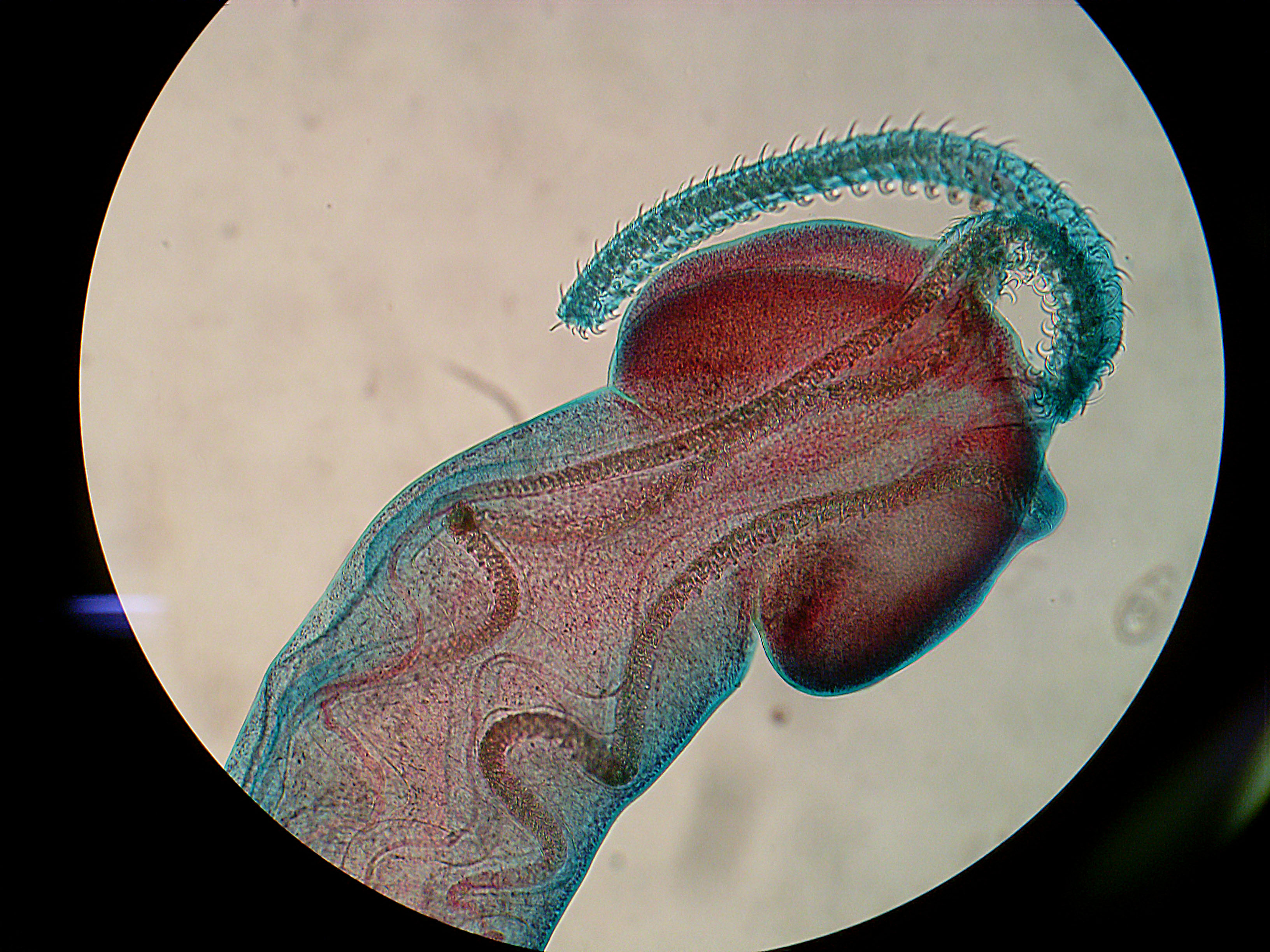
Photo by Caroline Wohlfeil
There were a number of interesting talks from the wildlife session, first up was a talk by Caroline Wohlfeil - a student from Michael Bull's lab. She gave a talk on sleepy lizards (see right) and the reptile tick, Bothriocroton hydrosauri. These ticks go through 3 stages in their life-cycle, alternating between feeding on a lizard and dropping off in a sheltered area to develop once they are fully engorged. It is in this latter stage that there ticks are transmitted - when lizards take shelter at refuges that have previously been used by infected lizards, they pick up ticks that were dropped off from the previous lizard. Using GPS loggers which continuously recorded the lizard's activity and location, Caroline was able to use that data to work out how often each of the tracked lizard had opportunities for infection. Her network analysis revealed that lizards that are highly-connected also had higher tick loads.

This was followed with a talk by Luz Botero Gomez, a student at Murdoch University, on trypanosome infections in little marsupial call the Brushed-Tail Bettong or Woylie. We have previously covered trypanosomes in another marsupials on this blog, namely the koala, but as it turns out, there is a great diversity of Trypanosoma in native marsupials - most of it still unknown. Woylie are known to be infected with 3 species - T. cruzi (the species which causes Chagas disease), T. copemani, and an as yet unnamed clade of Trypanosoma. Some of those Trypanosoma species are also found in other Australian marsupials but only the woylie is known to carry all three. Much like the koala-infecting trypanosome, T. copemani seems to only cause problem when it occurs in mixed infection with other Trypanosoma species - such co-infections can leads to inflammations and lesions in the tissue. In addition, these different trypanosomes also seem to have varying degrees of tissue specificity, with some species occurring in the blood, while other in muscle tissues, but overall mixed infections are more likely to occur in organs and muscles. Given the Woylie is currently critically endangered, it is very important to know what kind of diseases are induced by these trypanosomes and how it is affected by whether they are single or mixed infections.

Photo from Wikipedia by Helenabella
For a change of pace from parasites threatening a critically endangered mammal, Amanda Ash (also from Murdoch University) presented a talk call "Parasite: embrace not erase" which praised the important functional roles played by parasites in various ecosystems, and discussed the results of a study she conducted looking at the inestinal parasites of African Painted Dogs. She collected fecal samples from captive and wild Painted Dogs and compared the types of parasite eggs and cysts found in those sample. She found that the intestinal fauna of captive dogs was comparatively depauperated, populated only by Giardia whereas the wild dogs had a more eclectic mix of tapeworms, hookworms, and various protozoan parasites - in addition Giardia. Another stark contrast between the captive and wild dogs is that whereas parasitic infection was ubiquitous in the wild population, with 99% had some sort of parasite, only 15% of the captive dogs carried intestinal parasites of some sort.

This has enormous implications for conservation measures such as captive breeding programs - animals which have not been exposed to a wide range of parasites and pathogens can grow up to become immunologically naive so that when they are release into the wild, they may not be able to cope with the wide range of parasites they encounter. In addition, it is unknown what other physiological side-effects may result from lack of exposure to parasites. According to the hygiene hypothesis, the numerous types of allergies and auto-immune diseases which afflict some of us living in western societies have result from the lack of exposure to parasitic worms which are masters at manipulating and modulating our immune system. By limiting both the prevalence and variety of parasitic infections in those captive African Painted Dogs, are we consigning them to the same fate?

During the poster presentation, we saw some students who have come up with creative ways of presenting a 2 min talk - a student from James Cook University read a poem about whether wild dingoes pose a threat to the health of Indigenous communities in Queensland, while a student from University of Western Australia was literally singing the praises of using volatile chemicals for malaria parasite detection. Some of the most fascinating poster talks may present nightmarish scenarios to some people, but for different reasons.

Photo by Kate Hutson
For non-parasitologists, the tongue-biter seems like a one-off freak of nature. But in fact there are actually many species of tongue-biting isopods and other parasitic crustaceans which inhabit the mouth, gills, and branchial cavity of fish. One genus of tongue-biter isopod - Ceratothoa - encompasses 29 known species worldwide, 6 of which are found in Australia waters. However, a new study by Melissa Martin from University of Tasmania revealed that there at least 12 species of Ceratothoa (from 7 families of fish), and 4 of them are new to science.

Dinh Hoai Truong, a student from the Hutson lab at James Cook University presented a horror of a different kind - less visceral than having a parasite in your mouth, but more of a biosecurity nightmare to aquaculturists. He presented a poster on Neobenedenia - a hermaphroditic monogenean which infects the skin of Barramundi. His experiment showed that a single Neobenedenia is able produce eggs through self-fertilisation for consecutive generations without suffering any deleterious effects of inbreeding - each consecutive generation of inbred Neobenedenia are just as infective as the last. This means that even a single worm can start an entire sustained infestation at a fish farm. Unlike the widespread monogenean Gyrodactylus - a notorious aquacuture pest which has the viviparous "Russian Doll"-style "worm-within-a-worm" reproductive set-up (which allows them to swarm a fish like aphids on a rose bush) - Neobenedenia does what most monogeneans do and simply produce eggs. However because they are able to self-fertilise and have very short generation time, they can still become a serious pest to aquaculture.

In the next and final post on #ASP2012 (Australia), we will talk about how environmental factors can affect the generation time of Neobenedenia, and meet many other weird and wonderful marine parasites.

Next post: Swimming with the Parasites

July 11, 2012

Special Report: #ASP2012 (Australia) Part II: Parasites Gone Wild!

 This is Part 2 of my special report on #ASP2012 (Australia) - for part 1 see here.

The presentation on DFTD as the "perfect parasite" was followed with a talk by Andrew Thompson who holds a Chair in Parasitology at Murdoch University. He talked about how the presence of humans and our activities have often exposed wildlife to various infectious diseases. Wildlife are usually seen as a source of potentially harmful infectious diseases, and are often treated as the "bad guys" when it comes to pathogens. But in fact, sometimes wildlife have more to fear from us, and they act as sentinels, sinks, and sufferers of emerging infectious diseases which had been brought about through human action.

Tapeworm cysts in wallaby lung (photo from here)
For example, since the introduction of dogs and domestic livestock, macropods such as kangaroos and wallaby have become host to cysts of hydatid tapeworms (which Carl Zimmer wrote a post on about a month ago and was among the first batch of parasites to be featured on this blog). Hydatid infections in macropods come from eggs which are deposited in the environment by farm dogs which have the adult tapeworms living in their intestine. The dogs themselves acquire the worm from feeding on offal of infected livestock. Thus our canine companions are acting as the conduit for hydatid to jump from livestocks (one of their original hosts) to the likes of Skippy.

Bandicoot photo by JJ Harrison
from the Wikipedia
But hydatids are not the only introduced parasites which is afflicting Australia's marsupial fauna. Everyone's favourite cat parasite - Toxoplasma gondii - can also end up infecting the brain of bandicoots. However, this did not result from bandicoots coming into in close contact with cats (or rather, their feces). Instead, being such cute little mammals, people often leave food out for bandicoots in their backyard, and encourage them to enter into urban environments where they are more likely to be exposed to infective cysts.

In addition to introduced parasites, Australia's marsupials are home to all manner of little known vector-borne infections. There are multiple species of trypanosomes found in native marsupials (which I will discuss in more details in the next post), but there is very little information on their ecology and vectors. The vector for Trypanosoma cruzi (which causes Chagas disease) is a triatomine bug, but next to nothing is known about the Australian species and their potential role in vectoring those parasites. Thompson also discussed what appears to be a unique species of Leishmania in red kangaroos - which is transmitted via a midge (photo below right) instead of sandflies like other Leishmania.
Photo from this paper

Speaking of little known parasite fauna of Australian animals, Prof. Ian Beveridge - who is an absolute goldmine of knowledge on parasite biodiversity - gave a talk on that very topic. A fact he presented during his talk, which got retweeted a few times during the livestream, was that the average kangaroo is carrying 60000 nematode worms inside them. What I did not tweet at the time was that some individual kangaroos can be infected with up to half a million nematodes. Beveridge estimated that there are about 300 species of nematodes found in macropods. Traditionally horses and other equine are considered as particular "wormy" hosts (Sorry about that Bronies, but your Ponies be loaded with Wormies), harbouring a great diversity of nematode parasites - something which was remarked upon by Hippocrates. However, Beveridge estimated that macropods may in fact be equally "wormy" if not more so - there are so many nematode species which are yet to be found and described, and many host species which have not been properly examined for parasites (Beveridge mentioned road kills as an opportunistic sources of parasite samples - something which I have done on occasions.)

Even with the worms that are already known, it could be that they are even more diverse than we initially expected. Beveridge talked about a case where nematodes from rock wallabies which have previously been classified as 3 species (based on their morphological features) were later revealed by DNA analyses to be composed of 15 distinct genetic lineages (we have previously posted about cryptic species complex on this blog here and here).

And just bring it full circle and refer back to the previous post - the Tassie Devil is host to some unique parasites itself. Out of the two species of flukes, two species of tapeworms, and three species of nematode that it hosts, one of the flukes and two of the tapeworms are unique to the Tassie Devil and found in no other animals. If we lose the devils thanks to DFTD, we will lose those one-of-a-kind parasites too. Sometimes parasite extinctions can be brought about through the best of intentions (see the case of the Californian Condor) - when the devils are brought in for captive breeding or as an "insurance population", the vets treat them for parasites - so good-bye special worms! However, Dasyurotaenia robusta - a species of tapeworm unique to the Tassie Devil - is actually covered by the Threatened Species Protection Act in Tasmania.

Save the devil, save the D. robusta!

Coming up in the next part: Sleepy Lizards, Painted Dogs.