"So, naturalists observe, a flea has smaller fleas that on him prey; and these have smaller still to bite ’em; and so proceed ad infinitum."
- Jonathan Swift
Showing posts with label blood feeder. Show all posts

May 10, 2025

Rocinela sp.

The bonefish is a popular recreational species for catch-and-release fishing. It is targeted by anglers using fly rods or light tackle, and requires some skills to do so as they're easily startled, and once hooked can put up quite a struggle. But if you are wading on a beach while fly fishing for bonefish, you might in turn become the target, because one of the bonefish's parasites may have its eyes on you too.

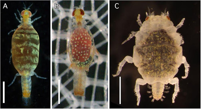
Left: Rocinela isopod feeding on a bonefish just above its right eye, Centre: Rocinela isopods on bonefish at the base of the dorsal fin and left flank, Right: Rocinela isopod dorsal view.
Photos from Figure 2 of the paper.

This blog has previously featured Cymothoidae isopods, which tend to be somewhat picky about what types of fish they parasitised But the isopod being featured in today's post isn't picky at all, in fact,  when it comes to its next meal, and it doesn't always have to be a fish. Rocinela is a genus of isopods that belongs to the Aegidae family, and unlike the cymothoids which tend to stay on their hosts for extended periods of time, these isopods are temporary blood feeders, rather like land-dwelling leeches or bed bugs. On rare occasions, they can even feed on human blood. But adopting this kind of free-wheeling blood-sucking can open yourself up to becoming an unwitting carrier of many microscopic passengers.

The study we're looking at in this post investigated the health and microbes of bonefish at Belize. The scientists in this study captured bonefish around Ambergris Caye, and examined each fish for scars and ectoparasites (such as Rocinela), then collected some blood samples for genetic analyses. The scientists also analysed the blood present in the gut of the isopods they collected, to identify what kind of fish they had been feeding on. Genetic analyses of blood-suckers' meals have previously provided valuable insights into the hosts of ectoparasites.

Two of the three sites the scientists sampled from were frequented by Rocinela, and about 70 percent of the isopods they found on the bonefish had plump bellies that were full of blood. As expected, most of the isopods were filled with bonefish blood, but one of the Rocinela also had blood from a type of small killifish called the mangrove rivulus, and somewhat alarmingly, there was an isopod in the sample which had fed on human blood at some point.

What's even more interesting were the plethora of virus sequences that were found. Possibly because of its indiscriminate feeding habits, Rocinela has inadvertently picked up about 11 different types of viruses. Most of those were viruses that usually infect arthropods. One of them, XKRV-2, is related to a group of viruses which have been previously reported from a range of crustaceans, including parasitic isopods, so its presence was to be expected. 

But one of the Rocinela also carried a less expected virus called XKRV-1, which is more related to a common genus of fish virus called Aquareovirus. None of the bonefish sampled had XKRV-1 in their blood, which means Rocinela has picked up the virus from one of other fish species that it had fed on. And rather than just being a transient, XKRV-1 has been persisting in the isopod's belly for a while - which is a common adaptation for vector-borne viruses such as those found in ticks and mosquitoes.

Given Rocinela can feed from a variety of fish, its payload of viruses may disembark into one of its hosts during feeding, so it could be transferring viruses between different species at sea. While Rocinela is also known to feed on humans, the likelihood of those fish viruses jumping into us is comparatively low - viruses that jump into humans tend to come from mammals and other warm-blooded animals, especially those that are evolutionarily closer to us, such as non-human primates. But a much bigger concern is that since Rocinela harbours so many different viruses and it is so indiscriminate about the type of hosts that it feeds on, it might end up acting like a transmission hub for viruses to jump from wild fish into aquaculture species.

Most studies looking at vector-transmitted diseases focus on land-dwelling arthropods such as ticks, fleas, and mosquitoes, but crustaceans like Rocinela and other parasitic isopods might be overlooked vectors that are providing a taxi service for pathogens under the waves.

Reference:

November 16, 2022

Acanthobdella peledina

In the cold rivers and lakes of the arctic and subarctic region, there live some rather peculiar worms with a face full of tiny hooks and an appetite for blood. These worms live as ectoparasites of fish, and they belong to a group called Acanthobdellida - relatives of leeches that seem to have gone down their own evolutionary path. These worms have also been called "hook-faced fish worms" and the entire group consists of only two known species - Acanthobdella peledina and Paracanthobdella livanowi

Top: Acanthobdella peledina on a grayling.
Bottom: Scanning electron micrograph of the whole worm (left) and close-up of the anterior body region (right).
Photos from Figure 1, 6, and 7 of the paper

Their mouthpart has been described as being a less sophisticated version of a leech's mouthpart - they lack the saw-edged jaws or the extensible proboscis found in many leeches, nor do they have the muscular sucker which surrounds the mouth. Instead, they have a protrusible pharynx and a series of hooks on the first five segments of the body, which they use to attach themselves to their fishy hosts. 

They have previously been considered to be a "missing link" between leeches and the rest of the Clitellata - the group of segmented worms that also includes earthworms and tubifex worms - as they have certain features which are commonly found in other clitellate worms but are absent in leeches. This includes having tiny bristles (called chaeta) on their segments, and a reproductive system similar to those found in earthworms.

Acanthobdella peledina is found all across the subarctic, where they range from being relatively rare to being found on over two-thirds of the fish at a given location. Given it is so widely distributed, with populations scattered across different geographical locales, could each of those distinct populations actually be different species? A group of researchers set out to determine whether there are actually more species of these hook-faced worms than meets the eye. Furthermore, they also wanted to find out how closely related Acanthobdella and Paracanthobdella are to each other. They did so by comparing museum specimens of hook-faced worms which have been collected from sites across the subarctic, including Norway, Sweden, Finland, Alaska, and Russia. 

Aside from examining their anatomical features, the researchers also compared five different key marker genes from these worms. Some of those DNA segments came from the mitochondria, others from the cell's nucleus. The reason for comparing multiple genes is that each has their own histories, and may offer different perspectives on the organism's evolutionary history. It is like interviewing different witnesses at a crime scene.  Unfortunately, for whatever reasons, the DNA of these worms proved to be particularly challenging to amplify and sequence, so for most specimens they were only able to sequence up to four of the five genetic markers they were aiming for, with some specimens only yielding sequences for two of the genes. Despite that limitation, the researchers were able to use the sequences they obtained to resolve the hook-faced worm's evolutionary history.

Despite their wide distribution across the arctic and subarctic regions, Acanthobdella peledina does appear to be a single, widespread species. While the Alaskan population of worms are genetically distinct from the Nordic population, they are not dissimilar enough for them to be considered as separate species. Furthermore, based on their analysis, the two living species of hook-faced worms are quite closely related to each other. In fact, it seems they had only diverged from each other just prior to the last ice age. So, far from being some kind of "missing link" between leeches and other clitellate worms, these hook-faced worms belong to their own distinct group.

But while the two living species had shared a common history until relatively recently, the hook-faced worms as a group had evolutionarily split off from the leeches a long time ago. Based on available data on these worms, this might have occurred during the early Cenozoic as the ancestors of the hook-faced worms became specialised on arctic freshwater fishes that arose during that era, such as salmonids.

So it might have been the pursuit of salmonids that had sent these worms down their own distinct path - a story which is probably relatable to any fly fishers out there.

Reference:
de Carle, D. B., Gajda, Ł., Bielecki, A., Cios, S., Cichocka, J. M., Golden, H. E., Gryska, A. D., Sokolov, S., Shedko, M. B., Knudsen, R., Utevsky, S., Świątek, P. & Tessler, M. (2022). Recent evolution of ancient Arctic leech relatives: systematics of Acanthobdellida. Zoological Journal of the Linnean Society 196: 149-168.

August 14, 2022

Cyclocotyla bellones

At the top of this blog, there is a quote by Jonathan Swift about how fleas have smaller fleas that bite them. Indeed, parasites becoming host to other types of parasites is actually a rather common phenomenon in the natural world. Those who would parasitise the parasites are called "hyperparasites".

Left: Cyclocotyla bellones on the back of a Ceratothoa isopod, Right: C. bellones coloured red with Carmine staining.
Photos from Figure 1 and 5 of the paper.

The parasite featured in this post was once suspected of being a hyperparasite. Cyclocotyla bellones is a species of monogenean - it belongs to a diverse group of parasitic flatworms that mostly live on the body of fish, parasitising the fins, skins, and gills of their hosts. But unlike other monogeneans, C. bellones does not attach itself to any part of a fish's body, instead it prefers to stick its suckers onto the carapace of parasitic isopods, such Ceratothoa - the infamous tongue biter. Since Ceratothoa is itself a fish parasite, and C. bellones is routinely found attached to those tongue-biters, this has led some to think that it might be a hyperparasite of those parasitic crustaceans.

But it takes more than simply sticking yourself onto another organism to be considered as a parasite of it. After all, there are algae that grow on the body of various aquatic creatures, or barnacles that are found on the backs of large marine animals like whales and turtles. But those are not considered as parasites as they don't treat their host as a food source, merely as a sturdy surface they can cling to - they're known as epibionts.

So strictly speaking, for Cyclocotyla to be a parasite of the isopod, it needs to be feeding on or obtaining its nutrient directly from its isopod mount. When scientists examine the bodies of the tongue-biters with C. bellones on them, they seem to be pretty unscathed. There aren't any scratches or holes on the isopod's body which you'd expect if C. bellones had been feeding on it. Indeed, the monogenean's mouthpart seems ill-suited for scraping through the isopod's carapace.

Additionally, C. bellones' gut is filled with some kind of dark substance similar to those found in other, related monogenean species. This is most likely digested blood from the fish, which the monogenean has either sucked directly from the fish's gills, or indirectly via the feeding action of its isopod mount. Let's not forget that the isopod itself is a fish parasite that feeds on its host's blood, so if it gets a bit messy during mealtime, perhaps Cyclocotyla is there to suck up any spilled blood. Or it might be doing a bit of both.

The researcher noted that Cyclocotyla is not alone in its habit of riding isopods. Other monogeneans in its family (Diclidophoridae) have also been recorded as attaching to parasitic isopods of fish. And aside from riding isopods, they all share one thing in common - a long, stretchy forebody, looking somewhat like the neck of sauropod dinosaurs. Much like how the neck of those dinosaurs allowed them to browse vegetation from a wide area, the long forebody of Cyclocotyla allows it to graze on the fish's gills while sitting high on the back of an isopod. So fish blood is what C. bellone is really after - the isopod is merely a convenient platform for it to sit on.

But why should these monogeneans even ride on an isopod in the first place? Cyclocotyla and others like it have perfectly good sets of suckers for clinging to a fish's gills. Indeed, there are other similarly-equipped monogeneans that live just fine as fish ectoparasites without doing so from the back of an isopod. Well, that's because the fish themselves don't take too kindly to the monogeneans' presence. These flatworms are constantly under attack from the fish's immune system, which bombards them with all kinds of enzymes, antibodies, and immune cells. By avoiding direct contact with the fish's tissue, Cyclocotyla and other isopod-riders can avoid being ravaged by the host's immune system - which is something that other monogeneans have to deal with on a constant basis.

So it seems that Cyclocotyla and other isopod-riding monogeneans are no hyperparasites - they're all just regular fish parasites that happen to prefer doing so while sitting on the backs of isopods. Cyclocotyla bellones prefers to share in the feast of fish blood with its isopod mount, while sitting high above the wrath of the host's immune response.

Reference:

March 17, 2022

Thaumastognathia bicorniger

Gnathiidae is a family of parasitic isopods that can be considered as ticks of the sea. I make that comparison not only because gnathiids are blood-feeding arthropods, but like ticks, their life cycle involves going through a series of feeding and non-feeding stages. The blood-hungry fish-seeking stage is called a zuphea that, much like how a tick would on land, attaches itself onto passing fish and starts feeding to its heart's content. Once it is fully engorged with a belly full of blood, it becomes what's called a praniza, which drops off the fish to grow and moult into its next stage. Gnathiid isopods need to go through alternating between the zuphea and the praniza stage at least three consecutive times before they can reach full maturity.

Thaumastognathia bicorniger stripe (left) and spots (centre) pigemented third stage praniza, and adult male (right)
From Fig. 2. of the paper

The paper featured today is about Thaumastognathia bicorniger, a gnathiid isopod which has recently been described from the waters of Japan. The researchers who described this species found the isopod on various chimaera and sharks that were caught by fishing vessels operating in the waters of Suruga Bay and around Kumejima Island. Additionally, they were also able to obtain previously collected specimens of this isopod that had been stored at the laboratory of fish pathology at Nihon University. Those specimens were originally collected from various different cartilaginous fishes that were caught by fishing vessels off the southern coast of central Japan.

Based on their samples, this isopod has been recorded to feast on the blood of at least ten different species of cartilaginous fishes including nine species of sharks from six different families, along with one species of chimaera (also known as ratfish, in this case the Silver Chimaera). Thaumastognathia bicorniger larvae were always found in the gill chamber of their hosts, where they attached themselves to the blood-rich gill filaments. These isopods are tiny, with the third stage praniza larva measuring about 3.7-4.8 mm long, so having one or two of them would merely pose a minor inconvenience to the host. 

However, some sharks were found to be infected with dozens or even hundreds of those tiny blood-suckers. Of those, the Blotchy Swellshark (Cephaloscyllium umbratile), the Shortspine Spurdog (Squalus mitsukurii), and the Starspotted smooth-hound (Mustelus manazo) appeared to be among this gnathiid's favourite hosts, as they were commonly found to be infected with at least 50 T. bicorniger larvae and some even harboured hundreds of those blood-sucking isopods in their gill chambers. Additionally, much like how ticks are known to carry various pathogens, gnathiid isopods have also been implicated in the transmission of blood-borne parasites in coral reef fishes.

The juvenile stages of T. bicorniger seem to come in two different colour patterns - spotty and stripey. This was only visible in the live or freshly caught specimens as the colour faded rapidly when they are preserved in ethanol. Genetic analysis revealed that despite their superficial differences, those two colour morphs belong to the same species, and it is unclear whether the different colour patterns signify anything, as they're not associated with a particular haplotype, sex, nor host species.

The researchers kept some of the gnathiid larvae alive in captivity to see if any of them would metamorphose into an adult stage - but only one successfully moulted into an adult male. Among gnathiid isopods, there is a high degree of sexual dimorphism - the male gnathiids have squat body with big mandibles, while in contrast, female gnathiid have a larger rotund body for brooding eggs into larvae. Neither of which look anything like a "typical" isopod like a woodlouse or even the infamous tongue-biter parasite and its cymothoid relatives.

For other species of gnathiid isopods, metamorphosing from the third-stage praniza into a mature adult is a relatively brief process. After their last feeding session, some species would take just a week or two to mature into a reproductive adult, while others may take up to two months at most. However, T. bicorniger took a whooping 204 days to moult from a third stage praniza into an adult. So why does T. bicorniger take so long to mature compared with other species of gnathiid isopods?

Gnathiid metabolism and growth is greatly affected by water temperature, and many of the gnathiids that have very short development time are found in warmer, tropical waters. In this study researchers kept their T. bicorniger at 10-20°C in their lab, which is slightly cooler than the water temperature that those other known gnathiids are regularly exposed to. However, there is a species of Antarctic gnathiid - Gnathiia calva - which only took 6 weeks to transform into an adult despite living in waters that were kept at 0 to -1°C.

Alternatively it might have something to do with the fishes that they were feeding on. Many sharks have high levels of urea in their blood, which may make their blood more difficult to digest for any would-be blood-suckers. Lamprey that feed on basking sharks are specially adapted to excrete large volumes of urea which is found in their host's blood. The need to detoxify your food would most likely complicate the digestion process, decrease the blood's nutritional value, which would result in cost to development time. But then again there is another gnathiid species - Gnathia trimaculata - which infects Blacktip reef shark (Carcharinus melanopterus) and it only takes 6 (for males) or 24 days (for female) to moult into an adult.

So for now, the reason(s) why T. bicorniger seems to take such a long time to grow into an adult compared with other species of gnathiid isopods, remains a unsolved mystery.

Reference:
Ota, Y., Kurashima, A., & Horie, T. (2022). First Record of Elasmobranch Hosts for the Gnathiid Isopod Crustacean Thaumastognathia: Description of Thaumastognathia bicorniger sp. nov. Zoological Science, 39: 124-139

July 14, 2021

Echinophthirius horridus

Lice are common parasites of mammals. Humans alone are host to three different species of lice, and it's not just humans or land mammals that can get infected with lice. Pinnipeds - seals and sea lions - also have to contend with being covered in those ectoparasites. Unlike many other ectoparasites in the sea which have been bestowed with the name of "lice" such as salmon lice, tongue-biter lice, or whale lice (all of which are crustaceans), seal lice are true lice, in that they are parasitic insects belonging to the order called Phthiraptera.

Left: An adult seal louse, Right: two opened seal lice eggs (nits) glued to a strand of seal fur hair
From Fig. 1 of the paper

When the ancestors of modern pinnipeds took to the sea some time in the Oligocene about 30 million years ago, the lice followed them into the water, and in the process, they have to deal with all the challenges associated with living on an aquatic host. Seal lice belong to a family of lice called the Echinophthiridae and they have some specialised adaptations for living on hosts that spend most of their time immersed in sea water. This include elongated spiracles (the opening insects use to breathe) with mechanism for closing, a dense covering of spines and scales, and stout clamp-like claws that allow them to grip tightly onto their hosts' fur.

Blood-sucking arthropods such as ticks, fleas, and lice are often responsible for transmitting a wide variety of parasites and pathogens. And it seems that seal lice can also play a similar role in the sea. While performing routine diagnostics on 54 harbour seals and a very heavily-infected grey seal pup that were hospitalised at the Sealcentre Piteterburn (a seal rehabilitation and research centre in Netherlands), a group of scientists were able to use that opportunity to collect a massive number of seal lice from those marine mammals. They ended up collecting 200 lice from the harbour seals, and another 1000 from that one very heavily infested seal pup.

Those researchers divided the lice into batches of 1-20 lice, based on the individual host that they came from (the lice from the heavily infected seal pups were divided into multiple batches of 15 lice), then grind them up, and examined the lice slurry by subjecting it to polymerase chain reactions that amplified the DNA of known seal parasites and pathogens.

The DNA analyses showed that the seal heartworm (Acanthocheilonema spirocauda) was the most commonly found parasite, with it being detected in about one-third of the lice samples. While most people would associate "heartworm" with the dog heartworm (Dirofilaria immitis), that species is just one out of many different filarial roundworms that live in the heart of mammals. The findings of this study corroborates with previously published research which have found heartworm larvae dwelling in the gut of seal lice, demonstrating that these ectoparasitic insects play a key role in the transmission and life cycle of these nematodes.

Alongside the heartworm, there were also some bacterial pathogens lurking in those lice. Some of the lice from the grey seal pup were also carrying Anaplasma phagocytophilum, the bacteria which causes tick-borne fever and as their name indicates, are usually carried by ticks. Additionally, a few of the lice from that seal pup and some of the harbour seals were also carrying a species of Mycoplasma bacteria. This microbe is commonly found in seals and other marine mammals, but when it gets transmitted to humans, it is also associated with a disease known as "seal fingers". However, unlike the heartworm, it is unclear if the lice actually play a role in the transmission of these bacterial pathogens, or if they were incidental infections that simply came with living on a seal host.

It is worth noting that while pinnipeds had retained an heirloom of their terrestrial ancestry in the form of lice, another group of marine mammals - the whales - have acquired their own unique suite of ectoparasites which are unlike that of any other mammals. They have "whale lice" which are actually crustaceans in the same group as sandhoppers, along with pennellid copepods - a family of parasitic copepods that usually infect fish, with the exception of one species which has evolved to parasitise whales.

So why are there no "true lice" on whales? Well, for all their adeptness at clinging to their host, lice ultimately depend on the presence of hair or similar structures to hang onto their host. When a seal dives underwater, the layer of fur forms a covering that the lice can shelter underneath. But no such shelter exists on the smooth, hair-free surface of a whale. As a result, while whales have escaped the lice (and have picked up other parasites in the process), pinnipeds have kept their fur, and along with it, their lice and the worms that they carry.

Reference:

April 21, 2021

Pterobdellina vernadskyi

If you've ever been out hiking in the wilderness, you would know that there is no shortage of tiny animals out there that love nothing more than to feast on your blood. They range from mosquitoes to midges, from fleas to ticks, and of course, let's not forget about leeches - a group of animals so synonymous with blood-sucking that its name is also used as a term for exploiting the life blood of others.

But leeches aren't just found out in the bushes, there are hundreds of species of blood-sucking leeches that are actually aquatic, feeding mostly on amphibians and fishes. In fact, spare a thought for the fishes, which have a whole family of jawless leeches called Piscicolidae that are after their blood.

Left (top) Antarctic toothfish with P. vernadskyi leeches on its body surface, and (bottom) in its mouth. Photos by Gennadiy Shandikov from Fig. 1 of the paper.
Right: Two live specimens of P. vernadskyi (the left leech has a spermatophore in its clitellum) from Fig. 2 of the paper
 
For fish, there is no escape from these leeches as they are found in a wide range of aquatic habitats, ranging from freshwater to the marine environment. They target a wide range of hosts, from trout and carp, to rays and sharks. Even in the inky depths of the deep sea, there are hungry leeches waiting for a tasty fish to swim by, and it is one of these deep sea leeches that is featured in today's post.

This post is about a newly described species of fish leech - Pterobdellina vernadskyi - which has been found parasitising the Antarctic toothfish (Dissostichus mawsoni) in the cold dark waters of Antarctica. The researchers who described this species collected them from fish that were caught by longline commercial fishing vessels - Antarctic toothfish are highly valued on the commercial market, where they are often sold alongside the Patagonian toothfish as "Chilean Sea Bass".

While most of the fish that the researchers encountered only had one or two leeches, some were afflicted with ten or more, and one unlucky fish was covered in twenty eight leeches all over its body. They tend to favour attaching to either the dorsal surface of the fish, or inside the mouth, where they are more sheltered. And P. vernadskyi can grow rather large compared to other fish leeches, reaching about 8 cm in length - so roughly the size of your finger.

Aside from its sheer size, another thing that differentiates it from other leeches are series of distinct, zig-zag ridges along its back and fin-like projections along its sides. It is not entirely clear what purpose those structures serve for the leech, though there are other deep sea ectoparasites which also have some unusual external structures. The researchers suggested that perhaps they serve some kind of sensory function that allows to leech to find their host, or they might be adaptations to the low oxygen levels of its environment, increasing the leech's surface area so it can absorb more oxygen from the surrounding waters.

In additional to those external features, it is worth mentioning that this leech's host, the Antarctic toothfish, is notable for producing antifreeze glycoproteins in its blood, which allows it to dwell in such frigid waters. But this additive would surely have some implications for the digestive system and physiology of P. vernadskyi compared with other fish leech that feed on hosts with more conventional blood.

Since the Antarctic toothfish can be found dwelling as deep as 2600 m below sea level, this would make P. vernadskyi the deepest Antarctican leech that has ever been recorded. However, it is NOT the deepest depths that a leech has ever ventured. That title goes to Johanssonia extrema which has been found in the hadal zone over 8700 m below sea level in the Kuril–Kamchatka Trench, where the waters are still and the pressures are crushingly immense.

Pterobdellina vernadskyi is just one out of two dozen different species of fish leeches that have been recorded from Antarctica, and there are a number of other leeches which have been reported from deep sea habitats. It would be safe to say that P. venadskyi, and other marine leeches that have been described in the scientific literature, represents only the tip of the iceberg. Where there are fish, there are leeches.

Reference:
Utevsky, А., Solod, R., & Utevsky, S. (2021). A new deep-sea fish leech of the bipolar genus Pterobdellina stat. rev.(Hirudinea: Piscicolidae) parasitic on the Antarctic toothfish Dissostichus mawsoni (Perciformes: Nototheniidae). Marine Biodiversity 51: 15.

January 17, 2020

Ceratophyllus (Emmareus) fionnus

When it comes to conservation and protecting threatened species, fleas would not usually be high on most people's list. Not only because most people are not fans of parasites, but also insects and just invertebrates in general gets little attention compared with charismatic megafauna, which attracts far more conservation resources. Additionally, there are comparatively less scientific research being conducted on invertebrates compared with vertebrate animals. So less is known about them, despite 99% of all animal life on Earth being invertebrates, and at least one fifth of them are under threat from extinction.
Adult Ceratophyllus (Emmareus) fionnus [insert: a Manx shearwater in flight]
Photos from Fig. 1 and 2 of the paper
Which brings us to the topic of the paper we are discussing in this post - a flea. But we're not just talking about any flea, we're talking about Ceratophyllus (Emmareus) fionnus which parasitises the Manx Shearwater (Puffinus puffinus). Like many other birds the Manx Shearwater is host to a wide range of parasites, both external and internal, but what makes C. (E.) fionnus special is that even though the Manx shearwater has a wide distribution across both the north and southern Atlantic ocean, this little flea seems to be found exclusively on an island off the coast of Scotland called the Isle of Rùm - and nowhere else. This alone earns it the distinction of being one of the few species of endemic Scottish insects.

The life cycle of fleas involves a non-parasitic larval stage that feeds on organic detritus in the surrounding environment. Only when the worm-shape larva pupates and emerges as an adult does it begin its vampiric life style. The Manx shearwater spend most of its life out at sea and only visits the Isle of Rùm to breed, and based on the life cycle of other seabird fleasC (E.) fionnus would breed in the nest and bedding. So when their hosts leave, the fleas stay and overwinter as pupal cocoons near the nests, and when spring comes, the blood-hungry adults emerge, eagerly awaiting the return of their hosts. While this arrangement seems to have worked well for C. (E) fionus, being restricted to a single island also makes it rather vulnerable to becoming extinct due to environmental changes.

There have been other cases of bird ectoparasites which have gone extinct in the relatively recent past due to various different reasons. The Huia louse, which only lived on the New Zealand bird Huia, is thought to have become extinct along with its host in early 20th century. And then there was the Californian condor louse - a species which was ironically (and unnecessarily) rendered extinct in an effort to conserve another (its host) during the Californian condor breeding program.

Those are just the cases that are better known - it can be safely assumed that throughout recent history, the extinction of many bird species around the world have been accompanied by an unnoticed wave of parasite co-extinctions. So how would one go about coming up a plan for conserving a species of flea? In a recently published paper, a group of researchers outlined a potential roadmap for protecting C. (E.) fionnus.

Like most invertebrates, there isn't much information on some of the most basic aspects of C. (E.) fionnus' biology, including their distribution and population level, so to start out with, we need to learn more about this flea species. But the usual methods for sampling and identifying insects and parasites will not be suitable since they often result in the death of the animal in question. So the researchers suggested that surveys of C. (E.) fionnus should use non-lethal methods for immobilising the fleas such chilling or carbon dioxide so that they can be identified using a field microscope.

While the Manx shearwater colony has been fairly stable on the Isle of Rùm, in more recent times their nest have come attack from introduced brown rats - and obviously if the shearwater colony disappear from the island, so will C. (E.) fionnus. So what can be done to safeguard a viable population of a flea species? Unlike other threatened animal species, captive breeding is not really an option for C. (E.) fionnus - raising a parasite species in captivity implicitly involves keeping its hosts in captivity and when the host in question is a migratory seabird, that's out of the question.

So the researchers suggested creating "insurance" populations of C. (E.) fionnus on some of the other Manx shearwater colonies within the British Isles. They nominated six potential sites to translocate founding populations. Translocation is a common strategy for conservation of vulnerable or endangered species. But this hasn't really been done before for parasites, so any such effort would require ongoing monitoring of both the host and parasite population to see if the translocation has been successful, or what effects this might have on the host population.

Aside from conserving parasites simply out of principle, there is also a more host-centric reason for protecting them. Exposure to parasites during early stages of the shearwater's life might be a vital step for them to develop a fully functioning immune system. So those fleas waiting in the nests could be giving the shearwater chicks a needed boost to their immune system early in life that allows them to survive into adulthood.

As mentioned above, there are other parasites that have already been driven to extinction right under our noses. The paper discussed in this post is one of the first to develop a conservation plan for a specific parasite species. Every single species of parasites are unique in their host preferences, life cycles, and distribution, so there won't be a one-size-fits-all plan that can possibly be applicable to all parasitic organisms. Especially when one considers the term "parasite" encompasses countless different phyla of animals, fungi, plants, and single-celled organisms.

Parasites are an integral part of biodiversity, and many of them are facing extinction in the foreseeable future. They deserve to be the target of conservation efforts just as much any other species. If our goal is to protect and conserve "wildlife", we shouldn't forget about the numerous wildlife which are small and hidden from plain sight.

Reference:
Kwak, M. L., Heath, A. C., & Palma, R. L. (2019). Saving the Manx Shearwater Flea Ceratophyllus (Emmareus) fionnus (Insecta: Siphonaptera): The Road to Developing a Recovery Plan for a Threatened Ectoparasite. Acta Parasitologica 64: 903-910.

November 12, 2019

Electrovermis zappum

Fish blood flukes are common parasites in the aquatic environment and many species have been described from all kinds of fish all over the world. However the full life cycle is only known for relatively few of such flukes, because while the adult parasite can be fairly common in the fish host population, the asexual stage living in the invertebrate host can be quite rare and difficult to find. The study featured in this blog post described the life cycle of Electrovermis zappum - a blood fluke that lives in the heart of the lesser electric ray, but spends part of its life cycle in a beach clam.

Left: An adult Electrovermis zappum, Right: the life cycle of E. zappum. From the Graphical Abstract of the paper
When it comes metamorphosis and transformation, most people usually think of caterpillars turning into butterflies, but such level of change pales in comparison to the different forms that digenean flukes take on at each stage of their life cycles. The adult E. zappum fluke is a long skinny worm about 1.5 mm long, living in the heart of an electric ray. Over half of its length is composed of reproductive organs, devoted to producing a steady stream of eggs. The eggs that manage to make their way out of the ray's body hatch into cilia-covered larvae called miracidia. This microscopic ciliated mote then infects a coquina clam.

It then undergoes another set of transformation as it enters the asexual stage of the life cycle. The lone miracidium turns itself into a clone army of self-propagating units call sporocysts which take over the clam's body. These sporocysts look like microscopic marbles, each measuring about one-tenth of a millimetre across, and packed within those translucent spheres are the next stage of the fluke's life cycle. Within each sporocyst are half a dozen skinny, tadpole-shaped larvae called cercariae - these develop and grow within the nurturing wall of the sporocysts until they are ready to be released into the water column, at which point the sporocyst will start growing the next batch of cercariae from its reserve of undifferentiated germinal cell balls.

A single infected clam can be filled with several hundred of those sporocysts, which occupy the space where the clam's gonads would have been, with some also spilling over into the digestive system. This process essentially turns the clam into a parasite factory that churns out thousands upon thousands of infective fluke larvae, saturating the surrounding waters. Both the bottom-dwelling electric ray and the coquina clam are found right next to each other in the swash zone of beach, so the cercariae are released right where the rays are likely to be.

Most of these short-lived, microscopic larvae will perish - eaten by other marine creatures or simply exhausting their energy reserves before encountering an electric ray. But enough of them will come into contact with an electric ray to continue the life cycle. When a cercaria comes into contact with a ray, it will discard its paddle-like tail, and burrow though the skin and into the blood vessels. It will then traverse the vast network of the fish's circulatory system until it finally settle within the heart's pulsating lumen, and start the cycle anew.

Because the asexual stage in the coquina clams allows E. zappum to continuously spam the water with waves of tiny baby flukes, this means it only takes a relatively small number infected clams for E. zappum to saturate the water with enough infective stages to maintain a viable population of the parasite in the ray hosts. Indeed, this was reflected in what the researchers found in this study - while the adult fluke was fairly common in the electric rays (fourteen of the fifty four rays the researchers examined were infected with adult E. zappum), infected beach clams were extremely rare - only SIX of 1174 clams that they examined at were infected.

On the beaches where these coquina clams and electric rays are found, each square metre of beach are densely packed with thousands of coquina clams. So looking for an infected clam amidst all that is like panning for gold - time-consuming and labour-intensive work which involves spending hours upon hours in front of a microscope with a bucket of shellfish. This is one of the reason why the full life cycle of so few of these flukes have been described.

Furthermore unlike most other digenean flukes that tend to infect mollusc (mostly snails) at their asexual stage - which narrows down the list of potential animals to examine, some fish blood flukes are known to infect some unusual invertebrates. While E. zappum is relatively conventional in that it still uses a mollusc for the asexual stage of its life cycle, there are some species which have really gone off the beaten evolutionary path and have evolved to infect polychaete worms.

Blood flukes have been reported from other species of rays in other parts of the world. Based on their DNA, the blood flukes that infect cartilaginous fish all belong to their own special evolutionary branch among the fish blood flukes, and that the common ancestor of all the living blood fluke lineages, including those that infect mammals and birds today, might have originated over 400 million years ago.

So long before there were dinosaurs, long before there were mammals, even before a lineage of fish began crawling onto land, and at around the same time as when the earliest iterations of sharks and ratfish were prowling the Silurian seas, the ancestors of these flukes were already going through their life cycles, and well-acquainted with the hearts of vertebrate animals.

Reference:
Warren, M. B., & Bullard, S. A. (2019). First elucidation of a blood fluke (Electrovermis zappum n. gen., n. sp.) life cycle including a chondrichthyan or bivalve. International Journal for Parasitology: Parasites and Wildlife 10: 170-183.

October 10, 2019

Dinobdella ferox

When it comes to parasitology, sometimes you have to get really up close with your study organism, as one researcher in Taiwan did in trying to figure out the behaviour of Dinobdella ferox - a species of leech that has a habit of getting into some uncomfortable (for its host) places.

Dinobdella belongs to a family of leeches call the Praobdellidae - unlike other leeches that simply latch onto their host's skin and start sucking, Dinobdella and most other praobdellid leeches attach themselves to and feed from the host's mucous membranes - which means they either crawl up the host's nose, or occasionally even up their urethra or anus. Because of their habit of hiding themselves in parts of the host where the sun doesn't shine, it is rather difficult to figure out just what exactly what they get up to when they are attached to the host (aside from sucking blood).

Top: a D. ferox leech poking out of Dr Lai's nose., Bottom: a D. ferox leech which has emerged after the infection period
From Fig. 1. of the paper
Dr Yi-Te Lai at National Taiwan University decided to put his body on the line in the name of science, and infected himself with some D. ferox leeches, diligently documenting his own health and the leeches' behaviour throughout entire duration. He conducted three trials, each time administering himself with a different D. ferox leech - and you can see him demonstrating his procedure for self-infection in this video.

During this period, in addition to documenting the leech's behaviour based on his first hand experience, Dr Lai also took regular trips to a local clinical laboratory to examine the leech via endoscopy, and take measurements of his red and white blood cell counts to see what effects the leech's feeding might have on his blood works.

Some of the symptoms he experienced during the leeches' residency were to be expected, including nasal congestion, mild stinging sensations and some nosebleeds. But despite the leech's feeding, he found that both his red and white blood cell count held steady during the infection period, and his body was able to compensate for the blood loss. Furthermore, despite their activities in the nasal passage, they can be remarkably camera shy and were pretty good at hiding from the endoscope.

And those leeches had a ravenous appetite - during the course of their stay (which can range from 24-75 days), they grew to five to ten times their original length, and increased their body mass by up to 380 times. The juvenile leech starts out as a tiny dark mote just 3-4 millimetres long, but by the end of their stay, they were big enough to be easily noticeable when they decide to poke their head out.

Cohabiting with a bunch of nose leeches allowed Dr Lai to make round-the-clock observations and record behaviours which might not have been previously documented. After about a month into the infection period, the leeches started getting restless and were looking for a new host, and this behaviour manifested itself in some disconcerting ways.

When D. ferox starts looking for a new host, it develops an attraction to darkness and water. According to Dr Lai's account, whenever he was in a dark place such as in the middle of watching a movie at a theatre, the leeches came poking out of his nose. But this wasn't the only time when they made their presence noticeable - they also got nosy when he went about some of his daily routines like showering or washing his face. This overlapped with the ceasing of bleeding-related symptoms - which meant the leeches had finished feeding.

With their cohabitation coming to an end, Dr Lai tested out some methods for removing such leeches which have been reported in the scientific literature. His self-experimentation showed that while the leech can be coaxed out with a bowl of water, this only worked at later stages of the infection, presumably after the leech has finish feeding and was ready to move on. Once they were out, they made one final contribution to science - they were preserved in a vial of 95% ethanol and are now held at the Academia Sinica collection in Taiwan.

There is a bit of a tradition among parasitologists to infect themselves with all manners of parasites to learn more about their study organisms or test out various techniques for treatment. In this case, through self-infection, one researcher was able to shine some light on a leech which usual prefers hanging out in dark places.

Reference:
Lai, Y. T. (2019). Beyond the epistaxis: Voluntary nasal leech (Dinobdella ferox) infestation revealed the leech behaviours and the host symptoms through the parasitic period. Parasitology 11: 1477-1485

June 11, 2019

Pennella instructa

Swordfish are one of the top predators of the ocean. They can swim through the sea at blistering speed, and slash at their prey with their long, flat bill. But no matter how fast you are, there's one thing you can never swim away from - and that's parasites. This is especially the case for big animal like swordfish as their anatomy provides a wide range of different habitat for all kinds of parasites.
They range from sea lice (caligid copepods) that cling to the swordfish's face, to tapeworm larvae which dwell in their muscle, to roundworms that lay eggs under their skin - just to name a few.

Pennella instructa adult with a cyst. From Fig. 4 of the paper

This post will be focused on a study that reported on the occurrence a parasitic copepod - Pennella instructa - on swordfish caught from the north-eastern Atlantic. The researchers in this study visited the fish auction market at Virgo, Spain, during March to September 2011, looking for the presence of P. instructa on swordfish which were brought in by Portuguese and Spanish long line fish boats over that period.

Even though P. instructa is classified as a crustacean, those who are familiar with this blog (and my Twitter feed) would know that when it comes to parasitic copepods, one should abandon any and all preconceptions they might have of what a crustacean is "supposed" to look like. Pennella instructa is shaped vaguely like a toothbrush - a long narrow body that ends with an abdomen covered in a brush-like plume. The adult parasite can grow to about 20 centimetres (or 7 inches) long. It spends its adult life with the lower half of the body protruding from the swordfish, while the front half is anchored deeply in the host's tissue.

Having a parasite that is half-buried in its host's flesh sounds gruesome enough, but P. instructa does something else which elevates it to Cronenberg-level body horror. See, the parasite has not merely stuck its head into the swordfish's flesh and sucking its blood, it is also wrapped in a kind of meat cocoon that the parasite has crafted out of the host's own tissue. Essentially this parasite has sculpted a cosy little bag for itself out of swordfish meat. This parasite-induced cyst is similar to what some other fish parasites, like the fluke that lives on sunfish (Mola mola) gills, can do with their host.

Of the 1631 swordfishes that the researchers looked at, 167 were found to have visible P. instructa infections, though they only occurred in low numbers on each fish, with the most heavily infected fish carrying 4 individual copepods. But being the kind of parasite that it is, even a single P. instructa can have some significant impact on the swordfish's overall health, depending on where it is located. Aside from drinking the host's blood, the meaty cyst that P. instructa forms around itself can put pressure on the surround tissues and organs. The researchers found that while P. instructa can be found all over the swordfish's body, for whatever reasons, most of them prefer the posterior part of the swordfish, mostly in the thick, meaty part of the tail.

It could be that those sturdy tail muscles provide the parasite with a good site to anchor itself in place. Furthermore, that part of the fish's body is made of the powerful muscle which allows the swordfish to propel itself so quickly through the water, thus they'd be constantly supplied with a steady flow of blood which P. instructa can drink from. But this comes at a significant cost to the host, because if the parasite's cyst is located near the vertebrate column - as they would be if they are embedded in the tail - it may affect the fish's nervous system and compromising its swimming ability.

While P. instructa doesn't infect or cause any health issues in humans, a piece of swordfish steak with a big hole through it and a weird worm thing dangling out the side would probably be off-putting to any would-be customers. But perhaps we might want to consider adding P. instructa to the menu?
Pennella balaenopterae - a related copepod which infect whales - is considered to be gastronomic treat by the Inuit people of the Canadian arctic. So instead of seeing them as a pest, perhaps Pennella might be reconsidered as added garnish for your swordfish steak?

Reference:
Llarena-Reino, M., Abollo, E., & Pascual, S. (2019). Morphological and genetic identification of Pennella instructa (Copepoda: Pennellidae) on Atlantic swordfish (Xiphias gladius, L. 1758). Fisheries Research 209, 178-185.

May 8, 2019

Antarctophthirus microchir

Lice are common parasites on birds and mammals. They belong to the order Phthirapteraand this entire order of insects have dedicated themselves to living in the dense forest of feathers and fur on those warm-blooded animals. Aside from a few species of pelagic sea skaters, lice that live on pinnipeds (the group of mammals which includes sea lions, seals, and walruses) and sea birds can be considered as the only group of insects to have successfully made a living out in the open ocean.
Antarctophthirus microchir stages: (a) egg, (b) second-stage larva, (c) adult male, (d) adult female.
Photos from Fig. 4 of the paper

Living on a pinniped poses certain challenges which are unique to that particular environment. Any external parasites of such animals would have to withstand being frequently immersed in saltwater, and not get washed away when these marine mammals propel themselves through the sea. Lice found on birds and land mammals are commonly studied because they are fairly accessible. Studying sea lion lice such as Antarctophthirus microchir and their suite of unique adaptations is another matter.

Just collecting them in the first place is a challenge in itself. How does one collect lice from seals or sea lions? They are large, wild animals, and they spend a lot of their time at sea. Previously, pinniped lice can only be obtained from dead hosts - which is not ideal for a variety of reasons. But a team of researchers have come up with an ingenious but very simple solution - a lice comb, admittedly somewhat a modified one.

In the Chilean city of Valdivia, there is a small "urban" colony of sea lions. Those are a group of sea lions that hang out around the fish markets and piers of the Calle-Calle River and they are used to the presence of people. These sea lions present a valuable opportunity for researchers to study them in more details, including their ectoparasites. To collect lice from those marine mammals, the researchers made a "telescopic lice comb apparatus" - which is basically a lice comb taped to the end of a telescopic metal rod. They selected five individuals on the basis of their skin condition and temperament to try out their new device.

The "telescopic lice comb" being deployed and a close-up of the end of the comb. From Fig.1 and 2 of the paper

They carefully approached the sea lions with their telescopic lice comb and begin combing them for lice. All this took place under the sea lion's terms - when approaching the sea lions, the researchers maintain eye contact and avoid sudden movements, and the sea lions were allowed to inspect the telescopic lice comb before the researchers start applying it to their skin.

Each sea lions were combed for 15-45 mins, starting at their head, then moving further down the body. The researchers never tried to coax the sea lions with food, and they were free to leave if they ever felt uncomfortable about the whole process. And based on how the sea lion reacted to the experience of being combed, they seemed to have thoroughly enjoyed the process, in some cases changing position so that the researchers can scratch their itchier spots.

While the "telescopic lice comb apparatus" seems to have won the sea lions' approval, how well did it work for its original purpose of collecting parasites? Well, the researchers were able to successfully collect live lice from four of the five sea lions they combed, and every life stages of the sea lion louse were present in those samples - eggs, juveniles, and adults - the lot. So they were able to obtain the entire life cycle. And in the process, they were also able to pick up some samples from the sea lions themselves including hair and dandruff.

This opens up all manner of research possibilities into the life and adaptation of these otherwise difficult to access lice. These urban sea lions may have provide science with an opportunity to study an enigmatic parasitic insect, and all that was needed to make the most of it was a modified lice comb.

Reference:
Ebmer, D., Navarrete, M. J., Muñoz, P., Flores, L. M., Gärtner, U., Taubert, A., & Hermosilla, C. (2019). Antarctophthirus microchir infestation in synanthropic South American sea lion (Otaria flavescens) males diagnosed by a novel non-invasive method. Parasitology Research 118: 1353-1361

April 8, 2019

Ixodes holocyclus

There are 14000 known living species of blood-sucking animals, but while drinking blood has become a staple in many different lineages of animals,  some of nature's vampire can be quite picky about which animals they feed on. Even for those that drink from a variety of different animals, they might have preference for certain bouquets of blood over others.
Left: Female (top) and Male (bottom) Ixodes holocyclus, Right: Engorged female after feeding
Photos by Alan R Walker from here and here
Ixodes holocyclus is a species of hard tick native to Australia. It can infect a wide range of different animals including various Australian native marsupials, bird and reptiles. But over the last two hundred years, many other species of mammals have been introduced to the Australian continent, and I. holocyclus has eagerly taken to those new hosts as well. But while I. holocyclus is capable of drinking from both Australian native marsupials and the more recently introduced placental mammals, that does not mean that they are equivalents from the tick's perspective.

A group of researchers in Sydney conducted a study to look at the distribution of I. holocyclus on native and introduced mammals, in particular the long-nosed bandicoots and introduced black rats from areas around the Northern Beaches of Sydney, Australia. They captured these small mammals with cage traps, then briefly inspected them for ticks before letting them go free.

They found that on average, bandicoots had about three to four times as many I. holocyclus as rats, but most of those ticks were found on an unlucky few that were each infected with over 30 ticks. The ticks also distributed themselves different on the bodies of those animals. On the bandicoots, I. holocyclus spread themselves out pretty evenly across the host's body, clinging to the bandicoot's head, legs, belly, flanks, and there were even a few around the genital region. But on the rat they mostly hung around the head and neck region of the animal.

So even though I. holocyclus would happily drink blood from both bandicoots and rats, it seems they would much prefer a bandicoot. Compared with bandicoots which have co-evolved with I. holocyclus for a long time, rats are relatively recent interlopers. So while the ticks can infect them, rats are just not comparable to the native marsupials that they are more used to.

Ticks have specialised mouthparts for clinging to and feeding from their host, and even though I. holocyclus is a generalist that can drink blood from many different animals, its mouth part might not work equally well on them all. So whereas they can comfortably access all areas on the bandicoot, on a rat they stick to the sweet spot around the head to get their fill of blood.

This has important consequences when it comes to quantifying parasite abundance in a given environment. For example, if you are trying to find out about tick abundance in a given region, you might get vastly different results depending on which animals you decide to examine. Parasites are not evenly distributed across the landscape, across hosts, or even across different hosts' bodies. For a tick like I. holocyclus the host's body is an entire landscape in itself, and when in unfamiliar territory, it is better to stick to a well-trodden path.

Reference:
Lydecker, H. W., Etheridge, B., Price, C., Banks, P. B., & Hochuli, D. F. (2019). Landscapes within landscapes: A parasite utilizes different ecological niches on the host landscapes of two host species. Acta Tropica 193: 60-65

February 14, 2019

Petromyzon marinus (revisited)

Today we're featuring a guest post by Darragh Casey - a student from 4th year class of the Applied Freshwater and Marine Biology' degree programme at the Galway-Mayo Institute of Technology in Ireland. This class is being taught by lecturer Dr. Katie O’Dwyer and this post was written as an assignment about writing a blog post about a parasite, and has been selected to appear as a guest post for the blog. Some of you might remember Dr. O'Dwyer from previous guest post on ladybird STI and salp-riding crustaceans. I'll let Darragh take it from here.

What makes huge sharks jump skywards? Perhaps, the answer to this question is the ancient sea lamprey, Petromyzon marinus.

Image from Figure 1 of this paper
No one is quite sure about what makes the basking sharks of our oceans breach and leap like their predacious cousin, the great white shark. Many theorise this phenomenon is the shark’s action to rid itself of various menacing parasites from their bodies. It could be the case that the annoyingly adapted sea lamprey is proving one rowdy passenger too many, hence, pushing these sharks over the edge, or, in this case, the waterline.

Sea lampreys are one of the most noticeable and common ectoparasites observed on the second largest fish in the sea, the basking sharks. Interestingly, it’s not until the lampreys become adults that they begin to bother larger fish in the ocean, in fact, they don’t even enter the ocean until they’re adults.

Prior to becoming fully metamorphosed they will have spent the last 3 – 5 years of their lives burrowed in the sediment of rivers, filter feeding on organic matter in the water column, and then they transform to become parasitic wanderers. Once they find a suitable host they use their oval shaped sucking mouth and many small teeth to grasp on and feed on the tissues and blood of an unsuspecting donor.

When the victim is the basking shark, the lamprey show their unique abilities to full power. First off, they have to penetrate the hard dermal denticle armour of sharks, which is no mean feat! The next problem they face is the high urea levels in the tissues and the blood of basking sharks. To cope with this potentially toxic level of urea in their host’s blood, the lamprey has a fantastic capability to dispel the urea whilst feeding, using this ability for their survival as described by Wilkie and colleagues. The lamprey also use lamphredin, a chemical in their saliva with anti-clotting properties, to prevent wounds from healing while feeding.

A pair of sea lamprey feeding on a basking shark, from Fig. 1 of this paper
The resulting damage from sea lamprey, especially in great numbers, can be very negative on the basking shark. They deprive the sharks of some of their urea, which is vital for osmoregulation to keep constant pressure in their bodily fluids, and they leave the sharks with open wounds which can become infected, and who knows what could happen then? However, it is more likely, that the sharks, only experience minor lamprey-related health deficiencies.

After a few years, the lampreys will eventually jump ship from their aggravated marine host and return to riverine habitats to find a suitable ally to mate with, spawn, and die soon after. In doing so, they set the foundations for a new generation of lampreys to hassle the basking sharks of the oceans for many years to come.

Are the sea lamprey such a nuisance to these sharks that they decide to momentarily leave the water in an attempt to shake them off? It’s hard to know for certain but one thing is for sure, if blood draining parasitic fish were to latch on to me I would be trying to leave the ocean pretty fast too.

References:
Johnston, EM., Halsey, LG., Payne, NL., Kock, AA., Iosilevskii, G., Whelan, B. and Houghton, JDR. (2018). 'Latent power of basking sharks revealed by exceptional breaching events’. Biology Letters. 14: 20180537

Wilkie, M., Turnbull, S., Bird, J., Wang, Y., Claude, J. and Youson, J. (2004). ‘Lamprey parasitism of sharks and teleosts: high capacity urea excretion in an extant vertebrate relic’. Comparative Biochemistry and Physiology Part A: Molecular & Integrative Physiology. 138: 485-492.

This post was written by Darragh Casey.

October 8, 2018

Uncinaria sp.

Hookworms are long, skinny gut-dwelling vampires, and there are about 500 million people around the world who are infected with these parasitic roundworms. Hookworm infections cause chronic blood loss and iron deficiencies, which can lead to anemia, lethargy, and hinder childhood development. But it's not just humans who are afflicted by hookworms, there are 68 different described hookworm species and they infect over a hundred different species of mammals from around the world.
Right: Group of South American Fur Seals ( photo by Dick Culbert). Top Left: Intestine of fur seal pup filled with Uncinaria hookworms (from this paper), Bottom Left: The head and mouth of Uncinaria (from this paper)
The life cycle of the typical hookworm is fairly straightforward. Hookworm larvae are hatched from eggs that are deposited in the environment with the host's faeces. When the larval parasite hatches from the egg, it spends about a week developing in the soil and can survive for 3-4 weeks while waiting for a host. If it encounters an appropriate host, the parasite burrow through the host's skin, then take a journey around the body via the blood vessels, through the heart and lungs, and eventually settling down in the intestine where it matures into an adult worm. Much like the human-infecting species, those other wildlife-infecting hookworms can cause chronic illness from their blood-feeding. Additionally, the host is constantly exposed to new infections from other infected hosts in the area. While these parasites can be a debilitating burden on the host, hookworms by themselves aren't usually known to cause host death.

But the fur seal hookworm - Uncinaria - breaks that mould. The adult worm lives in the gut of seal pups, and unlike other hookworms that have a lifespan of at least a year or more, the adult stage of Uncinaria doesn't live that long - most seal pups are free of the hookworms within a month or two after the initial infection. But during that time, Uncinaria takes a massive toll on its host and it is one of the leading causes of pup mortality, responsible for 30-70% of pup deaths during breeding seasons. One of the reason for their lethality is their aggressive feeding habit. Unlike other hookworms which are content to simply hang on to the intestinal wall and steadily sip blood, Uncinaria is a glutton that digs deep into the intestinal wall to get their fill and churn out as many eggs as possible during its short life. Their motto is "live fast, die young, leave a whole lot of eggs".

Along with with its short lifespan and unusually aggressive feeding habit, Uncinaria also differs from other hookworm in having a very convoluted life cycle. Unlike human hookworms, seal pups do not acquire their infection from hookworm larvae in the surrounding environment - instead they get it directly from their mother's milk. Once Uncinaria enters the seal pup's gut, it mature into an adult worm within two weeks and starts producing eggs that are shed from infected pups and get spread all over the rookery grounds. After hatching, the hookworm larvae burrow into any seals that they encounter, and migrate to the belly blubber.
Life cycle of Uncinaria, from Fig. 1 of the paper
Once there, instead of developing any further, the parasite lay dormant until the next breeding season - when that eventually comes around, the Uncinaria larvae in female seals make their way to the mammary glands where they can be on stand-by to infect the next generation of seal pups. As for the adult male seals? Because the transmission cycle relies upon the mother's milk, male seals are effectively a dead end host for this parasite.

So why has Uncinaria evolved to live the way that it does? Well, unlike land-dwelling mammals which can deposit hookworm eggs into the soil for many years and get repeated exposed to new hookworm larvae from their surroundings, Uncinaria does not have those luxuries. Its hosts spend most of their time out at sea and when they do come onto land, they only do so temporarily. The breeding season is the only time when new hosts are around on land for long enough and congregating in sufficient numbers for the parasites to disperse and infect new hosts. Uncinaria only has that brief window of opportunity to complete its life cycle, and to do so successfully means it need to saturate the rookery soil with eggs. And the cost for all those eggs are paid for with the blood of seal pups.

There are a wide range of different factors that determine how harmful a parasite or pathogen is to towards its host. In contrary to popular misconception (or wishful thinking), a well-adapted and successful parasite is not necessarily one that has evolved to live harmoniously with its host, but one that has evolved to get the most out of its host. And under some circumstances, it might mean that the road to successful life cycle completion is one which is paved with dead hosts.

Reference:
Seguel, M., Munoz, F., Perez-Venegas, D., Muller, A., Paves, H., Howerth, E., & Gottdenker, N. (2018). The life history strategy of a fur seal hookworm in relation to pathogenicity and host health status. International Journal for Parasitology: Parasites and Wildlife. 7: 251-260