"So, naturalists observe, a flea has smaller fleas that on him prey; and these have smaller still to bite ’em; and so proceed ad infinitum."
- Jonathan Swift
Showing posts with label crustacean. Show all posts

June 10, 2025

Sacculina pugettiae

Sacculina pilosella is a parasite of spider crabs (Scyra ferox), and while it is technically a barnacle, you're going to have to abandon all your preconceived notion of what a barnacle, or for that matter, an animal looks like in order to understand these parasites. These parasitic barnacles are called rhizocephalans and they are sometimes visible as a blob poking out of a crab's belly. While that is already far from what a conventional barnacle looks like, that's just the parasite's reproductive organ - the rest of its body is composed of an extensive network of roots that spread deep into the crab's body.

Spider crabs infected with Sacculina pugettiae (left) and Parasacculina pilosella (right).
Photos from Figure 2 of the paper

It was previously thought that those spider crabs only have a single species of rhizocephalan barnacle parasitising them, the aforementioned Sacculina pilosella, but DNA analyses of rhizocephalan specimens revealed that the spider crab is actually being tag-teamed by TWO parasitic barnacles hiding in plain sight. Turns out that what scientists have been calling "Sacculina pilosella" is actually two entirely different rhizocephalan species - Sacculina pugettiae and Parasacculina pilosella. It shouldn't be a surprise that their differences have gone unnoticed considering the body of these barnacles is just a blob with a mass of fine roots. Both species share the same breeding season between June to September, during the summer months, and sometimes they even infect the same crab simultaneously.

They do have some minor anatomical differences on the blob-like reproductive organ, but even when compared side-by-side, they can be tricky to tell apart. There is another anatomical feature which might provide a more reliable clue to the parasite's true identity, but that is only visible on a microscopic level. As previously mentioned, the body of a rhizocephalan is a massive network of rootlets, but not all those roots are made the same. Some of the roots, called trophic roots, absorb nutrients and are situated in the host's body cavity.

But the barnacle also grows a different type of roots that invade the host's brain. And it is those brain-invading roots that offer a way of distinguishing those two different species. Sacculina pugettiae has roots that end in microscopic goblets whereas P. pilosella has regularly shaped tapered ends to their brain-invading roots. While the significance of those microscope goblets is not clear, their presence is a reliable way to tell those barnacle species apart.

Often, when two different species of parasites are sharing the same host, this can result in a turf war, especially if they are body-snatchers that take up much of the host's body. Some parasites have even evolved specialised stages to fight off competitors. Since rhizocephalans have extensive roots that proliferate throughout the host's body, you would think that two such parasites living in the same crabs would inevitably end up butting heads (well, roots) with each other. But that's not what the scientists found. Somehow, these barnacles were able to share the same crab without conflict, both being able to successfully grow and reproduce, their rootlets intertwined with each other as they tickled the host's brain stem and absorb the crab's lifeblood in harmony.

Reference:
Lianguzova, A. D., Poliushkevich, L. O., Laskova, E. P., Golubinskaya, D. D., Arbuzova, N. A., Petruniak, A. M., & Miroliubov, A. M. (2025). Two in one: A case study of two rhizocephalan species invading the nervous tissue of one host. Journal of Zoology 325: 185-195.

May 10, 2025

Rocinela sp.

The bonefish is a popular recreational species for catch-and-release fishing. It is targeted by anglers using fly rods or light tackle, and requires some skills to do so as they're easily startled, and once hooked can put up quite a struggle. But if you are wading on a beach while fly fishing for bonefish, you might in turn become the target, because one of the bonefish's parasites may have its eyes on you too.

Left: Rocinela isopod feeding on a bonefish just above its right eye, Centre: Rocinela isopods on bonefish at the base of the dorsal fin and left flank, Right: Rocinela isopod dorsal view.
Photos from Figure 2 of the paper.

This blog has previously featured Cymothoidae isopods, which tend to be somewhat picky about what types of fish they parasitised But the isopod being featured in today's post isn't picky at all, in fact,  when it comes to its next meal, and it doesn't always have to be a fish. Rocinela is a genus of isopods that belongs to the Aegidae family, and unlike the cymothoids which tend to stay on their hosts for extended periods of time, these isopods are temporary blood feeders, rather like land-dwelling leeches or bed bugs. On rare occasions, they can even feed on human blood. But adopting this kind of free-wheeling blood-sucking can open yourself up to becoming an unwitting carrier of many microscopic passengers.

The study we're looking at in this post investigated the health and microbes of bonefish at Belize. The scientists in this study captured bonefish around Ambergris Caye, and examined each fish for scars and ectoparasites (such as Rocinela), then collected some blood samples for genetic analyses. The scientists also analysed the blood present in the gut of the isopods they collected, to identify what kind of fish they had been feeding on. Genetic analyses of blood-suckers' meals have previously provided valuable insights into the hosts of ectoparasites.

Two of the three sites the scientists sampled from were frequented by Rocinela, and about 70 percent of the isopods they found on the bonefish had plump bellies that were full of blood. As expected, most of the isopods were filled with bonefish blood, but one of the Rocinela also had blood from a type of small killifish called the mangrove rivulus, and somewhat alarmingly, there was an isopod in the sample which had fed on human blood at some point.

What's even more interesting were the plethora of virus sequences that were found. Possibly because of its indiscriminate feeding habits, Rocinela has inadvertently picked up about 11 different types of viruses. Most of those were viruses that usually infect arthropods. One of them, XKRV-2, is related to a group of viruses which have been previously reported from a range of crustaceans, including parasitic isopods, so its presence was to be expected. 

But one of the Rocinela also carried a less expected virus called XKRV-1, which is more related to a common genus of fish virus called Aquareovirus. None of the bonefish sampled had XKRV-1 in their blood, which means Rocinela has picked up the virus from one of other fish species that it had fed on. And rather than just being a transient, XKRV-1 has been persisting in the isopod's belly for a while - which is a common adaptation for vector-borne viruses such as those found in ticks and mosquitoes.

Given Rocinela can feed from a variety of fish, its payload of viruses may disembark into one of its hosts during feeding, so it could be transferring viruses between different species at sea. While Rocinela is also known to feed on humans, the likelihood of those fish viruses jumping into us is comparatively low - viruses that jump into humans tend to come from mammals and other warm-blooded animals, especially those that are evolutionarily closer to us, such as non-human primates. But a much bigger concern is that since Rocinela harbours so many different viruses and it is so indiscriminate about the type of hosts that it feeds on, it might end up acting like a transmission hub for viruses to jump from wild fish into aquaculture species.

Most studies looking at vector-transmitted diseases focus on land-dwelling arthropods such as ticks, fleas, and mosquitoes, but crustaceans like Rocinela and other parasitic isopods might be overlooked vectors that are providing a taxi service for pathogens under the waves.

Reference:

January 14, 2025

Cymothoa indica (et al.)

Tongue-biters are among the most (in)famous parasites found in fish, but they aren't the only type of isopods that parasitise fish, nor is the mouth the only spot ripe for parasitism - there are many other parts of a fish's body where an isopod can make itself at home. Why, right behind the fish's mouth are its gills, and this cosy, well-aerated and blood-rich location is where some isopods reside. There are also others that cling to the fish's skin where they gnaw and suck on host tissue, and even some that just burrow into the fish's body cavity for extra coziness.

Photo collage showing a range of cymothoid isopods on various fishes: (a) Cymothoa indica male (smaller one in the photo) and female attaching to the buccal chamber of Datnoides polota; (b) Cymothoa indica attaching to the mouth of Jonhius sp.; (c) Nerocila loveni attaching to the skin in the ventrolateral region of Deveximentum Interruptum; (d) Nerocila orbignyi attaching to the tail skin of Mugil cephalus; (e) Agarna malayi attaching to the gill cavity of Nematolosus nasus; (f) Joryma sawayah male (smaller one in the photo) and female attaching to the gill cavity of Nematolosus nasus.
From Figure 1 of the paper

So there are many different ways to parasitise a fish and cymothoid isopods are particularly adept at doing so. But some isopods are pickier than others when it comes to which fish they parasitise, and it seems to have something to do with where they live on a fish. The study featured in this post looked at factors that may have driven the preference of these parasites. To do this, the researchers studied fish collected from commercial trawlers at harbours and fish landing centres along the north-eastern coast of India, from Petuaghat down to Gopalpur.

The researchers examined a total of 5798 fish, of which 923 (from 59 fish species) were parasitised by 21 different species of cymothoid isopods. With this massive dataset, they were able to compare the host preference of tongue-biters, gill-biters, and the skin-biters, noting how many different species of fish each of them parasitise, and the characteristics of the fish they infect. From their analyses, it seems that generally speaking gill-biters tend to be most specific - they stick to a single fish species and are mostly found in pelagic schooling fish. In contrast, tongue-biters tend to infect fish that hang out near the seafloor, and are less selective about their host species. And the skin-biters are happy to just go after whatever fish they come across.

This trend might have something to do with the life histories of those isopods. The gill-biters have free-swimming larvae that reach their host by getting sucked into the respiratory current of fish swimming through the water column, and if those fish are in a school, there would be plenty of hosts available nearby for the next generation of gill-biters. On another hand, tongue biters have larvae that hang out on the seafloor, waiting to ambush any foraging fish that come near, so they are more likely to encounter a wider range of fish. But even though tongue-biters can infect more fish species than the gill-biters, each species of tongue-biter definitely has a "type".

For example, take Cymothoa indica, a tongue-biter which is found in a wide range of fish species across seven different families - while that seems like it has pretty broad taste, its hosts all tend to be shallow water fish that live and feed near the seafloor. Similarly, another tongue-biter - Catoessa boscii - infect seven different fish species, but all those fishes are similarly shaped, as they are mostly deep-bodied fishes such as jacks and scads. Meanwhile, the skin-biters have larvae that roam freely around the water, and can launch its attack from the seafloor or while rapidly looping in the water column. Essentially if it runs into a fish, it just latches on and starts gnawing.

Parasitic isopods are found in/on fish all over the world, and they have significant impact on fisheries and aquaculture. But despite their ubiquity, they are relatively under-studied, with most of the published research on their taxonomy, biogeography and patterns of host associations coming from only a handful of specialist researchers across the globe. Studies like the one featured in this post can provide us with some much needed insight into the secret lives of these widely found parasites.

Reference:


October 7, 2024

Nectonema sp

Sometimes a new scientific discovery comes about while one is doing the most mundane things, and it might not even be a scientist who happens to be doing it. Last year, an unusually high number of tanner crabs started showing up in the waters off the southern coast of Hokkaido. While these crabs are a bane for flounder fishermen as they have a habit of tearing up their nets, tanner crabs are easy to catch and they taste good, so numerous crabs have ended up in markets all over Japan, being sold for a relatively low price.

Top right: a cooked tanner crab with a coiled-up Nectonema worm inside of it. Top left: another cooked crab with a smaller Nectonema worm inside of it. Bottom: A Nectonema worm extracted and unraveled from the first tanner crab (scale bar = 2 cm). Photos from Figure 1 of the paper, taken by Rieko Yamamoto 

So what does this have to do with parasitology? Well, earlier this year a woman named Rieko Yamamoto had bought and boiled up some tanner crabs for a meal, but upon opening her would-be crab dinner, she discovered that one of the crabs came with an extra helping of worm, all coiled up like a bundle of cables. This worm was about 82 centimetres long and took up a lot of space in the crab's body. But instead of doing what some people might do, which is to toss the crab out the window in disgust, she calmly placed the parasitised crab in the freezer and contacted Dr. Keiichi Kakui, an invertebrate zoologist at Hokkaido university, who was able to identify the worm as Nectonema.

Nectonema is a genus of horsehair worm, and while horsehair worms are more commonly known from land-dwelling arthropods such as crickets and praying mantis, there is one offshoot lineage of horsehair worms that have taken up life within the denizens of the seas. Nectonema has previously been reported in many types of crustaceans, including rock crabsshrimpslobsterssquat lobsters, and even marine isopods, but this is the first time that it has been found in a tanner crab.

A week after this discovery, Ms Yamamoto bought another eight crabs and found one of those crabs also came with a worm, which means this parasite might not be all that uncommon among tanner crabs. Fortunately, Nectonema doesn't cause any harm to humans, so there are no public health issues here, though it might be an alarming sight to those who are unfamiliar with these worms.

The life cycle of these marine horsehair worms is a mystery, though if their more well-studied relatives is anything to go by, it might involve the larva infecting a smaller invertebrate first, before being eaten by the final host where it can grow to its full adult size. While horsehair worms in land-dwelling hosts are known for altering the behaviour of their hosts, such behavioural manipulation is due to the worm needing to move its terrestrial host into a water body to complete its life cycle. This is not necessary for Nectonema since it is already surrounded by water in the sea.

Nature is full of surprises, but if you are prepared and observant, you might come across a scientific discovery while having your next meal. So if you ever find a worm in your dinner - don't panic! It might turn out to be an important scientific discovery.

Reference:
Kakui, K. (2024). Nectonema horsehair worms (Nematomorpha) parasitic in the Tanner crab Chionoecetes bairdi, with a note on the relationship between host and parasite phylogeny. Diseases of Aquatic Organisms 159: 153-157.

September 8, 2023

Rhizolepas sp.

Parasitism has evolved a few different times in barnacles. Most parasitic barnacles belong to a group called the rhizocephalans, which are body-snatchers of decapod crustaceans like crabs and shrimps. Aside from them, there are two other known genera of parasitic barnacles: Anelasma squalicola - which is the bane of deep sea Squaliform sharks, and then there's the barnacle being featured in today's post - Rhizolepas, a rare little crustacean that parasitises seafloor-dwelling aphroditid scale worms. Both of them belong to a group called Thoracicalcarea, which happens to be a sister group to the rhizocephalans.

Left: Rhizolepas in situ attached to its scale worm host. Right: Rhizolepas removed from the host, showing its entire anatomy.
Photos from Figure 1 of the paper.

Rhizolepas has a general shape that broadly resembles typical stalked barnacles that can be found attached to piers or drifting debris, but it lacks the feeding legs that those barnacles use to filter food particles out of the water. Instead, it has a dense network of roots at its base that extend deep into the host's body which it uses to suck up nutrients directly from the host.

This blog post covers a recent study on Rhizolepas, and it's about time too because the last time anyone managed to collected a specimen of this little barnacle was back in 1960. The Rhizolepas specimen in this study was collected during a trawl in the seas off Kagoshima, southern Japan. Out of the ten Laetmonice scale-worms that were collected by the trawl, only ONE of them was infected with Rhizolepas. This provided an amazing opportunity to find out more about this rare little barnacle, so the scientists carefully removed the barnacle from its scale worm host and preserved it in high-grade ethanol for further DNA analyses.

How did Rhizolepas get to be the way it is now? Looking at its morphology is of relatively limited value - evolving towards parasitism does weird things to an organism's body. It is a process that turns copepods into fleshy blobs, and transform snails into sausages. So trying to work out the evolutionary origin of something like Rhizolepas based on its anatomy is an exercise in futility. But while its anatomy may have been modified beyond recognition, its evolutionary history is recorded in its DNA.

DNA analysis revealed that Rhizolepas' closest relatives are Octolasmis - a genus of goose barnacles that spend their lives attached to all kinds of different animals, including the shells and gills of crabs and the skin of sea snakes. The study also found another barnacle called Rugilepas, is actually nested among the various species of Octolasmis, and it provides a perfect transitional model for how Rhizolepas might have evolved from a regular stalked barnacle into a fully committed parasite.

Rugilepas lives on sea urchins, but they don't simply attach to their host, their presence induces a gall on the sea urchin's body which snugly encases the barnacle. However, unlike other gall-inducing animals in sea urchins, Rugilepas is walled off from the urchin's internal anatomy, and doesn't draw any nutrients from its host. Furthermore, while its feeding limbs are significantly reduced, they are not completely useless like those in Rhizolepas and Anelasma. So between Octolasmis and Rugilepas, we can get an ideal of the evolutionary steps that Rhizolepas might have taken on its path to becoming a parasite of scale worms

Based on its level of DNA divergence from other barnacles, Rhizolepas is estimated to have originated about 19 million years ago, during the Miocene. Given the external part of this barnacle no longer performs its ancestral function of feeding, the potential next step in their evolution would be to get rid of any dangly parts altogether, and become completely internalised within the host like their rhizocephalan cousins.

Barnacles are particularly pre-adapted for flirting with or even becoming completely committed to a parasitic lifestyle. Even among non-parasitic barnacles, these crustaceans are remarkably versatile in attaching to different living substrates, from sponges and corals, to whales and turtles. Perhaps this versatility gives barnacles an advantage in taking the next step from a mere hitch-hiker into a full-blown parasite. Since the oldest known barnacles date back to the mid-Carboniferous period around 330 million years ago, who knows what other marine animals they might have attached to or even parasitised throughout Earth's history?

Reference:

May 13, 2023

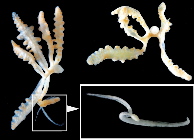
Dendrogaster nike

Parasitic crustaceans can evolve into some pretty funky forms and they have been featured multiple times on this blog. These crustaceans don't so much flaunt, but completely toss out all your expectations of what a crustacean or even an arthropod is "supposed" to look like. And among the best examples of that is Dendrogaster.

Left: Female Dendrogaster nike side view, with attached male (bracketed in the square), Top right: Female D. nike frontal view, Bottom right: Male D. nike. Photos from Fig. 2. of the paper

Dendrogaster is a genus of crustaceans that live as internal parasites of sea stars, nestled snugly within the body cavity of its host. So much so that their body shape seems to have evolved into somewhat resembling that of its host. In contrast to other crustaceans, Instead of having hard carapaces, segments, or jointed legs, Dendrogaster has multiple branching lobes, like some kind of fleshy, parasitic antler. They belong to a group of crustaceans called Ascothoracida - a sister group to the barnacles, who themselves are no strangers to the way that evolving towards parasitism can warp their body.

Dendrogaster rivals those parasitic barnacles in the "WTF Evolution?" department, and despite how bizarre they may look to us, they are not some rare oddity lurking in an obscure corner of the world. There are 35 known species of Dendrogaster and they have been found parasitising eighteen different families of sea stars all over the world, ranging from those dwelling in the shallows, to those inhabiting the deep sea over 2500 m below sea level. It seems that wherever sea stars went, Dendrogaster followed.

The paper featured in this blog post adds another species to this roster of evolutionary weirdos. This newly described species was found from sea stars living 1970 m below sea level, collected during a biodiversity survey at the An'ei Seamount, an offshore marine protected area off the eastern coast of Japan. The host was Asthenactis agni - a sea star which itself was newly discovered and described just late last year. This parasitic crustacean has multiple, wing-like branches protruding from its body, and it is this appearance which inspired its scientific name, Dendrogaster nike, named after Nike, the Greek winged goddess of victory.

But that's only how the female of the species looks like. The male is less than a quarter the size of its partner, and unlike the female Dendrogaster with its multiple protruding branches, the male is comparatively unremarkable, with a simple ovoid-shaped body and a pair of long thin testes dangling from it. It is usually found attached to its much larger and more flamboyant partner, floating inside the body cavity of a sea star.

Dendrogaster nike is just one of many new species of Dendrogaster that have been described over the last few years. In 2020, there were three other species of Dendrogaster which had been discovered from sea stars collected from the depths of the bathyal zone. It seems that sea stars from the deep sea are particularly favoured by this parasitic crustacean, and there are probably many other species of Dendrogaster yet to be discovered which are lurking in the abyss.

When scientists compared the DNA sequences of different Dendrogaster species, they found that the genus seems to be divided into two main sub-groups - those who stuck to the shallows, and those who ended up partying in the deep. While the evolutionary pathways of many parasites somewhat parallel that of their hosts, for Dendrogaster, it followed the hosts' habitats instead. This may provide some insight into the evolutionary origin of this bizarre, but widely found group of parasitic crustaceans.

When life hands you a sea star, sometimes it comes with a free Dendrogaster.

Reference:
Jimi, N., Kobayashi, I., Moritaki, T., Woo, S. P., Tsuchida, S., & Fujiwara, Y. (2023). Insights into the diversification of deep-sea endoparasites: Phylogenetic relationships within Dendrogaster (Crustacea: Ascothoracida) and a new species description from a western Pacific seamount. Deep Sea Research Part I: Oceanographic Research Papers 196: 104025.

March 17, 2023

Inodosporus fujiokai

A few years ago, rainbow trout at a trout farm in the Shiga prefecture, Japan, were being struck down by a mysterious illness. The flesh of the dead fish were speckled with red dots and white cysts. It turns out the disease was caused by a type of previously unknown microsporidian parasite. Microsporidians have been reported from other farmed fish in Japan, where they are locally called "beko disease". It was suspected that the trout might be getting infected from their food, and during feeding trials it was found that trout fed with fresh or chilled prawns developed the disease, while those fed frozen prawns stayed healthy. This shows that prawns were somehow involved in the life cycle of this parasite.

Left: Prawn infected with Indosporus fujiokai (indicated by red arrow), Centre: Electron microscopy of spores from muscles of an infected prawn (top), and a spore from the muscles of an infected trout (bottom). Right: An infected trout showing signs of hypoxia associated with infection by I. fujiokai (top), muscles of infected trout with red specks and white cysts of the parasite as indicated by arrows (bottom).
Photos of prawns + spores from Fig. 1, 7, and 9 of the paper, Photos of infected trout + their flesh from Fig. 3 and 6 of this paper

Microsporidians are single-celled parasites which are related to fungi. There are 1500 known species, though the actual number of microsporidians out there is likely to be much higher. For most of them, relatively little is known aside from how they look like and what they infect. About half of all known microsporidians are parasites of aquatic animals (and their parasites), and their life cycles can vary considerably between different species. Despite their importance as parasites of fish and crustaceans in aquaculture, the life cycles of many microsporidians are unknown. 

In the study featured in this blog post, researchers set out to find samples of the Shiga trout farm parasite out in the wild - and they found it amidst some prawns from Lake Biwa. Microsporidian-infected prawns are easy to spot because in contrast to healthy prawns which are translucent, infected prawns become opaque white as the parasite proliferates in their muscles. But surprisingly, despite the numerous spores filling up their flesh, infected prawns seemed rather healthy and were able to live for several weeks in the lab. Some of them even managed to produce eggs despite being parasitised! This is in stark contrast to the effect that this parasite has on its trout hosts.

The researchers named this microsporidian Indosporus fujiokai - after a parasitologist who, back in 1982, suggested the involvement of prawns in the transmission of microsporidian parasites. But that is not the entire story, because those prawns were harbouring a lot more than just I. fujiokai. The researchers actually found FOUR different types of microsporidians in those prawns, including the one that they eventually named Indosporus fujiokai. These microsporidians all differ in their spore sizes and shapes, and all of them were entirely new to science. Three of the microsporidians, including I. fujiokai, belong to a group called "Marinosporidia'' which are usually found infecting fish and aquatic invertebrates - this was to be expected since they were examining prawns. However, one of the microsporidians was more unusual, as it hails from an entirely different part of the microsporidian tree called "Terresporidia", which is composed of species that usually infect insects.

The results of this study suggests that prawns and other crustaceans could be harbouring a rich array of microsporidian parasites that are currently unknown to science, and there might be many more of them out there which are infecting fish by the way of crustacean hosts. While the researchers in this study were able to resolve the life cycle for I. fujiokai, mysteries continue to surround the life cycles of the three other microsporidians that they found - what hosts they might infect in the next stage of their respective life cycles are anyone's guess at this point.

As is often the case with parasites, just as you manage to answer one question, three (or more) others pop up in the process. So if life gives you a raw prawn, you should examine it for parasites.

Reference:

August 14, 2022

Cyclocotyla bellones

At the top of this blog, there is a quote by Jonathan Swift about how fleas have smaller fleas that bite them. Indeed, parasites becoming host to other types of parasites is actually a rather common phenomenon in the natural world. Those who would parasitise the parasites are called "hyperparasites".

Left: Cyclocotyla bellones on the back of a Ceratothoa isopod, Right: C. bellones coloured red with Carmine staining.
Photos from Figure 1 and 5 of the paper.

The parasite featured in this post was once suspected of being a hyperparasite. Cyclocotyla bellones is a species of monogenean - it belongs to a diverse group of parasitic flatworms that mostly live on the body of fish, parasitising the fins, skins, and gills of their hosts. But unlike other monogeneans, C. bellones does not attach itself to any part of a fish's body, instead it prefers to stick its suckers onto the carapace of parasitic isopods, such Ceratothoa - the infamous tongue biter. Since Ceratothoa is itself a fish parasite, and C. bellones is routinely found attached to those tongue-biters, this has led some to think that it might be a hyperparasite of those parasitic crustaceans.

But it takes more than simply sticking yourself onto another organism to be considered as a parasite of it. After all, there are algae that grow on the body of various aquatic creatures, or barnacles that are found on the backs of large marine animals like whales and turtles. But those are not considered as parasites as they don't treat their host as a food source, merely as a sturdy surface they can cling to - they're known as epibionts.

So strictly speaking, for Cyclocotyla to be a parasite of the isopod, it needs to be feeding on or obtaining its nutrient directly from its isopod mount. When scientists examine the bodies of the tongue-biters with C. bellones on them, they seem to be pretty unscathed. There aren't any scratches or holes on the isopod's body which you'd expect if C. bellones had been feeding on it. Indeed, the monogenean's mouthpart seems ill-suited for scraping through the isopod's carapace.

Additionally, C. bellones' gut is filled with some kind of dark substance similar to those found in other, related monogenean species. This is most likely digested blood from the fish, which the monogenean has either sucked directly from the fish's gills, or indirectly via the feeding action of its isopod mount. Let's not forget that the isopod itself is a fish parasite that feeds on its host's blood, so if it gets a bit messy during mealtime, perhaps Cyclocotyla is there to suck up any spilled blood. Or it might be doing a bit of both.

The researcher noted that Cyclocotyla is not alone in its habit of riding isopods. Other monogeneans in its family (Diclidophoridae) have also been recorded as attaching to parasitic isopods of fish. And aside from riding isopods, they all share one thing in common - a long, stretchy forebody, looking somewhat like the neck of sauropod dinosaurs. Much like how the neck of those dinosaurs allowed them to browse vegetation from a wide area, the long forebody of Cyclocotyla allows it to graze on the fish's gills while sitting high on the back of an isopod. So fish blood is what C. bellone is really after - the isopod is merely a convenient platform for it to sit on.

But why should these monogeneans even ride on an isopod in the first place? Cyclocotyla and others like it have perfectly good sets of suckers for clinging to a fish's gills. Indeed, there are other similarly-equipped monogeneans that live just fine as fish ectoparasites without doing so from the back of an isopod. Well, that's because the fish themselves don't take too kindly to the monogeneans' presence. These flatworms are constantly under attack from the fish's immune system, which bombards them with all kinds of enzymes, antibodies, and immune cells. By avoiding direct contact with the fish's tissue, Cyclocotyla and other isopod-riders can avoid being ravaged by the host's immune system - which is something that other monogeneans have to deal with on a constant basis.

So it seems that Cyclocotyla and other isopod-riding monogeneans are no hyperparasites - they're all just regular fish parasites that happen to prefer doing so while sitting on the backs of isopods. Cyclocotyla bellones prefers to share in the feast of fish blood with its isopod mount, while sitting high above the wrath of the host's immune response.

Reference:

October 15, 2021

Caledoniella montrouzieri

Mantis shrimps (Stomatopoda) are some of the most formidable crustaceans in the sea; armed with trinocular colour vision, and a pair of powerful raptorial limbs that punch so hard, it generates supercavitating bubbles which collapse with such energy, the vapour within them briefly turns into white-hot plasma. Arguably, one of the most impressive animals in the sea. But to Caledoniella montrouzieri - a mantis shrimp is simply a big juicy host. 

Underside of a parasitised mantis shrimp, showing the male, female, and egg capsules of Caledoniella montrouzieri
Photo from Fig. 1 of the paper, taken by Ryutaro Goto.

Most parasitic snails belong to the Eulimidae or Pyramidellidae family, and both of them parasitise slow-moving or sedentary invertebrates such as echinoderms, molluscs, and polychaete worms. But Caledoniella has taken a different, independent route down to parasitism town. It doesn't belong to either of those families, and instead of a slow life feeding on some barely mobile hosts, it lives life in the fast lane, clinging to the belly of a nimble, predatory crustacean.

Such a lifestyle requires some specialised anatomy. In most snails, the foot is a flat muscular organ that is used for crawling over various surfaces. But in Caledoniella, the foot has been transformed into a big suction disc that allows it to cling firmly onto its very agile host. During the course of its evolution, it has also lost one of the key diagnostic characteristics of molluscs - the rasp-like radula in the mouth which snails use to scrape bits of food, be they algae or the flesh of other animals, into their mouth. Instead, it has a mouth that is more suited for suction feeding, and has highly developed salivary glands to facilitate its liquid diet. This snail is a vampire of mantis shrimp, sucking on their host's gill filament for that sweet, sweet hemolymph.

Caledoniella has some noticeable sexual dimorphism with the female snail being much larger than the males. When these snails mature, they pair up as a monogamous couple, living out their lives together on the underside of a mantis shrimp. But this happy couple likes to keep themselves to seperate parts of the mantis shrimp, with the female living near the tail of the shrimp, and the male living near the middle of the abdomen. Sitting between them are all the egg capsules they have been busily making together. This gastropod couple takes their toll on the mantis shrimp, which experience stunted growth, reduced moulting, and infertility.

So how did Caledoniella ended up with its unique way of life? The closest living relatives of Caledoniella are snails that live as roommates with mantis shrimps, hanging on the walls of the crustacean's burrow. While these snails are frequently in the presence of the burrow's main tenant, that's as far as their relationship with the mantis shrimp goes. They are strictly commensals that never lay their foot on the mantis shrimp, and it is likely that was the lifestyle of Caledoniella's ancestors. But at some stage, after living in such close quarters with mantis shrimps for so long, some of those meek wallflower snails just couldn't resist getting more intimate and started taking a bite of its crustacean roomie, thus giving rise to the clingy blood-sucking Caledoniella

But when you trace its evolutionary history even further back, it seems those mantis shrimp roommates have themselves evolved from snails that originally lived in the burrows of an entirely different animal - spoon worms! So the ancestors of Caledoniella switched from sharing quarters with spoon worms, to living with mantis shrimp, to living on mantis shrimps

Perhaps somewhat surprisingly, Caledoniella is not the only mollusc that spend their lives clinging to the mantis shrimp - there are also a few species of tiny clams from the Galeommatoidea family called yoyo clams that live attached to the mantis shrimp's belly.  But unlike Caledoniella, they don't go as far as to feed on their host's blood. These clams receive protection from living on the belly of this heavily-armed crustacean, and the host's agile movements provide it with plenty of water flow for all their respiratory and filter-feeding needs. While they aren't blood-suckers, they seem to have followed the same evolutionary pathway as Caledoniella, evolving from ancestors that originally lived as commensals in the burrows of mantis shrimps.

For molluscs, it seems that sharing room with a marine benthic invertebrate is a surefire gateway to becoming a clingy parasite.

Reference:
Goto, R., Takano, T., Eernisse, D. J., Kato, M., & Kano, Y. (2021). Snails riding mantis shrimps: Ectoparasites evolved from ancestors living as commensals on the host’s burrow wall. Molecular Phylogenetics and Evolution, 163:107122.

June 16, 2021

Allokepon hendersoni

Crabs have some pretty scary parasites infecting them. They range from worms that use them as vehicles to complete their complex life cycles, to parasitic dinoflagellates that turn their muscles into bitter slurry, and on top of those, there are also other crustaceans that can take over their body, and in some cases, castrate them in the process. These body-snatching crustaceans come in two main types - bopyrids and rhizocephalans. 

Top: Bopyrid isopod and an infected crab, Bottom: Rhizocephalan barnacle with infected crab.
Photos modified from the graphical abstract of the paper

Bopyrids are parasitic isopods in the same suborder as the infamous tongue biter parasite, but instead of going into a fish's mouth, they go inside the body of crabs and make themselves at home, often causing a characteristic bulge on the infected crab's carapace. And then, you have the rhizocephalans, which are freaky barnacles that have a body composed of a network of roots which wrap themselves around the crab's internal organs.

Each of them inflict their own respective flavour of pain on their crab hosts.

This study looked at the effects that these parasitic crustaceans have on the two-spotted swimming crab (Charybdis bimaculata), which is host to both parasitic isopods and barnacles. Here representing the bopyrid isopods, we have a species named Allokepon hendersoni. And fronting for the barnacles, is an as yet undescribed species of rhizocephalan. As hinted at earlier, these two body-snatcher parasites seem to have different effects on the crabs - but what exactly are they?

When scientists compared infected crabs with uninfected crabs, they found the effects to be most pronounced in male crabs, with both species of parasites causing a reduction in weight and claw size of their hosts. This is most likely due to the energetic drain associated with hosting these crustaceans, since they can grow to alarmingly large sizes when compared with their hosts. 

While reducing the claw size may leave the host crab less able to compete with uninfected males, on another hand (or claw as the case may be), it would not be in the interest of the parasite for its host to be getting into too many fights and risk injuries anyway, so it can be a beneficial side-effect from the parasite's perspective

But there were some changes which were more specific to particular parasite species. Male crabs infected with Allokepon had a narrower abdominal flap (the triangular flap on the "belly" of a crab). In male crabs, this flap would usually serve to protect the gonopods - which are specialised appendages that arthropods use in reproduction - but given the crab is already hosting such a demanding resident in its body, it wouldn't be getting up to any of that any time soon.

In contrast, the rhizocephalan barnacle had the opposite effect on the crab and widened that flap - this is part of a whole suite of changes that these parasites induce in male hosts. Male crabs that are infected by rhizocephalans develop characteristics which are associated with female crabs in both appearance and behaviour. In female crabs, the wider abdominal flap serves to cradle and brood the eggs before they hatch. So the barnacle essentially "feminise" the male crabs so that they can become better babysitters for the barnacle's offspring.

Fortunately for this crab population, infection rate was very low. Of the 2601 crabs the scientists examined, only 14 were infected with the isopod, and 21 infected with the rhizocephalan barnacle, though the isopod seems to have a preference for infecting male crabs, whereas the barnacle was less discriminate.

But if you are the unfortunate crab that gets infected, you are in for a bad time either way.

Reference:
Corral, J. M., Henmi, Y., & Itani, G. (2021). Differences in the parasitic effects of a bopyrid isopod and rhizocephalan barnacle on the portunid crab, Charybdis bimaculata. Parasitology International, 81: 102283.

June 11, 2019

Pennella instructa

Swordfish are one of the top predators of the ocean. They can swim through the sea at blistering speed, and slash at their prey with their long, flat bill. But no matter how fast you are, there's one thing you can never swim away from - and that's parasites. This is especially the case for big animal like swordfish as their anatomy provides a wide range of different habitat for all kinds of parasites.
They range from sea lice (caligid copepods) that cling to the swordfish's face, to tapeworm larvae which dwell in their muscle, to roundworms that lay eggs under their skin - just to name a few.

Pennella instructa adult with a cyst. From Fig. 4 of the paper

This post will be focused on a study that reported on the occurrence a parasitic copepod - Pennella instructa - on swordfish caught from the north-eastern Atlantic. The researchers in this study visited the fish auction market at Virgo, Spain, during March to September 2011, looking for the presence of P. instructa on swordfish which were brought in by Portuguese and Spanish long line fish boats over that period.

Even though P. instructa is classified as a crustacean, those who are familiar with this blog (and my Twitter feed) would know that when it comes to parasitic copepods, one should abandon any and all preconceptions they might have of what a crustacean is "supposed" to look like. Pennella instructa is shaped vaguely like a toothbrush - a long narrow body that ends with an abdomen covered in a brush-like plume. The adult parasite can grow to about 20 centimetres (or 7 inches) long. It spends its adult life with the lower half of the body protruding from the swordfish, while the front half is anchored deeply in the host's tissue.

Having a parasite that is half-buried in its host's flesh sounds gruesome enough, but P. instructa does something else which elevates it to Cronenberg-level body horror. See, the parasite has not merely stuck its head into the swordfish's flesh and sucking its blood, it is also wrapped in a kind of meat cocoon that the parasite has crafted out of the host's own tissue. Essentially this parasite has sculpted a cosy little bag for itself out of swordfish meat. This parasite-induced cyst is similar to what some other fish parasites, like the fluke that lives on sunfish (Mola mola) gills, can do with their host.

Of the 1631 swordfishes that the researchers looked at, 167 were found to have visible P. instructa infections, though they only occurred in low numbers on each fish, with the most heavily infected fish carrying 4 individual copepods. But being the kind of parasite that it is, even a single P. instructa can have some significant impact on the swordfish's overall health, depending on where it is located. Aside from drinking the host's blood, the meaty cyst that P. instructa forms around itself can put pressure on the surround tissues and organs. The researchers found that while P. instructa can be found all over the swordfish's body, for whatever reasons, most of them prefer the posterior part of the swordfish, mostly in the thick, meaty part of the tail.

It could be that those sturdy tail muscles provide the parasite with a good site to anchor itself in place. Furthermore, that part of the fish's body is made of the powerful muscle which allows the swordfish to propel itself so quickly through the water, thus they'd be constantly supplied with a steady flow of blood which P. instructa can drink from. But this comes at a significant cost to the host, because if the parasite's cyst is located near the vertebrate column - as they would be if they are embedded in the tail - it may affect the fish's nervous system and compromising its swimming ability.

While P. instructa doesn't infect or cause any health issues in humans, a piece of swordfish steak with a big hole through it and a weird worm thing dangling out the side would probably be off-putting to any would-be customers. But perhaps we might want to consider adding P. instructa to the menu?
Pennella balaenopterae - a related copepod which infect whales - is considered to be gastronomic treat by the Inuit people of the Canadian arctic. So instead of seeing them as a pest, perhaps Pennella might be reconsidered as added garnish for your swordfish steak?

Reference:
Llarena-Reino, M., Abollo, E., & Pascual, S. (2019). Morphological and genetic identification of Pennella instructa (Copepoda: Pennellidae) on Atlantic swordfish (Xiphias gladius, L. 1758). Fisheries Research 209, 178-185.

February 12, 2018

Neocyamus physeteris

Today we're featuring a guest post by Sean O’Callaghan - a student from 4th year class of the Applied Freshwater and Marine Biology' degree programme at the Galway-Mayo Institute of Technology in Ireland. This class is being taught by lecturer Dr. Katie O’Dwyer, who has previous written guest posts about salp-riding crustaceans and ladybird STI on this blog. This post was written as an assignment on writing a blog post about a parasite, and has been selected to appear as a guest post for this blog. Anyway, I'll let Sean take it from here.

Sperm whales are the largest toothed animal alive and they are capable of diving down to depths of 1200 m to feast on cephalopods (including the planet's largest cephalopods, the colossal and giant squids), but despite their size and abilities, these leviathans can fall victim to a range of cunning ectoparasites, including…Whale Lice!

Line drawing of adult female Neocyamus physeteris from Fig. 2 of this paper, SEM photograph from Fig. 2 of this paper
Three species of whale lice are known to target sperm whales, and from this trio there is a divide of preference between male and female whales. Neocyamus physeteris is one such example - they would rather live on a female whale than a male one. While the exact reasoning behind why there is such a divide in parasite species targeting opposite sexes, the answer may be due to the habits of male whales, which frequent the polar waters more often than the females who seek out the warmer waters around temperate zones.

Whale lice are not really lice in a taxonomic sense. Instead, they are classed as amphipods, crustaceans related to the so-called "lawn shrimps" which are found in some back gardens, but with more specialised features for hanging on to a free-swimming whale. Neocyamus physeteris’ body is flattened like a leaf but largely segmented and have legs tipped with hooked edges that act like crustacean crampons to ensure a consistently ample footing. Otherwise the lice would find itself cast adrift without a home or food supply to die alone in the deep. They also possess sharpened mandibles to munch through the host whales epidermis (top skin layer) while for breathing it has two pairs of gills lining its underside towards the front half of the body. Neocyamus physeteris’ head is quite small in comparison to the rest of its body and is dotted with a pair of tiny eyes along with two antennae. Their white colouration almost gives off a dandruff-like appearance against the whale’s darker complexion (though they would be well camouflaged on Moby Dick if it had existed and was also female!).

They are so intertwined with their host that their life cycle that they lack a free-swimming larval phase or active transmission to other whales, offering limited opportunities to move between hosts (unless during social activities where the whales may rub against one another). So it is fair to say that they live, feed and breed on top of their own biological ark, from the sea's clear surface waters to dark depths of the twilight zone, quite a dependent but extreme lifestyle!

Like most whale lice, little is known about the habits of N. physeteris, but it is so specialised for its life-style that whenever the whale dies, the lice would also kick the can as they require a live host. Hanging onto a host may not seem like an exciting lifestyle, but it is a highly beneficial strategy (for the lice at least). Given its tendency to devour sperm whale skin mainly in areas that are sheltered from water movements like the genital slits, body creases or injured skin, this allows the lice to take advantage of a lifetime supply of renewable food. In other words, the lice won’t starve while on a whale, however there will be an increase demand for firm footholds as the parasite population increases, so the species' overall success is not necessarily always good for the individual louse. The whale probably doesn’t suffer too badly when only a handful of lice are present however a colony must surely be highly irritating to say the least.

The strain imposed on N. physeteris at different depths due to the varying degrees of pressure imposed between the surface and abyss would far exceed our own limits. Undoubtedly there must be a risk posed by potential fishy predators on occasion given the lack of cover afforded by a whale’s skin. However, the benefits appear to outweigh the risks - otherwise they would cease to exist as a species. There is still much to learn about these fascinating parasites but until new means of studying the movements and behaviours of these small, somewhat inconspicuous amphipods on top of a large mobile host like a sperm whale are developed, it could take a while to unravel the intricacies of this skin serrating invertebrate!

References
Hermosilla, C., Silva, L.M.R., Prieto, R., Kleinertz, S., Taubert, A. and Silva, M.A. (2015). Endo- and ectoparasites of large whales (Cetartiodactyla: Balaenopteridae, Physeteridae): Overcoming difficulties in obtaining appropriate samples by non- and minimally-invasive methods. International Journal for Parasitology: Parasites and Wildlife. 4, 414-420.

Leung, Y. (1967) An illustrated key to the species of whale-lice (Amphipoda, Cyamidae), ectoparasites of Cetacea, with a guide to the literature. Crustaceana 12, 279-291.

Oliver, G. and Trilles, J.P. (2000). Crustacés parasites et épizoítes du cachalot, Physeter catodon Linnaeus, 1758 (Cetacea, Odontoceti), dans le golfe du lion (Méditerranánée occidentale). Parasite. 7, 311-321.

This post was written by Sean O’Callaghan

January 11, 2018

Riggia puyensis

It is no secret that I am a big fan of parasitic isopods, especially those in the Cymothoidae family - the most well-known of which is the tongue biter parasite, and my love for these adorable crustaceans has even manifest itself in some of my artwork. But while the tongue-biters are no doubt the most (in)famous representatives of that family, to the extent that they even made an appearance on an episode of the Colbert Report, it is their less well-known cousins - the belly-dwellers/burrowers - that turn the horror factor up a notch (or four, or eleven) and as a result, really earned my adoration.

Left: Adult female Riggia puyensis (scale bar = 10 mm), Right: Adult make Riggia puyensis (scale bar = 1 mm)
From Fig. 3 and Fig. 9 of the paper

Imagine if the chest-burster xenomorph from Aliens didn't just explode through your ribcage and leave you for dead - instead, it stays inside your torso for the rest of your life, laying a steady stream of eggs that trickle out through a small(ish) hole in you belly. That's how these belly-dwelling isopod live their lives. So let's kick off the year with a recently described species of these belly-dwellers!

I've previously written a post about a species of belly-dweller call Artysone trysibia which lives in the body cavity of an armoured catfish from the Amazon. This post features Riggia puyensis, which is quite similar to A. trysibia in that it was also found to be parasitising armoured catfish, specifically two species from the Bobonaza River and Puyo River in central Ecuador - Chaetostoma breve and Chaetostoma microps - both of which are better known as suckermouth armoured catfish.

Most of the R. puyensis specimens that the scientists found in this study were females, but the scientists did come across three male specimens which were clinging to the limbs of the female isopods. These male isopods are comparatively tiny reaching only one-tenth the length of the adult female R. puyenesis. The small size and relative rarity of males is par for the course for Riggia. In other studies on this genus of parasite, male isopods are rarely found, if at all. It is possible that this is because the mating strategy of the male isopod is to scoot in, mate with the larger female, then go off and find another infected host.

Riggia puyensis inside its host, from Fig. 2 of the paper
In this study, each infected fish was only parasitised by a single female isopod - which is probably just as well since R. puyensis is quite large in relation to the host. The female R. puyensis reaches over an inch in length and considering one of the host catfish is a species that grows to about four inches long at most, that parasite is a hefty load to be carrying around. It would be like having a corgi living inside you.

So it may seem rather surprising that the survival of these fish does not seem to be compromised by the parasite. In fact, a previous study have shown that the parasite may in fact enhance the infected fish's growth. But this parasite-induced growth spurt comes at a price - after all, there is no free lunch in nature and for the gain in body growth, the parasite incurs a severe penalty on the fish's reproductive functions. A study on bonefish parasitised by Riggia paranensis found that infected fish has reduced level of sex hormones and undeveloped gonads.

So Riggia render its fish host impotent in order to free up more resources for body growth, and a bigger host means more for the parasite to consume. So while a chest-bursting xenomorph invokes a more immediate visceral reaction, the way that R. puyensis and other parasitic castrators modify their hosts' body to fuel their own reproduction presents a more existential form of lingering horror.

Reference:
Haro, C. R., Montes, M. M., Marcotegui, P., & Martorelli, S. R. (2017). Riggia puyensis n. sp.(Isopoda: Cymothoidae) parasitizing Chaetostoma breve and Chaetostoma microps (Siluriformes: Loricariidae) from Ecuador. Acta Tropica 166: 328-335.

September 8, 2017

Sylon hippolytes

Some of you might have heard of the infamous parasitic barnacle Sacculina carcini which infects crabs and take over their bodies. These barnacles are true body-snatchers in every sense - they divert the host's resources for their own growth and reproduction, and by doing so they end up castrating their host. Additionally, some can also can alter their host's behaviour, making them unwitting babysitters for their eventual spawn.

(A) Shrimp infected with Sylon hippolytes; (B) Internal structure of infected shrimp show its nervous system (n) and the interna (i) and externa (e) of Sylon; (C) Close-up internal view; (D) Close-up internal view with colour-marking.
Photos from Fig 2 of the paper
While it sounds like a gruesome fate for the host, parasitic castration is a very clever way for a parasite to get the most out of the host without killing it. While the host can no longer reproduce, and thus dead from a evolutionary perspective, it doesn't need its reproductive organ to stay alive, but instead it now serves as a walking life support system for the parasite - a walking dead. So just how big can these body snatchers get in comparison with their host?

Sacculina carcini and related parasites belong to a group of very unusual parasitic barnacles call Rhizocephala. Their bodies consist of a network of roots call the interna which wrap around the host's organs, and a bulbous reproductive organ call the externa which sticks out of the host's abdomen. In a previous post I wrote about a study which used micro-CT scans to look at how the these parasites' roots are distributed around the host's organs. In the study featured in this post, a group of scientists compared the anatomy of two different rhizocephalan species and how it relates to their reproductive strategies

The two species they compared were Sylon hippolytes which infects the shrimp Pandalina brevirostris, and Peltogaster (featured in a previous post on this blog) which infect hermit crabs. They collected specimens of both parasites (and their hosts), and prepared them for scanning. After putting the prepared specimens through the micro-CT scanner, they used special software to calculate the parasites' volume and were able to construct a 3D computer model of each parasite along with the internal anatomy of their hosts.

Additionally, they also counted the number of eggs produced by each parasite, and for both species, it seems bigger hosts means more parasite eggs. Both Peltogaster and Sylon grew to about the same size in proportion to their respective hosts (17.78% for Peltogaster, 18.07% for Sylon). But the key difference lies in how much of that mass is distributed between reproductive externa versus the interna root system in the host's body.

The shrimp-infecting Sylon devoted the bare minimum to its interna which is only about 2.5% of the volume of its externa. In contrast, the interna root system of the hermit crab-infecting Peltogaster was about one-fifth of the volume of its externa. So why is there such a massive difference between those two species since they parasitise the host in a similar way? The answer lies in their respective reproductive investments. The Sylon specimens measured in this study had about 1400 to over 22000 eggs, and to produce all those eggs Sylon has to devote a lot more of its mass to its reproductive tissue. In contrast, Peltogaster produced a comparative modest number of eggs, only 371 to 4580.

But why does Sylon put so much into egg production while leaving the bare minimum to the part of its body which is actually embedded in the host? The main reason is that Sylon only gets one shot at breeding - it only ever produce a single brood in its lifetime before it withers away, so it has to make the most of it by having a massive externa. In contrast, once Peltogaster becomes established in a host, it spawns repeatedly and grow a new externa each breeding season, and in order to do so, it needs to invest in a robust network of tendrils which will stay in the host for good.

In this sense, Sylon has a "YOLO" approach to host exploitation and reproduction, whereas Peltogaster is in it for the long haul and so devote more of itself to establishing an extensive root system inside the host. This also has important consequences for the host as well since both parasites places such a massive burden on their hosts - while the demanding presence of Sylon will eventually come to pass, Peltogaster is a persistent body-snatcher that's going to stick around for quite a while.

Reference:
Nagler, C., Hörnig, M. K., Haug, J. T., Noever, C., Høeg, J. T., & Glenner, H. (2017). The bigger, the better? Volume measurements of parasites and hosts: Parasitic barnacles (Cirripedia, Rhizocephala) and their decapod hosts. PloS One 12(7): e0179958.

March 25, 2017

Balaenophilus manatorum (revisited)

At some stage of their lives, parasites need to move from one host to another - some move around a lot throughout their lives, staying just briefly on a given host before moving onto another. While others only do it once during their larval stage - once they reach their host, they are there for life. Either way, they still need to make a perilous journey to their host.

Top right: newly hatched nauplii, Top left: Copedpodite V stage, Bottom: Adult female with eggs
Image composited from photos from Fig.1, 5, and 6. of the paper

This post is about study on Balaenophilus manatorum - a tiny parasitic copepod that lives on sea turtles. How does a tiny crustacean like that manage to find their way onto a turtle in the wide expanse of the sea? Do they jump on board when the turtle come into contact with each other, or can the larval stage swim on their own? Obviously they have managed to find a way because this copepod is very common among the juvenile loggerheads in the western Mediterranean, with over 80 percent of loggerhead turtles infected with B. manatorum. Given how small they are (the adult copepod is only about a millimetre long), it seems as if they would be barely a nuisance to their host. But when they occur in large numbers, they can be an serious menace.  And they seem to have a very particular taste. It was thought that B. manatorum feed mostly (if not exclusively) on sea turtle skin.

To find out more about how B. manatorum infect their hosts and what they feed on, a team of scientists did a series of studies on some B. manatorum which were removed from a batch of sea turtle hatchlings. These hatchlings were being reared at the Sea Turtle Rescue Centre (ARCA del Mar) - a rescue and rehabilitation for sea turtles in Spain. They came from a brood of eggs that was removed from a beach frequent by tourist to ensure their safety, but during their stay at the centre, many of them develop symptoms of infestation by B. manatorum, each of them infected with about 300 B. manatorum and one unlucky turtle was hosting over 1400 copepods. While removing the copepods from the turtles, the research team collected some of the egg-bearing female copepods that were on the turtles, and reared them until their eggs hatched into larvae for the further study.

In the feeding trials, the copepods were offered a menu selection consisting of: flakes from the baleen plates of a fin whale, fish skin (from a blue whiting), green alga, loggerhead turtle skin flakes (from some hatchlings that had succumbed to B. manatorum infestation). All those items were dyed with a stain to track if they get ingested. They confirmed that these copepod only ate turtle skin flakes and didn't touch the other items on the menu. Other species of Balaenophilus have been recorded from the baleen plates of whales, but B. manatorum feed exclusively on turtle skin. From the moment it is born, B. manatorum is equipped with mouthparts which are well-suited for scrapping flakes from hard flat surfaces, such as the skin of a turtle. So it is no wonder heavy infestations of B. manatorum can cause severe lesions and skin erosions in turtles, especially for the more vulnerable hatchlings

But B. manatorum still need to reach the turtle in the first place. When placed in a dish of seawater, newly hatched copepods (called nauplii) seemed rather helpless, only able to crawl around. But if they manage to survive to grow into the subsequent stages called copepodite, they will develop legs that would allow them to swim for a bit - just barely, and once they grow past a stage call Copepodite IV, they can swim well enough to reach another turtle on their own. It seems that this parasite relies mostly on the social behaviour of the turtle for transmission. Newly hatched B. manatorum nauplii cannot swim and would have to wait for the turtles to touch each other (for example during mating) to climb onboard another host (rather like how human lice are transmitted), whereas the copepodites and adults can just swim across if another turtle comes close enough

Therefore, these parasitic copepods may present as a kind of social cost to these turtles, since not only is a social communicable parasite, it can also be a sexually transmitted infection. For B. manatorum, their entire world really is found on the back of a turtle.

Domènech, F., Tomás, J., Crespo-Picazo, J. L., García-Párraga, D., Raga, J. A., & Aznar, F. J. (2017). To Swim or Not to Swim: Potential Transmission of Balaenophilus manatorum (Copepoda: Harpacticoida) in Marine Turtles. PloS One 12(1), e0170789.