"So, naturalists observe, a flea has smaller fleas that on him prey; and these have smaller still to bite ’em; and so proceed ad infinitum."
- Jonathan Swift
Showing posts with label trematode. Show all posts

April 14, 2025

Biospeedotrema spp. (et al.)

The deep sea is home to unique communities of organisms, and wherever there is life, there are parasites, and deep sea habitats such as hydrothermal vents with their multitude of species can provide the conditions to support parasites with complex, multi-host life cycles

But not all such parasites are equal in their requirements, for example digenean flukes can be demanding prima donnas when it comes to the necessary hosts for their complex life cycles.  Not only does the adult fluke needs a vertebrate host to live in, they also have an asexual stage that needs to go into a specific type of invertebrate, usually some kind of snail, and maybe even other small animal to act as go-betweens to carry the larval stage from the snail to the vertebrate host. So while habitats like hydrothermal vents are teeming with life - do they have what it takes to support those fastidious flukes?

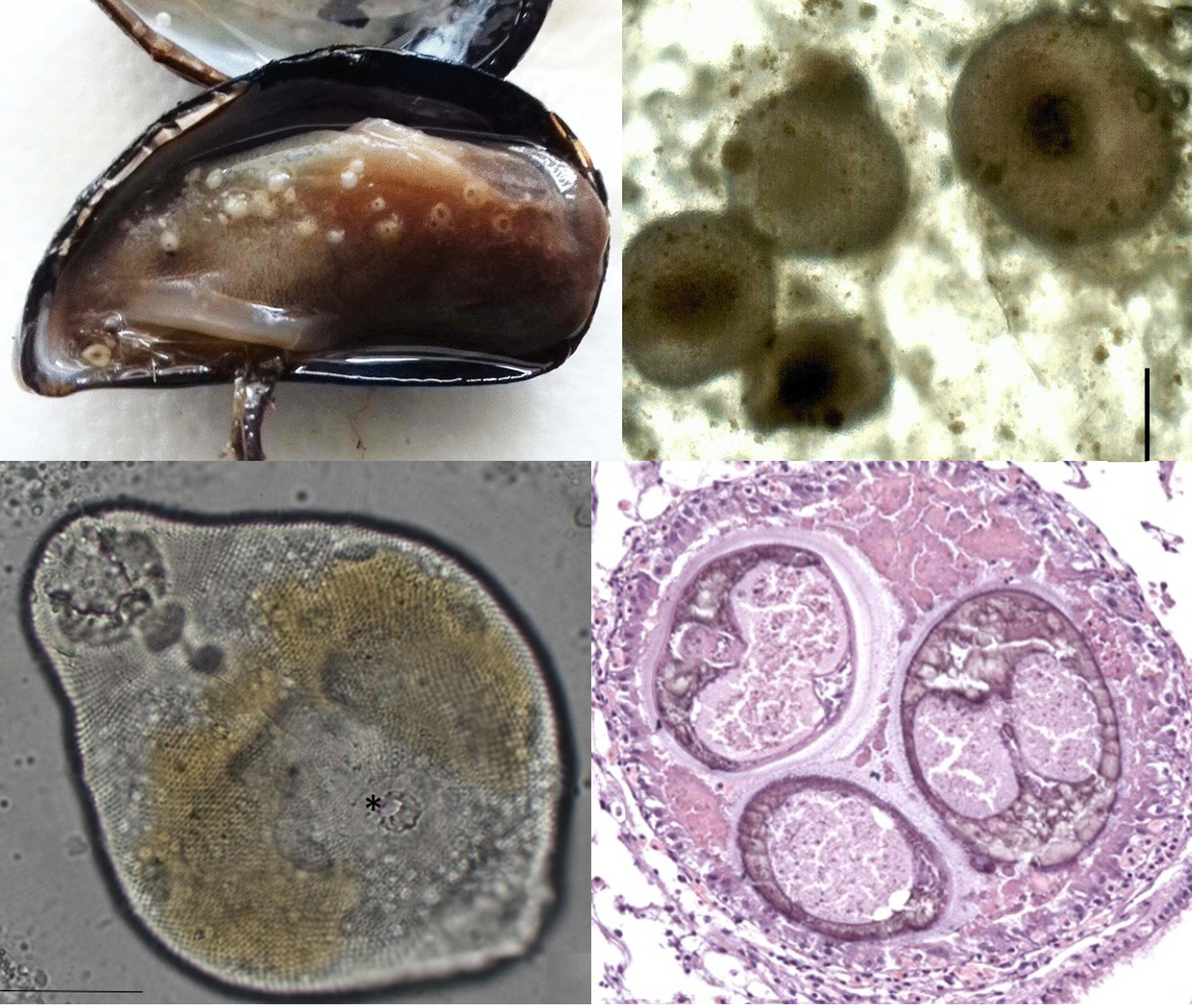
Left: Different types and stages of digenean flukes found at hydrothermal vents Including adult and sporocysts of Neolebouria (top left), adult and metacercariae of Biospeedotrema (top right), adult and metacercariae of Caudotestis (bottom), and unknown cercaria (bottom insert) All scale bars: 500 μm. Right: Proposed life cycle of digenean flukes at hydrothermal vents, with the adult living in vent fish, snails as first intermediate hosts, shrimps and other invertebrates as the second intermediate host. Photos of flukes from Fig. 5 of the paper, life cycle diagram from Fig. 1 of the paper. 

To answer that question, scientists from the Woods Hole Oceanographic Institution looked for parasites in samples of organisms collected from hydrothermal vent sites located about 2500 metres below sea level, along the East Pacific Rise. They ended up with a mixed bag of different animals composed of various fish and invertebrates, and when the scientists dissected those deep sea creatures, it turns out many of them were filled with all kinds of flukes at various stages of their life cycles. This range from adult flukes in the guts of vent fishes, to the sausage-like asexual stages in snails, to larval cysts embedded in the bodies of invertebrates, and even the short-lived, free-roaming cercaria stages crawling about in the samples.

Seven different types of flukes were present in the guts and gall bladder of vent fishes such as the pink vent fish (Thermarces cerberus) and viviparous brotulas (Thermichthys hollisi). And by sequencing selective sections of their DNA, the scientists were able to match those adult flukes with corresponding larval stages in a wide range of vent animals including shrimps, crabs, snails, and polychaete worms. For example, the fluke asexual stages found in glass limpets turned out to be a match with the adult flukes found in one of the vent fish.

In total, the researchers were able to identify three distinct genera - Biospeedotrema, Caudotestis, and Neolebouria - but they also found free-living cercaria stages of an unknown fluke. These cercariae have a stubby, sucker-like tail and while they superficially resemble the cercariae produced by the fluke which was found in glass limpets, DNA analysis shows that it does not genetically match with any of the other flukes in the samples. The life cycle and hosts of those peculiar cercariae are currently unknown, but their presence indicates that there many other infected hosts yet to be discovered at those hydrothermal vent communities.

So while this study has managed to fill some gaps in our knowledge about parasitism in the deep sea, there are still many mysteries. Digenean flukes need rich communities to complete their life cycles, and their presence at hydrothermal vents tells us that even though such vent sites are short-lived, these habitats can support a very rich community of different organisms. But many of these biodiverse habitats are under threat from a wide range of current and planned human activities. There is so much more that we need to learn about these biomes of the deep, and we also need to learn to value them, lest they become casualties in the face of ceaseless demands for minerals and other commodities.

Reference:

November 14, 2024

Saccularina sp.

The bay scallop (Argopecten irradians) is a highly prized shellfish, but it has suffered through a rough history from overharvesting, habitat loss, and natural enemies. This culminated in a massive population decline in the 1980s that led to the closure of all its fisheries across certain regions along North Carolina. With the depletion of its wild populations, efforts are being made to raise the bay scallops in aquaculture to meet demands. But now a new woe has fallen upon this besieged bivalve with the appearance of a never-before-seen parasite.

Left: Bay scallop infected with Saccularina, the red arrows indicating the parasite's sporocysts in the gills. Right: the cercaria stage of Saccularina which is a "cystophorous"-type cercariae.
From Fig. 1 and 2 of the paper

In 2012, a researcher started noticing a gill-dwelling parasite in both caged and wild bay scallops along the coast of North Carolina, and later at the Gulf Coast of Florida. These parasites are readily visible in the shellfish’s gills as they become swollen with the parasite's presence. Examination under a microscope revealed the parasites to be a species of parasitic fluke which is using the scallop for the asexual stage of its life cycle. Essentially, these trematode flukes are converting the shellfish into a parasite clone factory that pumps out a stream of free-swimming larvae to infect the next host in the life cycle.

Given such an operation consumes a lot of the host’s resources, this can interfere with the scallop’s growth and survival, and thus it is a major concern to the scallop fisheries in North Carolina. The key to managing any parasitic infection is an understanding of its natural history and life cycle, and unfortunately, given its relatively recent discovery, the life cycle of this parasite is almost entirely unknown. However, related fluke species can give us some clues, and as it turns out, this bay scallop parasite is no ordinary fluke.

First of all, DNA analysis showed that one of this fluke's closest relatives is Saccularina magnacetabula, a trematode species found on the other side of the world in Australia. While it is genetically similar enough to the bay scallop parasite for them both to be in the same genus (Saccularina), there is enough geographical and genetic distance between them that they are clearly different species. Furthermore, instead of scallops, S. magnacetabula infects the Sydney cockle (Anadara trapezia) as the chosen bivalve for its asexual stage. As for the parasite's next stops, it's a multi-part journey involving tiny crustaceans, followed by a type of smallish Australian fish called whiting (Sillago sp.), and finally the adult fluke completes its life cycle nestled in the fin membranes of the giant herring (Elops hawaiensis).

Saccularina magnacetabula, and by extension, the bay scallop parasite, belongs to a family of flukes called Didymozoidae - a flukey group of flukes with some very unusual anatomy and habits. While the adult stages of most trematodes are generally leaf-shaped, didymozoids come in all kinds of shapes and sizes. And they are found in a wide range of different bony fishes all over the world, mostly marine species. Additionally, instead of living in the final host’s gut like most flukes do, didymozoids cram themselves into all kinds of nooks and crannies such as the muscles, the gills, or even the fin membranes as is the case for S. magnacetabula.

While there are many known species of didymozoids, the life cycles for most of them are a mystery, with the hosts for the asexual stage known only for a few species. But those handful of species alone showed didymozoids to have quite the eclectic range. Aside from bivalves like cockles and scallops, the asexual stage of other didymozoids infect snails, but not just any regular snails - one species is known to use pelagic sea snails (which are also called “sea elephants”), while another species infects worm snails which are peculiar sea snails with twirly shells that encrust on rocks and other hard surfaces.

So, based on the information above, we can make some inference about the likely source of the bay scallop parasite. It’ll have to be some kind of predatory sea-dwelling fish harbouring the adult stage of the fluke, and given S. magnacetabula completes its life cycle in the giant herring, the bay scallop parasite is most likely completing its life cycle in some kind of predatory herring-type fish in the region, which means ladyfish or Atlantic tarpon

While we may have some clues about the bay scallop parasite’s life cycle, how they might have gotten there is more of a mystery though. This parasite was first seen in bay scallops in 2012, but if the disseminator of this parasite is really a local fish species such as the tarpon, why has it only been noticed now? Whatever its origin turns out to be, it seems the bay scallop parasite is not ready to give up its (many) secrets.

Reference:
Boggess, H. F., Varney, R. L., Freshwater, D. W., Ben-Horin, T., Preister, C., McCurry, H., Wilbur, A. E. & Buck, J. C. (2024). A newly discovered trematode parasite infecting the bay scallop, Argopecten irradians. Aquaculture 589: 740960.

July 11, 2024

Parvatrema spp.

Parasites are known for their complex life cycles, especially among parasitic flatworms such as flukes. And the flukes that are being featured in today's post have life cycles that make them a fluke among flukes. This blog post is about an extensive study that culminated from 25 years of work, where a group of researchers were able to identify and describe five different fluke species with a peculiar life cycle adaptation.

Parvatrema sp. “quadriramis” cercaria stage (left), young metacercaria (centre), parthenogenetic metacercaria containing fully-formed metacercariae (right). Insert: Parvtrema parthenogenetic metacercariae in the hepatopancreas of a limpet.
Photos from Fig. 4 and Fig. 11 of the paper

In the cold waters of the northern European seas and the Sea of Okhotsk, there is a group of parasites with life cycles that defy the conventions of its class. They are five closely related species of flukes in the genus Parvatrema, and they spend parts of their lives lurking quietly in the bodies of clams and snails. In many ways, they're just like other digenean flukes, with multi-host life cycles that involve turning their first mollusc host into a clone factory, producing clonal larvae which go off to infect a second host, and culminating in sexually mature adults living in the gut of vertebrate animals. But these Parvatrema flukes have evolved to do some things differently once they reach their second host.

These flukes infect sea snails such as limpets and periwinkles as their second host. Some species sit in the extrapallial space - the fluid-filled gap between the snail's fleshy mantle and the shell, others get into the gonads and digestive organs. Usually, this is a relatively dormant stage of the fluke life cycle, where they literally sit and wait to be swallowed by the appropriate final host. But with these Parvatrema flukes, instead of simply sitting around waiting to be eaten by a bird like other flukes would, they have another round of asexual reproduction, as a treat.

Each of those immature flukes become filled with numerous miniature clones of itself until it is stuffed to the point of exploding. Some of them take it further, with unlimited consecutive generations of parthenogenetic clones, each fluke exploding into multiple clones and then each of those clones explodes into even more clones and so on, like a never ending series of Matryoshka dolls. On top of that, some of those Parvatrema doing unlimited fluke works are also able to produce cercariae - the free-swimming larval stages - which then go off to infect more sea snails to start the asexual cycle again. 

So why did they evolve this unique developmental stage? Parvatrema are tiny flukes, the adult stage only live for a few days in the gut of a bird, and they produce less than a hundred eggs - a relatively low number compared with other flukes which may produce thousands or even millions of eggs over their lifetime. To make things worse, the likelihood of any one fluke successfully infecting the right hosts at each consecutive stages of its life cycle is astronomically low, so they need to multiply their numbers at every chance they get.

Furthermore, the final hosts for these flukes are migratory birds which only come once a year - so they need to make the most of their brief stay by making sure that if they only eat one infected snail, instead of just getting a single or a dozen flukes in each snail, they're getting the whole gaggle of fluke clones arriving en masse into the bird's gut in their hundreds or thousands, ready to get on with the business of producing the next generation.

Essentially, these Parvatrema flukes recapitulate the process that most other digenean flukes only undergo in the first host. Asexual reproduction in the first host is arguably one of the key evolutionary innovation of digenean flukes, allowing them to offset the losses associated with the process of being transmitted from one host to the next. Since Parvatrema seems to do asexual reproduction at every possible opportunity, they can provide us with insights into how flukes evolved their one weird asexual trick that gave them an edge in the transmission game.

Reference:
Galaktionov, K. V., Gonchar, A., Postanogova, D., Miroliubov, A., & Bodrov, S. Y. (2024). Parvatrema spp.(Digenea, Gymnophallidae) with parthenogenetic metacercariae: diversity, distribution and host specificity in the Palaearctic. International Journal for Parasitology. 54: 333-355

January 7, 2024

Prosthogonimus cuneatus

Prosthogonimus is a genus of flukes that live in a special part of a bird's anatomy. It is usually found either in the Bursa of Fabricius, an organ that only birds have, or in the oviduct, and it's this latter location which lend this parasite its common name, the oviduct fluke. This fluke is found all over the world in many different species of birds, and while it doesn't seem to cause much issues for wild birds, it presents a major problem for the poultry industry.

Left: Dragonflies Sympetrum vulgatum (top) and Sympetrum depressiusculum (bottom), Right: A metacercaria cyst of Prosthogonimus cuneatus. Photos from Fig. 2 and Fig. 3 of the paper

Since this fluke lives by clinging to and feeding on the surface of mucosal membranes, its activities can leave lesions and cause inflammations, and heavy infection of Prosthogonimus can lead to all kinds of oviduct disorders in chickens. This includes leaking milky discharges from the cloaca, laying soft-shelled or malformed eggs, or even egg peritonitis, where egg yolk material gets displaced into the hen's body cavity, leading to secondary infections and death. In some cases, the fluke can even end up getting bundled into the egg itself, which seems pretty mild compared with what I have mentioned above, but it would nevertheless be a nasty surprise for anyone looking to make an omelette. To make matters worse, there are currently no effective treatments available for getting rid of this fluke once a bird is infected.

So how do birds end up with this peculiar parasite? Prosthogonimus has a multi-host life cycle that takes it across three very different animals - freshwater snails, dragonflies, and birds. In the dragonfly, the larval Prosthogonimus lies in wait as a dormant cyst called a metacercaria, waiting for its host to get eaten by a bird. That is why this parasite is usually associated with free-range chickens, as they have more opportunity to feed on a variety of things. Most studies on Prosthogonimus have focused on the effects it has on the bird hosts, but surprisingly fewer studies have investigated the source of the infection - parasitised dragonflies.

To rectify this oversight, a group of researchers undertook a truly herculean effort to investigate the presence of Prosthogonimus in dragonflies from the Heilongjiang province, China. The researchers collected over TEN THOUSAND dragonflies, composed of 12 different species from 41 locations. They identified each of the dragonflies before dissecting them for Prosthogonimus metacercariae, which are usually located in the abdominal muscles. The researchers noticed that infected dragonflies tend to have softer abdominal muscles, possibly due to injuries caused by the presence of the Prosthogonimus cysts.

They found three different species of Prosthogonimus in those dragonflies, of which Prosthogonimus cuneatus was the most common. Overall, about 20% of the dragonflies they examined were infected by Prosthogonimus, but it was more common in some species than others. The spotted darter (Sympetrum depressiusculum) was most frequently infected (28.53% prevalence), followed closely by the vagrant darter (Sympetrum vulgatum) (27.86% prevalence) and the autumn darter (Sympetrum frequens) (20.99% prevalence). The highest number of fluke larvae in a single dragonfly goes to an unlucky Sympetrum kunckeli which was packed with 157 Prosthogonimus metacercariae in its abdomen.

But dragonflies are aerial predators - how do they end up being infected with fluke larvae which are shed from freshwater snails? Well, before becoming acrobatic flying hunters, dragonflies spend their early life as underwater predators. But this aquatic life also expose them to Prosthogonimus' waterborne larvae, which are drawn into the dragonfly nymph's body through its respiratory current - in other words, they get sucked through the dragonfly nymph's butt whenever it takes a breath. Even as the dragonflies metamorphose into airborne adults, they carry the legacy from their youth in the form of Prosthogonimus cysts

Overall, the study found that Prosthogonimus was most common in Heihe, which might be due to the presence of large wetlands in the area. Those wetlands are home to high levels of biodiversity which help support the life cycle of this parasite - they provide habitats for numerous snails that can host the asexual stage of Prosthogonimus, along with wild birds that would usually act as the final host for this parasite. Just add dragonflies, which are always common around water bodies, and the circle of life is complete for Prosthogonimus.

Studying and elucidating the life cycles and ecological role of parasites in their natural context is an important part of disease ecology research. Understanding what these parasites actually do in nature can help us prevent them from causing problems in the animals that we raise.

Reference:
Li, B., Lan, Z., Guo, X. R., Zhang, A. H., Wei, W., Li, Y., Jin, Z. H., Gao, Z. Y., Zhang, X. G., Li, B., Gao, J. F., & Wang, C. R. (2023). Survey of the Prosthogonimus spp. metacercariae infection in the second intermediate host dragonfly in Heilongjiang Province, China. Parasitology Research 122: 2859-2870.

October 10, 2023

Atriophallophorus winterbourni

In Lake Alexandrina of New Zealand lives a species of tiny freshwater snail called Potamopyrgus antipodarum. These snails are capable of alternating between sexual and asexual reproduction and can be extremely abundant. So much so that they have become invasive in many other parts of the world. Outside of their original home, they are free to proliferate to their heart's content. But back in New Zealand, these snails don't always have things go their way. They are held back by a whole menagerie of flukes which parasitise them - at least 20 different species in fact.

Top: Photo of the snail Potamopyrgus antipodarum by Michal Maňas, used under Creative Commons (CC BY-SA 4.0) license. Bottom: The metacercariae cysts of Atriophallophorus winterbourni, from Figure 1 of the paper.

These flukes have a range of different life cycles, but all of them use P. antipodarum as a site of asexual reproduction - converting the snail's insides into a clone factory and rendering it sterile in the process. These flukes might be the reason why these snails continue to engage in sex every now and then, despite asexual reproduction being much more efficient. Sex is necessary to maintain genetic variations - the key ingredient in the evolutionary arms race against all those flukes.

Researchers who have been studying these snails and their flukes noticed that while all 20 species of those parasite are essentially body snatchers that take over the insides of their unwitting host, one species - Atriophallophorus winterbourni - goes beyond simply messing with their host's physiology and seems to be influencing the snail's behaviour too. Snails infected with A. winterbourni tend to be found in the shallow areas of the lake. Among snails collected from the shallow water margin of the lake, they represent 95% of the infections. Is this because those areas just happen to be hot spots for snails to get infected with A. winterbourni? Or are these flukes actually coaxing the snails into hanging out in the shallows?

To figure out if there's something special about A. winterbourni, researchers compared snails infected with A. winterbourni with those that were infected with a different species of fluke - Notocotylus - to see if such behavioural change is simply a side-effect of fluke infection, or if it is something specific to A. winterbourni. The researchers did this by setting up a series of ten 5 metres long tubular mesh cages that stretched across different depth clines in the lake, from less than 0.8 metres at the shallow end to 2.8 metres at the deep end, with different sections of the cage corresponding to different levels of water depth. Using snails collected from two high infection prevalence sites at the lake, they added about 800 snails to the deepest section of each cage, and the snails were allowed to freely roam between the different sections. After eleven days, samples of snails were randomly collected from each depth level and examined for parasites.

There are some key differences in the life cycles of A. winterbourni and Notocotylus that makes them good for comparisons. Just like other flukes, A. winterbourni undergoes asexual reproduction inside the snail host, producing a whole load of clonal larvae. But unlike many other flukes, these clonal larvae stay in the snail and transform into cysts, where they wait to be eaten by a duck hungry for snails. In contrast, snails that are infected with Notocotylus release those clonal larvae into the surrounding waters, and they do so continuously over the course of about 8 months. These larvae attach themselves to vegetation or the shells of other snails, and are transmitted to grazing ducks that accidentally ingest them. Therefore, unlike A. winterbourni, their transmission is largely decoupled from the snail's own movement and behaviour.

So after those eleven days of allowing infected snails to roam in the cages, what did the researchers find? Well, snails infected with A. winterbourni were heavily distributed towards the shallow end, with over a third of the snails in that section being infected, which is over three times higher than the expected background infection level (11%). In the deepest section of the cage, A. winterbourni-infected snails were rare, representing only 3-5% of the snails in that section, and some of them were immature infections. In contrast, those infected with Notocotylus were found to have distributed themselves fairly evenly across the entire depth cline. It is unclear what exactly A. winterbourni is doing to the snails that makes them favour shallow water, but more importantly, why would they do this? What's in it for the fluke? Well, the final hosts for A. winterbourni are dabbling ducks that only feed in the shallow parts of the lake. So in order for A. winterbourni to make a successful rendezvous with its final host and complete its life cycle, it will have to prod its snail host into the shallows.

Atriophallophorus winterbourni belong to a family of flukes called Microphallidae, and there are a few other species in this family which are also known host manipulators. For example, Gynaecotyla adunca is a species that infects marine mudsnails, and it coaxes its mudsnail host into stranding itself onto beaches, which brings them closer to the crustaceans that serve as the next host in the parasite's life cycle. There's also Microphallus papillorobustus, which infects little sand shrimps (amphipods), and it alters their behaviour in a number of different ways that makes them more visible to hungry birds. Even though not all members of Microphallidae are host manipulators, it's a trait that does seem pretty common in this fluke family. Sometimes, in order to complete a life cycle, you just have to drag that snail to where you need it to be.

Reference:

August 10, 2023

Bothrigaster variolaris

Student guest post time! One of the assessments that I set for students in my ZOOL329 Evolutionary Parasitology class is for them to summarise and write about a paper that they have read in the manner of a blog post. The best blog posts from the class are selected for re-posting (with their permission) here on the Parasite of the Day blog. So from the class of 2023, here’s a post by Nikita Sheelah, about a bird of prey with too many flukes.

To dare to do what hasn’t been done before has been the driving force behind many advancements in society, such as the creation of vaccines, anime, or the ground-breaking Reese’s Peanut Butter Cups. Being the first in recorded history to do something different essentially immortalises people in the history books, which often carries incredible pride and achievement. This seems to be the case for a group of trematode flukes (Bothrigaster variolaris) which infected a snail kite (Rostrhamus sociabilis), and made their way into the bird’s air sacs, causing the snail kite’s fatal end. 

Left: Snail Kite, photo taken by Bernard DuPont, used under Creative Commons (CC BY-SA 2.0) license. 
Right: Bothrigaster variolaris fluke from Fig. 6 of the paper. Centre Insert: Bothrigaster variolaris fluke on the pericardium of the Snail kite's heart from Fig. 1 of the paper

“Big deal,” you say, “trematodes infect air sacs in birds all the time.” And you’re right! Death from trematodes infecting air sacs is fairly common,  but this has mostly been reported in Passeriformes; birds known to be more susceptible to these parasites. It has even been reported in snail kites themselves, but that was in Florida rather than South America. Every continent needs its own firsts, after all. 

So how did this even happen? Let me explain. Snail kites, as you might have ingenuously guessed from the name, eat snails! Apple snails (Pomacea spp.), to be precise. Trematodes in the Cyclocoelidae family use snails as their hosts for the larval stage, meaning when those snails are eaten, little baby trematodes get to grow up into a mature adult in the body of whatever ate the snail (usually birds). So, much like eating too many candy apples can rot your teeth with cavities, the snail kite indulged in too many infected apple snails and rotted their insides. With flukes. Not cavities. And the insides weren’t rotten, just parasitised. That wasn’t that great of an analogy, actually. 

A wildlife rehabilitation hospital brought this male adult snail kite into their care and did their best to help him, but he passed shortly after arrival. Immediately afterwards, a necropsy was performed to poke and prod at his insides, taking tissue samples and collecting the flukes. Not the most dignified funeral rites, but it’s all in the name of science, because over 200 flukes were counted in the bird! Thirty-five were collected for DNA analysis and were identified to be in a distinct clade within the Cyclocoelidae family. The physical characteristics of the flukes backed this up, especially the ventral sucker, which is characteristic to the genus Bothrigaster within that family.

Researchers concluded that the bird most likely died from suffocation due to the obstruction by the parasites, as well as lesions in the respiratory tissue. They also noted a mature trematode in one of the wing bones, which is a pretty uncommon spot for a parasitic flukes to be. What an adventurer!

So, these ambitious Cyclocoelidae made history by being the first reported trematodes to have caused death by air sac infection in snail kites in south America. Realistically, this may happen more than we think, and has probably been happening for quite some time, but being the first trematodes to be written about in this sense is a pretty big feat! Their mothers must be so proud. 

References: 

This post was written by Nikita Sheelah

February 13, 2023

Parvatrema sp.

Pearls may look beautiful to us, but for some parasites, they represent a slow and claustrophobic death. Pearls are secreted by the soft and fleshy mantle, the part of a mollusc's body that also produces the shell. Indeed, pearls and shells are made from the same material - calcium carbonate. For the shellfish that produce them, pearls are battle scars of their fight against parasites.

Top left: Mussel infected with Parvatrema, Top right: Pearls from a mussel Bottom left: Parvatrema metacercaria stage from a mussel, Bottom right: Cross-section of a pearl showing three flukes trapped within.  
Top row of photos from Fig 1 of this paper. Bottom row of photos from Fig. 2 of the paper.

Bivalves are host to a wide range of different parasites that use them as a home, a site of propagation, or even as a convenient vehicle to their next host. One of the most common types of parasites that infect bivalves are trematode flukes. Some species embed themselves stubbornly in the mollsuc's tissue, others impair their ability to use parts of their body, and there are even some that end up castrating their shellfish host. Sometimes, these seemingly passive molluscs put up a fight against these tiny intruders, especially when they get into the mantle fold. And they do so by secreting calcium carbonate around the invading parasite, smothering these flukes alive - and the result of that gruesome interaction is a pearl.

The study being featured in this post looked at the frequency of pearls and parasites in mussels on the northwestern Adriatic coast. The flukes that are most commonly associated with pearls there are those from the Gymnophallidae family, and this study focus on one particular genus - Parvatrema. These flukes use mussels as their intermediate host, where the larvae temporarily reside and develop until they are eaten by shorebirds - this parasite's final host.

Out of the 158 mussels that the researchers examined, about two-thirds of them were infected, and most of the mussels had a mix of both live flukes and pearls.Their parasite load varied quite a lot, from some mussels with a few flukes, to one with over 3700 flukes. But on average, each mussel harboured about 200 flukes. The flukes were scattered throughout the mussel's body, but most were concentrated near the gonads, and some were found at the base of the gills. A few were squeezed in between the mantle and the shell - and it is those that are at the most risk of being turned into pearls. 

Speaking of which, about half the mussels that the researchers examined had pearls of some sort in them. But there were far fewer pearls than there were flukes. Each mussel had 35 pearls on average, but they were nowhere near the size of pearls most people associate with jewellery. These pearls were about the same size as fine sand grains, but they were pearls nevertheless - complete with entombed fluke(s) in each of them.

The high prevalence of Parvatrema in mussels from this area means that it could be risky to set up mussel farms there, at least near the coast where the parasite's bird hosts like to hang out. No one wants to buy mussels riddled with parasites, and while pearls are considered as valuable, the type of pearls found in these mussels only decrease their market value. That is one of the reasons why some mussel farming operation are located offshore where they won't be exposed to Parvatrema and other parasitic flukes. 

Based on the results of the study, pearl formation seems a bit hit-or-miss as a defensive mechanism. The majority of flukes get away with living rent-free in the mussels without setting off the pearly deathtraps, and it's not entirely clear why some of them trigger pearl formation, while most flukes are left alone. Despite this, some recent studies indicate that bivalves are not the only molluscs that can entomb their parasites that way. Some land snails are also capable of sealing away various parasites such as flukes and roundworms into their shell. 

So it seems the molluscs have evolved a general two-in-one defensive package that can potentially protect them against both predators and parasites. While neither shell nor pearls offer guaranteed protection against predators and parasites respectively, it's still better than having nothing at all.

Reference:
Marchiori, E., Quaglio, F., Franzo, G., Brocca, G., Aleksi, S., Cerchier, P., Cassini, R. & Marcer, F. (2023). Pearl formation associated with gymnophallid metacercariae in Mytilus galloprovincialis from the Northwestern Adriatic coast: Preliminary observations. Journal of Invertebrate Pathology 196: 107854.

January 15, 2023

Leucochloridium passeri

Leucochloridium paradoxum is one of those parasites which is immediately recognisable on sight. Commonly known as the "zombie snail parasite", its habit of turning the eyestalks of snails into pulsating candy canes has also earned it the name "green-banded broodsac", and it has appeared in various forms of media, including the opening of the Chainsaw Man anime. But far from just being a bizarre one-of-a-kind oddity, L. paradoxum is just one out of ten known species in the Leucochloridium genus which infect amber snails and produce these "broodsacs" structures. And these colourful, pulsating sacs are the key for distinguishing different species of Leucochloridium.

Left: Snail infected with Leucochloridium passeri collected from Hemei Township (Changhua County) by Jui-An Lin, photo from Fig. 1 of the paper. Top right: Labelled L. passeri broodsac from Fig. 1 of the paper. Bottom right: A trio of L. passeri broodsacs with metacercariae removed from an infected snail, from Supplementary video 3 of the paper

The adult stage of Leucochloridium are found in birds where they dwell in the cloaca or a special organ called Bursa of Fabricius. While parasite identification is usually based upon the various anatomical features of the adult parasite, in the case of Leucochloridium, the adult flukes of different species all look rather similar to each other. In contrast, the broodsac stages come in a wide variety of colours and patterns that are extremely noticeable and unique to each species. So short of comparing their DNA sequences, the colours and stripes of the larval broodsacs are the most reliable way to tell apart the different species.

This blog post features a study on Leucochloridium passeri, a species that was first described as adult flukes from Eurasian tree sparrows in Guangdong, and has subsequently been found across the Indomalayan realm. It is one of five different Leucochloridium species found in Taiwan, but it is the only one for which their broodsac stage has been documented. While not as well known as L. paradoxum, its broodsacs nevertheless present an attention-grabbing sight. You might recognise it from this video, which has gone viral and been posted all over the internet, usually without credit or attribution of the original source.

It can be easily distinguished from L. paradoxum and other Leucochloridium species by a distinctive wide band of red-brown patches or longitudinal stripes in the mid-section of each mature broodsac. Many people who have some familiarity with this parasite would know about the pulsating sacs forcing their way into the snail's eye tentacles, but what they might not know is that those are only part of the entire parasite mass residing within the snails.

Those pulsating "broodsacs" are actually the parasite's asexual larvae. In addition to the very flamboyant mature broodsacs, there are also translucent immature broodsacs which are tucked away deeper in the snail's body. Digenean flukes have an asexual stage in their life cycle, and in most flukes they produce hundreds to thousands of sausage-shaped asexual larvae in the snail's body. Those wriggly sausages would then give birth to free-swimming larvae called cercariae that are release into the environment where they infect the next host in the life cycle. In the case of Leucochloridium, the cercariae stay in those wriggly sausages and develop into round, jelly-coated cysts within the snail. Each mature broodsac can contain up to two hundred cysts, so when a bird swallows one of these colourful wriggling sausages, they are inviting hundreds of flukes to take up residency in their cloaca.

The L. passeri broodsacs described in this study were found in Yilan County in Taiwan, and they look very similar to some Leucochloridium broodsacs which have been found in Okinawa, Japan. They both have the distinctive wide band of red-brown stripes and splotches, and when researchers compared their DNA sequences, they found that they both belong to the same species - Leucochloridium passeri.

Relatively little is known about the birds that can serve as the final hosts for L. passeri, but researchers have noticed that the distribution of various Leucochloridium species in different zoogeographical regions seems to be related to the distribution of birds and amber snails which are native to those particular regions. Since some of those birds are migratory, this provides Leucochloridium with the means to cross oceans while seated snugly in the butt of their feathery host, ready to settle down wherever there are amber snails to infect. 

Reference:
Chiu, M. C., Lin, Z. H., Hsu, P. W., & Chen, H. W. (2022). Molecular identification of the broodsacs from Leucochloridium passeri (Digenea: Leucochloridiidae) with a review of Leucochloridium species records in Taiwan. Parasitology International 102644.

P.S. Leucochloridium is a very distinctive parasite and has been subjected to numerous artistic depictions, here's my own artistic depiction of Leucochloridium in the form of a Parasite Monster Girl.

July 18, 2022

Dolichoperoides macalpini

Australia has some of the most venomous snakes in the world, but the mouths of those reptiles are filled with more than just venomous fangs. In some cases, they are filled with tiny digenean flukes, specifically Dolichoperoides macalpini. This species of fluke was first reported from the lowland copperhead snakes in the 1890s, but it wasn't until 1918 that it was formally identified and described, and in 1940 it was placed in its own genus when it was recognised that it was specifically associated with elapid snakes. Since then, there hasn't been much further studies on this fluke, and the research team behind the paper in this post seeks to fill in that knowledge gap.

Left: Dolichoperoides macalpini in a snake's mouth, Right: Dolichoperoides macalpini in a snake's lungs
Photos from Fig. 1 of the paper

For this study, the researchers collected snakes from parts of Tasmania and Western Australia.
In Tasmania, they collected roadkills composed of Tiger Snake (Notechis scutatus) and Lowland Copperhead (Austrelaps superbus). While in Western Australia, they obtained freshly caught and euthanised Western Tiger Snake (Notechis scutatus occidentalis) which were collected as a part of another, larger project examining tiger snakes from wetlands in and around Perth. Dolichoperoides macalpini were mostly found in the snake's mouth, oesophagus, and stomach. And when the snake's mouth is open, the flukes are clearly visible as tiny black specks that clung to the roof of the snake's mouth (see accompanying photo). However, the snakes from Tasmania had D. macalpini in their lungs and intestine as well. So what's going on there? 

This could be because the snake specimens examined in Tasmania were roadkills. In some cases, after the host dies, its parasites may move from their usual location to different parts of the host's body, possibly due to some last ditch survival instincts. This phenomenon is well-known in anisakid nematodes, which is a major seafood-borne zoonotic parasite. After their fish host is caught, these worms often migrate from their host's viscera to its flesh. In the case of D. macalpini, once they sense that their host had died, perhaps they evacuated away from the mouth and throat to other, deeper parts of the body such as the lungs and intestine in a desperate bid for survival.

This may also explain some of the other differences the researchers found in the infection patterns of different snake populations. The Tassie snakes generally had fewer flukes than those which were caught around Perth. Since the Tasmanian snakes were found as roadkill, it is possible that the flukes which didn't crawl to the lungs or intestine had just ended up abandoning the snake altogether.

But this difference in fluke abundance may have also been influenced by other more innate factors of the snakes' ecologies. The encysted larval stage of D. macalpini are found in frogs, which the Perth snakes were particularly fond of, with frogs accounting for almost 90% of their diet. This provided them with ample opportunities to encounter the infective larval stages of D. macalpini through their food. In contrast, the Tassie snakes had a more varied diet consisting of rodents, birds, and lizards - but no frogs.

Additionally, there were also other differences among the flukes themselves. For example, while the snakes from Perth were more heavily infected, their flukes were only about half the size of those found in the Tasmanian snakes. While such size differences might have indicated that the flukes in those separate snake populations may in fact be different species, genetic analyses showed otherwise. The 18S rRNA gene and ITS gene sequences - which are key genetic markers for delineating different species among these parasites - were identical for the flukes from both Tasmanian and Perth snakes.

So there must be other reasons for such marked differences in their sizes. Perhaps in more heavily infected hosts, the crowded environment may have limited the flukes' growth? Studies on other species of flukes have found that those from more heavily infected hosts tend to be smaller on average than their counterparts from less parasitised hosts. This diminished growth may be the result of competition over limited resources, be it host nutrient, or simply available space for growth. Or perhaps there are slight variations between the biology of different snake species that can influence the fluke's growth?

The result of this study offers a brief glimpse into the distribution and infection patterns of D. macalpini in Australian snakes, and it raises some tantalising questions about the parasite's ecology. But there are many other reptile parasites in Australia for which little is known about them outside of a taxonomic description. Despite having one of the world's richest reptile fauna, the parasites fauna of Australian reptiles are relatively understudied. Not only are they an integral part of Australia's biodiversity, understanding these parasites can also tell us about how their reptile hosts are connect with the rest of the ecosystem.

Reference:

November 14, 2021

Fasciola nyanzae

Lake Kariba is the largest artificial lake in the world - Initially created in the late-1950s and early-1960s, it has since become inhabited by a wide variety of both endemic and introduced species. One of those introduced species is the water hyacinth (Eichhornia crassipes) which have made themselves quite at home in this giant artificial lake. These floating plants can proliferate at an alarming rate, clogging up the shorelines and sucking up vast quantity of water, nutrient and oxygen. They in turn are prime real estate for a variety of aquatic snails, and serves as breeding ground for many native and introduced snail species in the region.

Left: (a) Liver fluke Fasciola nyanzae, and (b and c) stomach flukes from hippos, from Fig. 1 of the paper.
Right: Photo of hippopotamus in Maasai Mara by Markrosenrosen, used under Creative Commons (CC BY-SA 4.0) license

So what does that have to do with parasites? For those who have been reading this blog for a while or know something about fluke biology would know the vital role snails play in the life cycles of digenean flukes - specifically the asexual part of their life cycles. Snails can get commandeered by flukes to act as parasite factories that churn out a stream of free-swimming clonal fluke larvae, and an infected snail can end up with 14-39% of their body mass being converted into parasite tissue. This means any place that is hosting an aquatic snail party would inadvertently become a fluke party too.

This study looked at how Lake Kariba has affected the transmission and infection of flukes in hippopotamus. Like any other large animals, hippos are host to a wide variety of parasites, but because they spend so much of their lives in water, this makes them an especially attractive host to a wide range of different flukes. In this study, researchers collected a variety of different aquatic snails from the northeastern shoreline of Lake Kariba, and examined them for fluke infections. They ended up finding six different species of snails that harboured hippo-infecting flukes.

Additionally, they were able to collect flukes directly from a male hippo which was culled near Kariba Town as part of the local wildlife management program. That hippo turned out to be home to a trove of flukes, ranging from long-bodied liver flukes dwelling in the bile ducts, to several hundred bright red stomach flukes that crowded the hippo's stomach. Using some good old-fashion morphological comparisons combined with DNA sequencing, they were able to identify those flukes and match them with the asexual stages which were found in some of the snails they collected.

The liver flukes, which were particularly large for flukes measuring at about 5 centimetres long, were identified as Fasciola nyanzae, a species known to occur in the region and are commonly recorded from hippos. But what's significant here is that whereas the usual snail host for F. nyanzae is Radix natalensis, a snail species which is native to the region, at Lake Kariba this liver fluke has also recruit two additional introduced snail species to serve as parasite factories to do its asexual reproductive biddings. This includes Radix plicatula from Asia, and Pseudosuccinea columella from North America. In fact, the invasive P. columella snail seems to be an even more receptive host to the hippo liver fluke than the native snails. What's more, F. nyanzae is not the only fluke that has taken a liking to the introduced snails. It seems that those bright red hippo stomach flukes also readily use both native and introduced snails for their asexual reproduction.

In addition, by examining the aquatic snails, the researchers were able to detect other flukes species which they didn't record from that male hippo they dissected, but are likely to be circulating in the local hippopotamus population. One of which was a species of hippo blood flukes (Schistosoma edwardiensis). Unlike the liver fluke or the stomach fluke, this blood fluke has only been found in native snails so far, but two of those snails were from the Bulinus genus, which has never recorded as hosts for S. edwardiensis before. This indicates that the fluke may be more prevalent and flexible about what snails it uses as hosts than previously thought.

While the human-infecting species of blood flukes such as Schistosoma mansoni and Schistosoma haematobium are extensively studied due to their public health significance, very little is known about blood flukes which infect wildlife, and information on species such as the blood flukes of hippos, including their ecology and life cycles, are very limited.

The conditions at Lake Kariba have created a haven for snails, which in turn makes it a hotspot for fluke infections, and the presence of introduced snail species exacerbates that. Because on top of contending with the native snails pumping out fluke larvae as they usually have, the hippos now have to deal with the flukes coming from the introduced snails as well. The introduced snails increase the overall parasite load in the environment - this phenomenon is known in disease ecology as "spillback", where an exotic host organism that was introduced to a new area might have started out largely free of its original parasites, but over time, they pick up some of the local parasite species and turn out to be even better hosts for them than their original hosts, thus amplifying the amount of parasite propagules in the environment.

Data and records on how environmental changes affect the epidemiology of wildlife diseases is severely lacking. And in order to obtain such information, it would require collaborations of researchers from many different fields including parasitology, veterinary science, ecology, and conservation biology. This study is but one small piece in a much larger story of how human activities impact the spread and transmission of infectious diseases on this planet.

Reference:

September 20, 2021

Unikaryon panopei

Like any living things, parasites can themselves become host to other infectious agents as well, and parasites that specialise in parasitising other parasites are called hyperparasites.  The paper we will be looking at today is about some microsporidian parasites that have evolved to parasitise flukes. Microsporidian are a group of single-celled parasites which are somewhat related to fungi, and they infect a wide range of invertebrate animals - including many parasitic animals.

Left: Fluke metacercaria infected with Unikaryon panopei surrounded with smaller, uninfected flukes
Right: Swollen fluke cell, filled with spores. Photos from Fig. 1. of the paper.

The species featured in this post - Unikaryon panopei - infects flukes which parasitise crabs. More specifically, these flukes were found in black-clawed mud crabs from Tampa Bay, Florida. The researchers who conducted this study collected a relatively small number of crabs - fifteen in total - but that was more than enough to find some which were infected with flukes, because all of them were absolutely loaded, with some crabs harbouring up to 250 fluke larvae. 

The fluke's free-swimming larval stages are able to get through the crab's tough exterior with a microscopic, scalpel-like structure called a stylet, which they use to slice their way through the vulnerable parts of the crab's cuticle, such as the leg joints and gill filaments. Once inside, they crawl to the hepatopancreas (also known as the digestive glands), where they curl up and transform into spherical cysts called metacercariae, and wait for the crab to be eaten by a bird. The flukes essentially use the crab as a temporary stopover and transport to get a ride into shorebirds.

At least that was the plan - until Unikaryon came along to completely ruin their lives, and some unlucky flukes found themselves becoming incubators for microsporidian hyperparasites. Fluke larvae which are heavily infected with Unikaryon swells to twice their usual size, and become filled with spores which are packaged in brown ovoid throughout the fluke's body. While in moderately infected flukes, the spores are mostly concentrated in the intestine and the still developing reproductive organs, in heavily-infected flukes, the hyperparasite replaces all of the fluke's internal tissue and organs, turning it a spore-filled husk.

When the researchers examined the evolutionary lineage of U. panopei in relation to other microsporidian parasites, they found that these hyperparasites might have evolved from microsporidians that originally parasitised crustaceans. For whatever reason, over time, they switched to targeting the parasites of said crustaceans instead. In addition to U. panopei, a handful of other Unikaryon species have also been reported from various species of flukes, and even one species from fish tapeworms. 

In addition to infecting the metacercariae cyst stages as found in this study, Unikaryon has also been found infecting other life stages of flukes, including the asexual stages in snails, and the free-swiming stages which are produced by infected snails. Yet despite being present in those other life stages, Unikaryon has never been found to infect adult flukes.

Given how Unikaryon has been able to insinuate itself into different parts of the fluke life cycle, while remaining strangely absent in the adult stage, this raises the question of how the flukes even get infected with these hyperparasites in the first place. Do they pick it up from the environment? If so, how - given the fluke stages they infect are situated deep in their host's bodies? How do they get released into the surrounding environment, and how are they transmitted to new hosts? Or is the hyperparasite inherited at birth, and just gets passed down each subsequent generation? If so, how could that be possible since it is absent from the adult stage of the fluke's life cycle? 

There are so many questions relating to some of the most basic aspects of this hyperparasite's ecology. Since most groups of parasites are severely under-studied, it is not surprising that we know even less about some parasites' own hyperparasites. These microsporidians are single-celled mysteries, packed in the bodies of animals, which themselves dwell in the armoured bodies of unassuming crustaceans.

Reference:
Sokolova, Y. Y., Overstreet, R. M., Heard, R. W., & Isakova, N. P. (2021). Two new species of Unikaryon (Microsporidia) hyperparasitic in microphallid metacercariae (Digenea) from Florida intertidal crabs. Journal of Invertebrate Pathology, 182, 107582.

August 16, 2021

Heterobilharzia americana

Heterobilharzia americana is a species of blood fluke which is native to North America. It is mainly a parasite of raccoons, but is also capable of infecting a wide range of other mammals. Its broad taste for different hosts brings them into contact with various domesticated animals, in particular, dogs. Indeed, it is more commonly known as the canine schistosome.

Left: Cercaria of H. americana, Right: Adults (left = male+female pair, right = single female fluke) 
Photos from Fig. 2 and Fig. 4 of the paper

When the aquatic larvae of these flukes come into contact with humans, much like those of bird schistosomes,  they get intercepted and killed by the immune system as they burrow into the skin, and the death throes of these larval parasites manifest themselves as an itchy rash. But in dogs, not only is the parasite able to establish itself and grow to sexual maturity, it also causes far more severe symptoms than merely an itchy rash.

Dogs infected with H. americana exhibit a host of serious pathologies including vomiting, bloody diarrhoea, weight loss, and lethargy. Additionally, the vast number of microscopic eggs produced by the adult flukes are transported via the circulatory system to various internal organs where they can cause inflammation, and form pockets of mineralization. While the parasite can be treated with prazinquantel and fenbendazole, their presence often go under-diagnosed as the disease they manifest are rather nonspecific, and the existence of this parasite is not as commonly known. 

Some of those eggs that are carried in the circulatory system eventually make their way to the outside world via the host's faeces. If an egg reaches a water body such as a pond, it hatches into a ciliated larva which seeks out a suitable snail host. Snails play an important role in the life cycle of digenean flukes like H. americana, for this is where asexual reproduction takes place. Through commandeering much of the snail's internal organs, the parasite raises an entire clone army of free-swimming larval stages called cercariae. A single infected snail can produce and release hundreds of infective cercariae into the surrounding waters on a regular basis.

The usual snail host for H. americana is Galba cubensis, a pond snail mostly found in the warmer parts of the Americas including Mexico, South America, and south-eastern parts of the United States. But a new study indicates that it has recruited a new snail host for the asexual stage of its life cycle, one which would allow it to spread further across North America. 

This study was based on a two-year long investigation into a small outbreak of H. americana in east-central Moab, Utah, where two severely ill dogs were euthanised after exhibiting symptoms associated with canine schistosomiasis. A necropsy revealed many of their internal organs were riddled with inflammation and mineralization caused by the presence of blood fluke eggs. Examination of faecal samples from other dogs in the neighbourhood found that some of them also contained the parasite's eggs.

Given the life cycle of H. americana, the researchers determined that the most likely source of the infection was a nearby irrigation pond which was regularly visited by dogs living in the neighbourhood, including the two deceased dogs. The pond was filled with many different species of aquatic snails, but there was just one species that was shedding schistosome cercariae - an amphibious snail called Galba humilis, which lived along the banks and waterline of the pond.

Galba humilis is widely known to serve as a host for liver flukes (Fasciola hepatica),  but this is the first time it has been recorded to host the canine schistosome as well. While these tiny snails are barely a centimetre in length, each can produce thousands of infective cercariae over its lifetime. The researchers found that on average, an infected snail can release about 800 cercariae during each shedding period. Furthermore, to increase their chances of encountering a host, they mostly come out of the snail at night between 6:00 pm and 7:30 pm, which overlaps with the active period of their main mammalian hosts - raccoons. 

Based on the faecal samples the researchers found in the area, the animals which introduced H. americana to the snails in that pond in Moab were most likely to be the aforementioned raccoons. And they were able to confirm this via experimental infection of snails from a captive-raised colony.

The most worrying implications from this study is that by acquiring a snail like G. humilis as a host, H. americana would be able to spread to more temperate regions. Galba humilis is widely found across the United States and is a common snail in human-built habitats like irrigation dams and ponds. These habitats also attract various animals like raccoons and other mammals which are viable hosts for this parasite. Thus these water bodies can bring together everything H. americana needs to complete its life cycle and reproduce.

With landscape changes due to agriculture, urbanisation and climate change, raccoons have become more abundant and are expanding their range across North America. In addition to raccoons, dogs are also common in urban areas and can serve as key reservoirs and means of dispersal for the parasite. Whenever they visit a pond with snails, infected dogs can introduce and establish a new H. americana hotspot for the local mammal population.

All these factors contribute to further the spread of this parasite across North America, and possibly elsewhere too. Raccoons have become a prolific invasive species in many parts of the world, and since H. americana has already switched its snail host once, it may do it again to whatever amphibious or aquatic lymnaeid snail it encounters. For the canine schistosome and other parasites, what played out at that pond in Moab is a sign of things to come in our changing world.

Reference:

November 12, 2019

Electrovermis zappum

Fish blood flukes are common parasites in the aquatic environment and many species have been described from all kinds of fish all over the world. However the full life cycle is only known for relatively few of such flukes, because while the adult parasite can be fairly common in the fish host population, the asexual stage living in the invertebrate host can be quite rare and difficult to find. The study featured in this blog post described the life cycle of Electrovermis zappum - a blood fluke that lives in the heart of the lesser electric ray, but spends part of its life cycle in a beach clam.

Left: An adult Electrovermis zappum, Right: the life cycle of E. zappum. From the Graphical Abstract of the paper
When it comes metamorphosis and transformation, most people usually think of caterpillars turning into butterflies, but such level of change pales in comparison to the different forms that digenean flukes take on at each stage of their life cycles. The adult E. zappum fluke is a long skinny worm about 1.5 mm long, living in the heart of an electric ray. Over half of its length is composed of reproductive organs, devoted to producing a steady stream of eggs. The eggs that manage to make their way out of the ray's body hatch into cilia-covered larvae called miracidia. This microscopic ciliated mote then infects a coquina clam.

It then undergoes another set of transformation as it enters the asexual stage of the life cycle. The lone miracidium turns itself into a clone army of self-propagating units call sporocysts which take over the clam's body. These sporocysts look like microscopic marbles, each measuring about one-tenth of a millimetre across, and packed within those translucent spheres are the next stage of the fluke's life cycle. Within each sporocyst are half a dozen skinny, tadpole-shaped larvae called cercariae - these develop and grow within the nurturing wall of the sporocysts until they are ready to be released into the water column, at which point the sporocyst will start growing the next batch of cercariae from its reserve of undifferentiated germinal cell balls.

A single infected clam can be filled with several hundred of those sporocysts, which occupy the space where the clam's gonads would have been, with some also spilling over into the digestive system. This process essentially turns the clam into a parasite factory that churns out thousands upon thousands of infective fluke larvae, saturating the surrounding waters. Both the bottom-dwelling electric ray and the coquina clam are found right next to each other in the swash zone of beach, so the cercariae are released right where the rays are likely to be.

Most of these short-lived, microscopic larvae will perish - eaten by other marine creatures or simply exhausting their energy reserves before encountering an electric ray. But enough of them will come into contact with an electric ray to continue the life cycle. When a cercaria comes into contact with a ray, it will discard its paddle-like tail, and burrow though the skin and into the blood vessels. It will then traverse the vast network of the fish's circulatory system until it finally settle within the heart's pulsating lumen, and start the cycle anew.

Because the asexual stage in the coquina clams allows E. zappum to continuously spam the water with waves of tiny baby flukes, this means it only takes a relatively small number infected clams for E. zappum to saturate the water with enough infective stages to maintain a viable population of the parasite in the ray hosts. Indeed, this was reflected in what the researchers found in this study - while the adult fluke was fairly common in the electric rays (fourteen of the fifty four rays the researchers examined were infected with adult E. zappum), infected beach clams were extremely rare - only SIX of 1174 clams that they examined at were infected.

On the beaches where these coquina clams and electric rays are found, each square metre of beach are densely packed with thousands of coquina clams. So looking for an infected clam amidst all that is like panning for gold - time-consuming and labour-intensive work which involves spending hours upon hours in front of a microscope with a bucket of shellfish. This is one of the reason why the full life cycle of so few of these flukes have been described.

Furthermore unlike most other digenean flukes that tend to infect mollusc (mostly snails) at their asexual stage - which narrows down the list of potential animals to examine, some fish blood flukes are known to infect some unusual invertebrates. While E. zappum is relatively conventional in that it still uses a mollusc for the asexual stage of its life cycle, there are some species which have really gone off the beaten evolutionary path and have evolved to infect polychaete worms.

Blood flukes have been reported from other species of rays in other parts of the world. Based on their DNA, the blood flukes that infect cartilaginous fish all belong to their own special evolutionary branch among the fish blood flukes, and that the common ancestor of all the living blood fluke lineages, including those that infect mammals and birds today, might have originated over 400 million years ago.

So long before there were dinosaurs, long before there were mammals, even before a lineage of fish began crawling onto land, and at around the same time as when the earliest iterations of sharks and ratfish were prowling the Silurian seas, the ancestors of these flukes were already going through their life cycles, and well-acquainted with the hearts of vertebrate animals.

Reference:
Warren, M. B., & Bullard, S. A. (2019). First elucidation of a blood fluke (Electrovermis zappum n. gen., n. sp.) life cycle including a chondrichthyan or bivalve. International Journal for Parasitology: Parasites and Wildlife 10: 170-183.

January 11, 2019

Polypipapiliotrema stenometra

Corals are host to a wide range of pathogens and one of the most unusual is a type of parasitic fluke which cause the polyps of Porite corals to become pink and puffy. Parasitic flukes (trematodes) have complex life cycles and are known to use a wide variety of different animals as temporary hosts in order to complete their life cycles. The fluke larvae that infect coral polyps complete their life cycle in coral-eating butterfly fishes, and their existence have been known for decades.
Left: taxonomic drawing of an adult Polypipapiliotrema stenometra from Fig. 2 of the paper.
Right: Pink, swollen Porites coral polyps infected with Polypipapiliotrema larvae (photo by Greta Aeby).
For quite a while, they were considered to be just another species within a genus call Podocotyloides, specifically Podocotyloides stenometra. But a recent study by a group of researchers found that not only are these coral-infecting flukes distinctive enough to be placed into its own genus called Polypipalliotrema, but that the flukes which have previously been classified collectively as "Podocotyloides stenometra" is in fact a whole conglomerate of different species, infecting coral polyps far and wide.

In this study, researchers examined 26 species of butterfly fishes collected from the French Polynesian Islands, and O'ahu, Hawai'i, and found 10 species which were infected with Polypipaliliotrema. Upon examining the DNA and the physical features of those flukes, they discovered that what was thought to be a single species turns out to be at least FIVE different species of coral-infected flukes, and there are variations in their geographical distribution.

Butterfly fish species that are found across different locations were sometimes found to have different species of Polypipapiliotrema at each location, so it seems some fluke species were localised to particular island groups. This means there might be more unique species of coral-infected flukes that remain undiscovered and undescribed from other coral reefs around the world.

In order for Polypipalliotrema to complete its life cycle, it needs the host polyp to be eaten by a butterfly fish. While coral polyps are stable food for some fish, they can be small and finicky to handle - you have to be quick and precise in picking the coral polyp lest it retreats back into its skeleton. Also, corals usually occur in vast colonies composing of hundreds and thousands of polyps, so the chances that the infected polyp would be among the ones eaten by a butterfly fish would be quite slim. On top of that, the polyps of Porite is consider to be poor quality food for most coral-eating fishes - their polyps are tiny and quick to retracts into its skeleton - so even fish that feed almost exclusively on coral polyps prefer species other than Porites.

But Polypipalliotrema has a clever way of stacking the odds in its favour, and it does what many parasites do - by manipulating its host. Coral polyps infected with Polypipalliotrema become swollen and bright pink, in complete contrast to the tiny uninfected polyps. Not only does the colouration draws the attention of butterfly fish, the swollen polyp also can't retract into the coral skeleton, making it easier to the butterfly fish pick them up and get more coral flesh for every mouthful.

But why should the butterfly fish eat something that is filled with parasites? Shouldn't they try to avoid parasitised prey, especially when the infected polyps are so easy to distinguish? Since this fluke is commonly found in butterfly fish, it is clear that they make no attempt at avoiding the fluke-laden polpys.

This could be that while Polypipapiliotrema is technically a parasite, it doesn't really harm the fish host that much, and because of what the fluke larvae do to coral polyps, the fish have an easier time getting its meal. As such, the relationship between Polypipapiliotrema and butterfly fishes is closer to a form of mutualism - by altering the coral polyp, the fluke helps butterfly fish get more to eat for less effort, and for its side of the bargain, butterfly fish allows the fluke to complete its life cycle.

Reference:
Martin, S. B., Sasal, P., Cutmore, S. C., Ward, S., Aeby, G. S., & Cribb, T. H. (2018). Intermediate host switches drive diversification among the largest trematode family: evidence from the Polypipapiliotrematinae n. subf.(Opecoelidae), parasites transmitted to butterflyfishes via predation of coral polyps. International Journal for Parasitology 48: 1107-1126.