"So, naturalists observe, a flea has smaller fleas that on him prey; and these have smaller still to bite ’em; and so proceed ad infinitum."
- Jonathan Swift
Showing posts with label mollusc. Show all posts

March 18, 2026

Pinnotheres pholadis

Pea crabs are a type of tiny crabs that live mostly in bivalve shellfish such as mussels, oysters, and scallops. Scientifically known as pinnotherids, they hang out in the mantle cavity, a muscular bag which these molluscs use to pump water in and out of their body. Nestled in this flesh chamber, pea crabs get to enjoy a steady stream of aerated water and delicious phytoplankton, while protected by the bivalve's hard shell (although this does make hooking up a bit tricky). While some of these crabs can take up a fair bit of room in their host's body, they don't tend to feed on the shellfish's tissues. So are they just innocuous house guests hanging out with their bivalve homies?

Top left: A Pinnotheres pholadis pea crab in a scallop's mantle cavity, Top right: A female pea crab,
Bottom left: A male pea crab, Bottom right: A female pea crab with eggs.
Photos from Fig. 2 and 3 of the paper.

Well, the study featured in this post shows that at least when it comes to Pinnotheres pholadis, their presence can have a detrimental effect on their hosts, especially among juvenile shellfish. Researchers in Japan collected both wild and farmed Yesso Scallops from twelve different locations across Mutsu Bay in Japan and examined them for pea crabs. They looked through 881 scallops, and found that close to one-third of them carried pea crabs. When they compared the conditions of the scallops with and without crabs, they found that the crab-endowed juveniles tend to be skinnier and have smaller shells, especially if they housed more than one crab. 

So if the crabs aren't feeding on the scallop's tissue, why would they affect the host's condition? Like other bivalves, scallops feed on algae and other organic detritus in the water, which they suck up and trap in strings of mucus for easy swallowing. But this bundle of nutrient-rich mucus also happens to be the pea crab's favourite food, and the resident crab can intercept the mucus strings before they can be swallowed by the shellfish. So these crabs are literally snatching food out of the scallop's mouth, making them a kind of internal kelptoparasite.

While older and larger scallops might be able to offset their tenant's appetite due to their greater filtration capacity and larger reserves of energy,  juvenile scallops need all the nutrients they can get to fuel their growing body. So they are less able to afford the crab skimming off the top from their phytoplankton slime smoothee. The researchers also found that crabs living in larger scallops tend to be bigger, possibly due to a combination of having more room in the mantle cavity, and also because bigger hosts are able to gather more food from the surrounding water. In this system, the available space with the bivalve places an absolute limit on how big the crabs can get.

So while for humans, having crabs may simply be itchy and embarrassing, for scallops, having crabs when you are young can potentially ruin you for life.

Reference:

December 13, 2024

Dicyema japonicum

On this blog, we like to revel in the obscure and bizarre, and it doesn't get much more obscure and bizarre than dicyemids - a group of parasites/symbionts which live exclusively in the kidneys of cephalopods like octopus, squid, and cuttlefish. But aside from its unusual habitat, the exact nature of dicyemids themselves has been a vexing mystery. Evolution has stripped them down to the bare minimum, they are bizarre animals with bodies that are composed only of 8-40 cells, with a significantly reduced genome. This also makes classifying them difficult, because they just don't have many of the usual features that scientists would use to work out an organism's evolutionary origin. There is some evidence to indicate that they might be relatives of flatworms (but perhaps not?), while other studies point to the so-called "jawed worms" as their relatives.

Left: Stained micrograph of Dicyema japnonicum, Right: East Asian Common octopus
Dicyema and Octopus photos taken by Dr. Hidedaka Furuya, used under Creative Commons (CC BY-SA 4.0 and 2.5) license

While dicyemids have very simple bodies, their life cycle is rather complicated. They have worm-like "vermiform" stages that live in the host's kidneys, and proliferate by giving birth to clones of themselves. Once their population reaches above a critical threshold, they get frisky and switch to sexual reproduction, producing stubby, ciliate-covered larvae called "infusoriform" that swim out to sea in search of a new host. Because of the different roles those stages play in the parasite's life cycle, they also exhibit very different behaviours.

A group of researchers in Japan investigated the behaviour of dicyemids from the East Asian common octopus (Octopus sinensis), a well-known and commercially-fished octopus species. Using octopuses obtained from commercial catches, the researchers were able to isolate both the vermiform and infusoriform stages of the parasite, and put them through a series of tests. The parasites were exposed to various different stimuli including light, drops of octopus fluids, pieces of coconut gel to mimic the interior of an octopus' kidneys, and flowing artificial sea water in a serological pippette to simulate the flow of urine through the kidneys.

They found that the wormy vermiform stages responded to flowing water by swimming against the current, like fish in a stream, and upon contact with the coconut gel, they stick their mushroom-shaped front end (called a "calotte") into the gel and hung on tight, as they would to the walls of an octopus' kidneys. In contrast, the infusoriform stage seems to swim around in a random manner, and tend to ignore or stayed away from the coconut gel. And even though they are supposed to go and infect a new host, they were not particularly attracted to any of the octopus fluids that the researchers presented them with, in fact, they were actually repelled by octopus blood.

The behavioural contrast between the two stages reflects their respective role in the parasite's life cycle. The vermiform is the reproductive stage that exists to populate the host's kidneys and give birth to the next generation. There is a lot of urine flowing through that part of the body, so if you actually want to stay there, you need to either keep swimming in place, or hang on tight. Because if you just go with the flow, you'll be on a one-way journey to the outside world. That's why in addition to swimming against the flow, when the vermiform stage touches something that feels like the interior of an octopus' kidney, it clings on for dear life. 

In contrast, the infusoriform is the dispersal stage which is meant to leave the octopus, so just going with the flow is a good way to achieve that end. But if their goal is to reach an octopus, why do they have an aversion to their host's blood? The researchers behind this study suggested that free flowing blood might be a sign of an injured or dying host which is not worth entering. This also tells us that these tiny parasites don't reach the kidneys through entering the host's circulatory system. 

But if that's the case, how do they reach the octopus' kidneys then? Also, if they are so indifferent to the scent of host material in the water, how do they even find their hosts? This is particularly puzzling as dicyemids are very picky about the host species they infect. But just as puzzling is how they are able to sense changes in their surrounding environment in the first place, since no sensory organs have been identified on these parasites. Dicyemids may seem like simple animals, but there's clearly a lot more to them than meets the eye.

Reference:
Hisayama, N., Takeuchi, Y., & Furuya, H. (2024). Taxes of Dicyemids (Phylum Dicyemida). Journal of Parasitology 110: 506-515.

November 14, 2024

Saccularina sp.

The bay scallop (Argopecten irradians) is a highly prized shellfish, but it has suffered through a rough history from overharvesting, habitat loss, and natural enemies. This culminated in a massive population decline in the 1980s that led to the closure of all its fisheries across certain regions along North Carolina. With the depletion of its wild populations, efforts are being made to raise the bay scallops in aquaculture to meet demands. But now a new woe has fallen upon this besieged bivalve with the appearance of a never-before-seen parasite.

Left: Bay scallop infected with Saccularina, the red arrows indicating the parasite's sporocysts in the gills. Right: the cercaria stage of Saccularina which is a "cystophorous"-type cercariae.
From Fig. 1 and 2 of the paper

In 2012, a researcher started noticing a gill-dwelling parasite in both caged and wild bay scallops along the coast of North Carolina, and later at the Gulf Coast of Florida. These parasites are readily visible in the shellfish’s gills as they become swollen with the parasite's presence. Examination under a microscope revealed the parasites to be a species of parasitic fluke which is using the scallop for the asexual stage of its life cycle. Essentially, these trematode flukes are converting the shellfish into a parasite clone factory that pumps out a stream of free-swimming larvae to infect the next host in the life cycle.

Given such an operation consumes a lot of the host’s resources, this can interfere with the scallop’s growth and survival, and thus it is a major concern to the scallop fisheries in North Carolina. The key to managing any parasitic infection is an understanding of its natural history and life cycle, and unfortunately, given its relatively recent discovery, the life cycle of this parasite is almost entirely unknown. However, related fluke species can give us some clues, and as it turns out, this bay scallop parasite is no ordinary fluke.

First of all, DNA analysis showed that one of this fluke's closest relatives is Saccularina magnacetabula, a trematode species found on the other side of the world in Australia. While it is genetically similar enough to the bay scallop parasite for them both to be in the same genus (Saccularina), there is enough geographical and genetic distance between them that they are clearly different species. Furthermore, instead of scallops, S. magnacetabula infects the Sydney cockle (Anadara trapezia) as the chosen bivalve for its asexual stage. As for the parasite's next stops, it's a multi-part journey involving tiny crustaceans, followed by a type of smallish Australian fish called whiting (Sillago sp.), and finally the adult fluke completes its life cycle nestled in the fin membranes of the giant herring (Elops hawaiensis).

Saccularina magnacetabula, and by extension, the bay scallop parasite, belongs to a family of flukes called Didymozoidae - a flukey group of flukes with some very unusual anatomy and habits. While the adult stages of most trematodes are generally leaf-shaped, didymozoids come in all kinds of shapes and sizes. And they are found in a wide range of different bony fishes all over the world, mostly marine species. Additionally, instead of living in the final host’s gut like most flukes do, didymozoids cram themselves into all kinds of nooks and crannies such as the muscles, the gills, or even the fin membranes as is the case for S. magnacetabula.

While there are many known species of didymozoids, the life cycles for most of them are a mystery, with the hosts for the asexual stage known only for a few species. But those handful of species alone showed didymozoids to have quite the eclectic range. Aside from bivalves like cockles and scallops, the asexual stage of other didymozoids infect snails, but not just any regular snails - one species is known to use pelagic sea snails (which are also called “sea elephants”), while another species infects worm snails which are peculiar sea snails with twirly shells that encrust on rocks and other hard surfaces.

So, based on the information above, we can make some inference about the likely source of the bay scallop parasite. It’ll have to be some kind of predatory sea-dwelling fish harbouring the adult stage of the fluke, and given S. magnacetabula completes its life cycle in the giant herring, the bay scallop parasite is most likely completing its life cycle in some kind of predatory herring-type fish in the region, which means ladyfish or Atlantic tarpon

While we may have some clues about the bay scallop parasite’s life cycle, how they might have gotten there is more of a mystery though. This parasite was first seen in bay scallops in 2012, but if the disseminator of this parasite is really a local fish species such as the tarpon, why has it only been noticed now? Whatever its origin turns out to be, it seems the bay scallop parasite is not ready to give up its (many) secrets.

Reference:
Boggess, H. F., Varney, R. L., Freshwater, D. W., Ben-Horin, T., Preister, C., McCurry, H., Wilbur, A. E. & Buck, J. C. (2024). A newly discovered trematode parasite infecting the bay scallop, Argopecten irradians. Aquaculture 589: 740960.

February 13, 2023

Parvatrema sp.

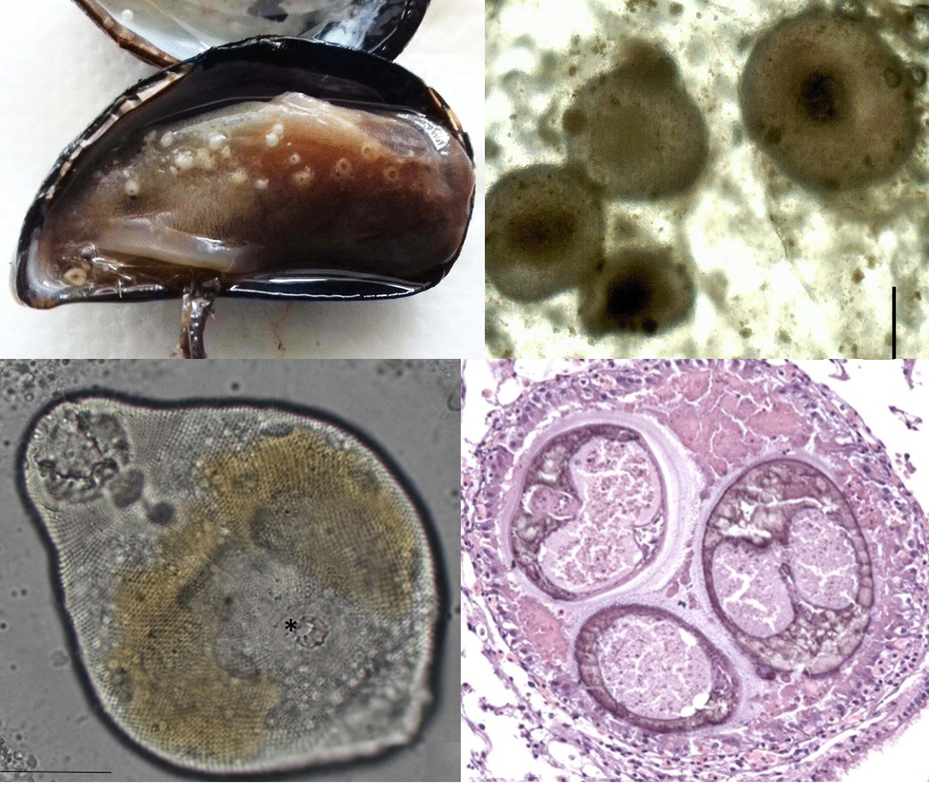
Pearls may look beautiful to us, but for some parasites, they represent a slow and claustrophobic death. Pearls are secreted by the soft and fleshy mantle, the part of a mollusc's body that also produces the shell. Indeed, pearls and shells are made from the same material - calcium carbonate. For the shellfish that produce them, pearls are battle scars of their fight against parasites.

Top left: Mussel infected with Parvatrema, Top right: Pearls from a mussel Bottom left: Parvatrema metacercaria stage from a mussel, Bottom right: Cross-section of a pearl showing three flukes trapped within.  
Top row of photos from Fig 1 of this paper. Bottom row of photos from Fig. 2 of the paper.

Bivalves are host to a wide range of different parasites that use them as a home, a site of propagation, or even as a convenient vehicle to their next host. One of the most common types of parasites that infect bivalves are trematode flukes. Some species embed themselves stubbornly in the mollsuc's tissue, others impair their ability to use parts of their body, and there are even some that end up castrating their shellfish host. Sometimes, these seemingly passive molluscs put up a fight against these tiny intruders, especially when they get into the mantle fold. And they do so by secreting calcium carbonate around the invading parasite, smothering these flukes alive - and the result of that gruesome interaction is a pearl.

The study being featured in this post looked at the frequency of pearls and parasites in mussels on the northwestern Adriatic coast. The flukes that are most commonly associated with pearls there are those from the Gymnophallidae family, and this study focus on one particular genus - Parvatrema. These flukes use mussels as their intermediate host, where the larvae temporarily reside and develop until they are eaten by shorebirds - this parasite's final host.

Out of the 158 mussels that the researchers examined, about two-thirds of them were infected, and most of the mussels had a mix of both live flukes and pearls.Their parasite load varied quite a lot, from some mussels with a few flukes, to one with over 3700 flukes. But on average, each mussel harboured about 200 flukes. The flukes were scattered throughout the mussel's body, but most were concentrated near the gonads, and some were found at the base of the gills. A few were squeezed in between the mantle and the shell - and it is those that are at the most risk of being turned into pearls. 

Speaking of which, about half the mussels that the researchers examined had pearls of some sort in them. But there were far fewer pearls than there were flukes. Each mussel had 35 pearls on average, but they were nowhere near the size of pearls most people associate with jewellery. These pearls were about the same size as fine sand grains, but they were pearls nevertheless - complete with entombed fluke(s) in each of them.

The high prevalence of Parvatrema in mussels from this area means that it could be risky to set up mussel farms there, at least near the coast where the parasite's bird hosts like to hang out. No one wants to buy mussels riddled with parasites, and while pearls are considered as valuable, the type of pearls found in these mussels only decrease their market value. That is one of the reasons why some mussel farming operation are located offshore where they won't be exposed to Parvatrema and other parasitic flukes. 

Based on the results of the study, pearl formation seems a bit hit-or-miss as a defensive mechanism. The majority of flukes get away with living rent-free in the mussels without setting off the pearly deathtraps, and it's not entirely clear why some of them trigger pearl formation, while most flukes are left alone. Despite this, some recent studies indicate that bivalves are not the only molluscs that can entomb their parasites that way. Some land snails are also capable of sealing away various parasites such as flukes and roundworms into their shell. 

So it seems the molluscs have evolved a general two-in-one defensive package that can potentially protect them against both predators and parasites. While neither shell nor pearls offer guaranteed protection against predators and parasites respectively, it's still better than having nothing at all.

Reference:
Marchiori, E., Quaglio, F., Franzo, G., Brocca, G., Aleksi, S., Cerchier, P., Cassini, R. & Marcer, F. (2023). Pearl formation associated with gymnophallid metacercariae in Mytilus galloprovincialis from the Northwestern Adriatic coast: Preliminary observations. Journal of Invertebrate Pathology 196: 107854.

April 21, 2022

Aggregata sinensis

Apicomplexa is a diverse phylum of single-celled parasites. They are found in a wide range of different animals, and includes some well-known species which can infect humans such as the malaria-causing Plasmodium, the infamous and widespread Toxoplasma gondii, and the gut-busting Cryptosporidium. But it is not as if this group has any particular affinity for humanity - humans are just one species among many across the animal kingdom that are hosts for apicomplexan parasites. Most of the more well-studied apicomplexans are those that infect terrestrial animals, especially domesticated species, but far less is known about apicomplexan parasites that are found in the marine realm.

Top left: Aggregata sinensis oocysts in the membrane between the arms of an octopus. Top right: Oocysts in the branchial heart.
Bottom left: Sporocysts found within an oocyst. Bottom right: Sporozoite released from a sporocyst.
Photos from Fig. 1 and Fig. 2 of the paper

Aggregata is a genus of apicomplexan which specifically targets cephalopods - mainly octopuses. Octopus can become infected from eating crustaceans such as shrimps which harbours the asexual stage of the parasite. Once they get into the octopus gut, the parasite takes over the digestive tract, and undergo sexual reproduction in the cells of the gut lining. There are twenty different known species of Aggregata, and it seems that for octopuses, there is no escape from this genus of parasite - even deep sea species living around hydrothermal vents are targeted by their own specialised species of Aggregata parasite.

So there are no doubt many other species of Aggregata out there which are still undiscovered. The paper featured in this blog post describes a species of Aggregata called Aggregata sinensis which has been found in octopus from the eastern-central coastal waters of China and the northern tip of Taiwan. The parasite was found infecting two species of octopus - the webfoot octopus and the long arm octopus - both of which are commercially important species that are caught by the local fishermen. 

The parasite was rather common, and depending on the location, between 20-100% of the octopuses that the researchers examined were afflicted with A. sinensis. Because the way an octopus becomes infected is from eating parasitised prey, Aggregata infection initially starts in the digestive tract, but it doesn't stay there for long. In heavy infections, the parasite spills over into other parts of the body in a very visible way. As Aggregata proliferates in the octopus, it leaves tell-tale signs of their presence in the form of white cysts that speckle the octopus' body. Those white cysts are called oocysts, which are the results of the parasite's sexual reproduction. Aggregata can wreak a destructive toll on the octopus's health. As the parasite proliferates, they smother the gut lining and destroy the submucosa cells, which compromise the octopus' ability to absorb nutrients. 

As if that's not enough, those white oocysts are filled with microscopic spheres called sporocysts which need to depart from the octopus' body to continue the life cycle, and they do so in a destructive manner. The release of those Aggregata oocysts necessitates the rupture and shedding of the surrounding hosts cells, resulting in ulcers and atrophy of the gut lining and connective tissues. Once free in the surrounding waters, should the sporocysts find themselves in an unlucky crustacean, they unravel to reveal their payload of worms-shaped sporozoites. These squirm out and settle in the crustacean's gut where they undergo asexual reproduction, and start the life cycle anew.

A recent study on the phylogeny of Apicomplexa suggests that Aggregata belongs to a group called the Marosporida - which occupies a key evolutionary position within Apicomplexa, separate from the rest of the phylum. Which means that understanding parasites like Aggregata may also help us understand the evolution of the Apicomplexa phylum as a whole, and how they became one of the most successful and ubiquitous group of parasites on the planet.

Reference:
Ren, J., & Zheng, X. (2022). Aggregata sinensis n. sp.(Apicomplexa: Aggregatidae), a new coccidian parasite from Amphioctopus fangsiao and Octopus minor (Mollusca: Octopodidae) in the Western Pacific Ocean. Parasitology Research 121: 373-381.

October 15, 2021

Caledoniella montrouzieri

Mantis shrimps (Stomatopoda) are some of the most formidable crustaceans in the sea; armed with trinocular colour vision, and a pair of powerful raptorial limbs that punch so hard, it generates supercavitating bubbles which collapse with such energy, the vapour within them briefly turns into white-hot plasma. Arguably, one of the most impressive animals in the sea. But to Caledoniella montrouzieri - a mantis shrimp is simply a big juicy host. 

Underside of a parasitised mantis shrimp, showing the male, female, and egg capsules of Caledoniella montrouzieri
Photo from Fig. 1 of the paper, taken by Ryutaro Goto.

Most parasitic snails belong to the Eulimidae or Pyramidellidae family, and both of them parasitise slow-moving or sedentary invertebrates such as echinoderms, molluscs, and polychaete worms. But Caledoniella has taken a different, independent route down to parasitism town. It doesn't belong to either of those families, and instead of a slow life feeding on some barely mobile hosts, it lives life in the fast lane, clinging to the belly of a nimble, predatory crustacean.

Such a lifestyle requires some specialised anatomy. In most snails, the foot is a flat muscular organ that is used for crawling over various surfaces. But in Caledoniella, the foot has been transformed into a big suction disc that allows it to cling firmly onto its very agile host. During the course of its evolution, it has also lost one of the key diagnostic characteristics of molluscs - the rasp-like radula in the mouth which snails use to scrape bits of food, be they algae or the flesh of other animals, into their mouth. Instead, it has a mouth that is more suited for suction feeding, and has highly developed salivary glands to facilitate its liquid diet. This snail is a vampire of mantis shrimp, sucking on their host's gill filament for that sweet, sweet hemolymph.

Caledoniella has some noticeable sexual dimorphism with the female snail being much larger than the males. When these snails mature, they pair up as a monogamous couple, living out their lives together on the underside of a mantis shrimp. But this happy couple likes to keep themselves to seperate parts of the mantis shrimp, with the female living near the tail of the shrimp, and the male living near the middle of the abdomen. Sitting between them are all the egg capsules they have been busily making together. This gastropod couple takes their toll on the mantis shrimp, which experience stunted growth, reduced moulting, and infertility.

So how did Caledoniella ended up with its unique way of life? The closest living relatives of Caledoniella are snails that live as roommates with mantis shrimps, hanging on the walls of the crustacean's burrow. While these snails are frequently in the presence of the burrow's main tenant, that's as far as their relationship with the mantis shrimp goes. They are strictly commensals that never lay their foot on the mantis shrimp, and it is likely that was the lifestyle of Caledoniella's ancestors. But at some stage, after living in such close quarters with mantis shrimps for so long, some of those meek wallflower snails just couldn't resist getting more intimate and started taking a bite of its crustacean roomie, thus giving rise to the clingy blood-sucking Caledoniella

But when you trace its evolutionary history even further back, it seems those mantis shrimp roommates have themselves evolved from snails that originally lived in the burrows of an entirely different animal - spoon worms! So the ancestors of Caledoniella switched from sharing quarters with spoon worms, to living with mantis shrimp, to living on mantis shrimps

Perhaps somewhat surprisingly, Caledoniella is not the only mollusc that spend their lives clinging to the mantis shrimp - there are also a few species of tiny clams from the Galeommatoidea family called yoyo clams that live attached to the mantis shrimp's belly.  But unlike Caledoniella, they don't go as far as to feed on their host's blood. These clams receive protection from living on the belly of this heavily-armed crustacean, and the host's agile movements provide it with plenty of water flow for all their respiratory and filter-feeding needs. While they aren't blood-suckers, they seem to have followed the same evolutionary pathway as Caledoniella, evolving from ancestors that originally lived as commensals in the burrows of mantis shrimps.

For molluscs, it seems that sharing room with a marine benthic invertebrate is a surefire gateway to becoming a clingy parasite.

Reference:
Goto, R., Takano, T., Eernisse, D. J., Kato, M., & Kano, Y. (2021). Snails riding mantis shrimps: Ectoparasites evolved from ancestors living as commensals on the host’s burrow wall. Molecular Phylogenetics and Evolution, 163:107122.

May 18, 2021

Anisakis physeteris

Being on top of the food chain sounds like it'd be pretty awesome - all the other animals in the ecosystem are potentially your food and nothing else hunts you. In reality, it also means that there are many parasites out there that see you as prime real estate, a nice place to settle down and start a family. And there's no way for you to avoid them since many of those parasites would be climbing their way up the food chain via the prey animals you have been eating. Nowhere is that more obvious than in the ocean.

The oceans are filled with parasites - not that you'd necessarily know since the vast majority of them are hidden out of plain sight within the body of their hosts. Many of them are parasitic worms that treat the oceanic food web like a transit system, using predator-prey interactions to get from one host to another. This post is about a study on two nematodes that cross path inside some oceanic squids

Left (a, c): Lappetascaris larva (top) embedded in squid mantle muscle, (bottom) viewed under the microscope.
Right (b, d): Anisakis physeteris larva (top) in squid testis, (bottom) viewed under the microscope
Photos from Figure 1 of the paper

A group of researchers from Italy looked at parasitic roundworms that are found in the umbrella squid and the reverse jewel squid. Both of them belong to a group of squid called the "cock-eyed squids", which are commonly found in the mesopelagic zone. The squid that the researchers examined were caught as by-catch from commercial trawling vessels that were operating off the coast of Italy and Naples, and every squid that they looked at were infected with some kind of nematode larvae. 

Most of the nematodes were of a genus called Lappetascaris, along with another species which was identified as Anisakis physeteris. While both of those parasites look superficially similar and sometimes co-infect the same squid, there are some key life history and life cycle differences between them. 

For parasites, a host is not a single homogenous entity, but a collection of different microhabitats, and each parasite species has their own taste when it comes to fine-scale real estate. In this case, the researchers found that A. physeteris mostly settled in the squid's testis whereas Lappetascaris preferred embedding itself in the firm mantle musculature (the part of squid which are sold on the market as "squid tubes").

But these worms don't just differ in the part of the squid they prefer, but also which species of squid they infect. While Lappetascaris was found in both the umbrella squid and the reverse jewel squid, A. physeteris was choosier, and was only found in the umbrella squid. Finally, the two worms complete their life cycles in totally different animals. Lappetascaris reaches maturity in the gut of large teleost fishes such as swordfish and billfish, whereas A. physeteris needs to get into the stomach of a sperm whale - as denoted by its species name (the genus name for sperm whale is Physeter).

This may explain why A. physeteris was only found in the umbrella squid. Compared with the reverse jewel squid, umbrella squid venture into much deeper water which overlaps with the sperm whale's usual hangouts. And this exposes them to infective stages that are being released from sperm whales which have hundreds and thousands of adult Anisakis worms in their gut.

While the popular perception of the sperm whale often depict them as duking it out with the giant squid, the majority of their diet is composed of more modestly sized cephalopods, and the umbrella squid seems to form a major part of their diet. That's not to say umbrella squid is not on the menu of other large oceanic predators like swordfish and billfish too (hence it is also infected by Lappetascaris larvae), but if you are a parasite that is looking for the ideal ride to get you into the belly of a sperm whale, you can't do much better than the umbrella squid. 

What about the Lappetascaris which are sharing that squid with A. physeteris? Well they better hope a swordfish would come along and snatched it up before it ends up in the belly of a marine mammal - an environment that it is ill-equipped to live in.

So while these two worms may sometimes meet in the same squid, they eventually have to go their separate ways - and reaching their respective final hosts would unfortunately spell doom for the other worm in the shared squid. As for the sperm whales, a belly full of yummy squid must inevitably lead to a stomach full of wriggly worms.

Reference:

August 13, 2019

Caulobothrium sp.

Scallops are highly prized as seafood because of their tasty adductor muscle and roe, but humans are not the only ones with a taste for scallops. These bivalves are on the menu for a wide range of marine animals including various crabs, snails, seastars, marine mammals, and fishes. And many parasites make use of these predator-prey interactions to complete their life cycles.

Scallops are an important part of the Peruvian aquaculture, but little is known about their parasites there. In the study we're looking at today, researchers collected samples of scallops from a scallop ranch in Sechura Bay over the course of three years between 2013 to 2015, to examine them for parasites. They ended up looking through a total of 890 scallops, and the parasite that they encountered most frequently were whitish cysts that turned out to be tapeworm larvae belonging to the genus Caulobothrium.
SEM and light microscopy photos of tapeworm larvae. The lower left photo shows the tapeworm's scolex
Photos from Fig. 1 and 2 of the paper
Those tapeworm larvae were embedded in the scallops' gonads, and their numbers ranged from just twenty to over two hundred per scallop. While the number of infected scallops varied each year, they were nevertheless consistently high, with about eighty to ninety percent of scallops harbouring tapeworms. While this level of prevalence may seem unusually high, this is actually comparable to previous studies on tapeworms in scallops from other regions, so this is nothing too out of the ordinary.

Ultimately, those tapeworms are waiting for a rendezvous with the final host which, based on what is known about other species of Caulobothrium around the world, is the most likely a ray of some sort. Tapeworm species in the Caulobothrium genus have been reported from eagle rays in the waters of United States and Chile, as well as stingrays on the coast of Australia. On the coast of Peru, the adult stages of Caulobothrium have been found in the gut of both eagle rays and cownose rays, and given the circumstances, it is likely that the tapeworms found in the scallop gonads represented the larval stage of those worms.

Rays have specialised jaws armed with heavy, rounded teeth that allow them to crunch through the shell of bivalves such as scallops, and this tapeworm make use of their taste for shellfish to complete their life cycle.

Tapeworm larvae are not the only parasites with an affinity for scallop roe. Flukes in the Bucephalidae family also infect the gonads of scallops and turn them into parasite factories that churn out streams of parasite larvae. Much like those flukes, the presence of so many tapeworm larvae in the scallop gonads can impair the scallop's reproductive capacity, which as you can imagine, would be a concern for scallop aquaculture since they can potentially reduce the number of scallop larvae produced during spawning season.

In terms of infected scallops' edibility, Caulobothrium is known for being host specialists which can only infect rays, so there is no real risk of these tapeworms infecting humans, but on an aesthetic level to most would-be consumers, scallops with tapeworm-filled roe simply look too gross to eat.

The life cycles of most marine tapeworms are not well understood, and of the over one thousands species of tapeworms which have been described from sharks and rays, the full life cycle is only known for a measly FOUR species. Finding and documenting the larval stage of such tapeworms in marine animals such as scallops can help us put together the biological puzzles that are their complicated life cycles, and work out the roles these parasite play in marine ecosystems.

Reference:
Castro, T., Mateo, D. R., Greenwood, S. J., & Mateo, E. C. (2019). First report of the metacestode Caulobothrium sp. in the Peruvian scallop Argopecten purpuratus from Sechura Bay, Piura, Peru. Parasitology Research 118: 2369–237.

December 12, 2017

Megadenus atrae

A few months ago, I wrote about a snail that forms galls in the spines of sea urchins, and while most people might not think of snails as parasites - let alone parasites that live on animals like sea urchins, sea stars, and sea cucumbers - the parasite-host relationship of snails and echinoderms actually goes back hundreds of millions of years. There are fossils of snail boreholes and galls on ancient echinoderms. In fact, they are probably one of the few examples of parasitism that leaves a clear trace in the fossil record. If a sea cucumber is to write a parasitology textbook, most of it would be devoted to snails.

(1) A pair of Megadenus atrae - female on the left, male on the right; (2) Drawing of a M, atrae showing the proboscis (pr) and the pseudopallium (pp) cut away to show the shell (sh); (3) The shell of M. atrae - the larger ones are the female snail
Photos from Fig. 1 of the paper

Most of these parasites are from a family of snails call Eulimidae and the study that this blog post is covering was focused on a species call Megadenus atrae.  This parasitic snail has a few peculiar features when compared with the kind of snails that most people would be more familiar with. The shell is mostly wrapped up in a fleshy hood call the pseudopallium with only the tip visible, and it also has a giant sucker-like proboscis which it uses to cling to its host.

While other parasitic snails may simply attach to the skin or reside in the spines of their echinoderm hosts, this snails hangs out at a very specific spot - M. atrae lives in the cloaca of Holothuria atra - the black sea cucumber.

As strange as it may seem to us land-lubbers, the sea cucumber's butt is a popular hangout or gateway for many animals. There's the pearlfish which inserts its slim body into the sea cucumber through the echinoderm's cloaca and uses it as a kind of living shelter (some species also nibble on the sea cucumber's gonads while it is in there). There are also various crustaceans that are perfectly at home in a sea cucumber's butt. It is at this prime piece of real estate that M. atrae spends its adult life

In this study researchers collected black sea cucumbers from the chain of islands known as the Nansei Islands which stretches from the southern tip of Japan to the north eastern part of Taiwan, and recorded the presence of this parasitic snail. The snail is not particularly abundant, it was only found at two of the seven island sites they sampled from, and even on a reef flat at Kuroshima where they were most common, it was only found in one out of every ten sea cucumbers. Megadenus atrae has also been reported from other parts of the world including New Caledonia, India, and Australia. And in those other studies, the prevalence of this snail range from one in ten sea cucumbers to as few as one in a thousand.

Given that this parasitic snail is sparsely distributed in the sea cucumber population, this presents some challenges when it comes to reproduction - the likelihood of a larval snail encountering a host which is already occupied by another M. atrae is low enough, but the chance of that snail being of the compatible sex is even lower. Unlike other symbionts like pea crabs which can leave their host for a booty call, the only mobile stage of M. atrae is when it is a free-drifting immature larva. Once they are in a sea cucumber's butt, they are there for life

While it is possible that the snail can send out some kind of pheromone to recruit other M. atrae to settle in their host, how can they guarantee the new arrival would be of the suitable sex? After all there's no dating apps for snails living in a sea cucumber's butt.

Despite such obstacles, the researchers noticed that these snails were always found in pairs, and always as a female-male pair. They suggested that that this parasitic snail might have a sex determination system which is similar to that of the tongue-biter parasite and a range of other animals call protandry. With a protandric system, the larva starts out life as an immature male. If it settles down alone, it grows into a mature female snail. But if the snail larva happens to settle in a sea cucumber which is already occupied by a mature female, it will grow into a mature male. That way, M. atrae ensures that it will end up with a suitable reproductive partner no matter the circumstance.

So life finds a way, even for a parasitic snail trying to find a life partner amidst a sea of unlikely butts

Reference:
Takano, T., Warén, A., & Kano, Y. (2017). Megadenus atrae n. sp., an endoparasitic eulimid gastropod (Mollusca) from the black sea cucumber Holothuria atra Jaeger (Aspidochirotida: Holothuriidae) in the Indo-West Pacific. Systematic Parasitology 94: 699-709.

August 16, 2017

Sabinella troglodytes

Snails are host to a wide range of parasites, especially parasitic flukes that turn snails into clone factories to pump out streams of parasite larvae. But there are species of snails which are parasites themselves, and many of them are parasites of echinoderms - the phylum of animal which includes the likes of seastars, sea urchins, and sea cucumbers.

Left: live adult Sabinella troglodytes, Centre top: snail eggs inside a spine gall, Centre bottom: snail feeding on spine gall,
Right top: Female snail feeding on top of a spine gall, Right bottom: A spine gall showing feeding scar from snail
From Figure 1 and 2 of the paper
This post features Sabinella troglodytes - a parasitic snail found off the coast of Brazil which lives on the body of the slate pencil urchin Eucidaris tribuloides. The paper featured in this post presented a description of its life-cycle and other natural history observation of this gastropod. Parasites tend to be very specific about what part of their host's body they live on, and if there's one thing that sea urchins are known for, it is their spines, and that's what S. troglodytes feed and live on

Most molluscs have a rasping organ call a radula which they use while feeding to scrape away at their food. In the vampire snail this has been modified into something like a syringe which they can use to stab into a fish to drink their blood. In predatory whelks, the radula is used like a file to rasp away at the hard shell of their prey (usually another mollusc) to access the soft, gooey centre. But that is not how S. troglodytes feed on its sea urchin host. Unlike most of its gastropod relatives, S. troglodyte has lost its radula - so how can it bore into the spine of a sea urchin to reach its tasty core? Based on their observations, the researchers who conducted this study concluded S. troglodyte is secreting some kind of corrosive substance to eat through the tough walls of the spine in order to gain access to all that soft internal spine tissue.

But this parasitic snail is not content to simply just feed on the sea urchin, they also alter the urchin's spines to make it a more comfortable home. Sabinella troglodytes is one of many species of gall-forming snails that parasitise echinoderms. As their name indicates, the slate pencil urchin is covered in straight, pencil-shaped spines - but the spines housing S. troglodytes look almost  like fattened tubers. Much like how gall-wasps can induce bulbous growths on their host trees, these gall-forming snails can cause growth abnormalities in the sea urchin's tissue. This is also somewhat comparable to Accacoelium contortum, a parasitic fluke that lives on the gills of ocean sunfish while wrapped in a cosy little flesh bag made out of the host's tissue.

It is currently unknown how S. troglodytes alters the sea urchin's spines, but it could be due to some other components in the snail's saliva - in addition to corrosive agent to erode the sea urchin's spine, it might also be spitting out growth factors that alters the tissue of the spine. In addition to being a cosy place to feed and hide from threats, these galls seem to be a bit of a love nest for S. troglodytes during the summer months. The researchers noted that between December and February, almost all the galls were mostly occupied by snail couples (consisting of a female and her smaller male mate) which have settled down to raise a brood of eggs. But for the rest of the year, the galls were filled with juvenile snails which had probably inherited the gall from their parents.

While the spines of many sea urchins are straight and narrow, they are not immune to tampering by the right parasite. Sabinella troglodytes shows that with a little biological renovation, one can turn even something like a sea urchin's spine into a cosy home suitable for raising a healthy brood.

Reference:
The gall-former Sabinella troglodytes (caenogastropoda: Eulimidae) and its association with Eucidaris tribuloides (Echinodermata: Echinoidea). Journal of Conchology 42: 371-377.

November 12, 2015

Colubraria reticulata

Vampires have undergone a lot of image change over the centuries and they are a common part of many culture's mythology. But vampires are also a common part of nature. Blood sucking is a life style found in over 14000 known living species. Even those vampires themselves have blood suckers that feed on them. But living as a blood-sucker require special adaptations, and one particularly unlikely vampire is Colubraria (formerly known as Cumia) reticulata, the vampire snail. It is a marine snail that feed on fish blood and it belongs to a family of vampire snails called the Colubrariidae - at least six species are known to feed on blood and it is quite likely that it is a trait shared by the entire family.
Image modified from Figure 2 of the paper

So just how does a snail feed on a comparatively agile animal like a fish? First of all, they feed at night when fish are asleep, a survival tactic shared by other blood-feeders like vampire bats. They also have modified mouthpart can can slice flesh like a tiny scalpel, which is mounted at the end of a long proboscis that can stretch to three times its body length. This enables it to bypass even a parrotfish's mucus sleeping bag which normally protects it against other nocturnal blood-suckers.

But those behavioural and anatomical adaptations are just the start, most of the tools C. reticulata brings to this blood feast exist on a molecular level. The vampire snail is able to secrete a range of specialised proteins, most of which have multiple effects on the host and overlap in their functions.

First of all when the snail is about to cut into the fish's flesh, it spits out an anaesthetic similar to compounds secreted by other blood suckers like mosquitoes, to numb the area of incision. Once C. reticulata gets access under the fish's skin, other types of compounds come into play. A major problem for any would-be vampire is the natural tendency for blood to clot. Imagine drinking a smoothie and suddenly it turns into a big block of solid curd. So during feeding, C. reticulata secretes a chemical cocktail that disrupts the process of blood clotting and wound healing. Furthermore, the anti-coagulant action needs to be active until the blood is fully digested, so the snail also have secondary glands in its oesophagus that secrete other types of proteins to keep the blood liquefied as it sits in the snail's gut.

In addition to anti-coagulants, C. reticulata also spits out vasopressive compounds that increases the fish's blood pressure. This is very important to the vampire snail's feeding style because its long proboscis is actually not very muscular - so it is not that good at sucking blood. Instead, the snail injects compounds that increase the fish's blood pressure so that it will actually be pumping blood into the snail's gut. When scientists looked into the vampire snail's molecular arsenal in more details, they found that many of the proteins secrete by the vampire snail can be considered as pretty standard fare for a vampire and are similar to those found in terrestrial blood-feeders like ticks and mosquitoes.

However, C. reticulata also has a few tricks up its shell which are unique compared with other vampires, in particular the complex of protein which it secretes to temporarily suppress the fish's coagulation and healing mechanism. This is actually quite a feat because comparing with other vertebrate animals, fish are very good at repairing vascular injuries, especially in delicate blood-rich organs like the gills which are exposed to the external environment.

Another substance unique to the vampire snail is turritoxin - which is also produced by the coneshell. At this point, scientists are unsure how vampire snail (or the cone shell) uses turritoxin in their hunting behaviour, though it is possible they release it as a way of lulling the fish into a compliant state. Scientists have observed that fish which are approached by the coneshell enters a kind of "hypnotic" state before they get stung with the coneshell's highly lethal neurotoxin. Perhaps the vampire snail also release turritoxin to coax its victim into a deeper state of sleep.

By investigating the molecular arsenal of the vampire snail, scientists can gain insight into how the vampire snail evolved to be a blood-feeder. In addition, some of compounds secreted by C. reticulata can finely manipulate the physiology of their host, and examining them in detail may lead to the development of compounds with useful medical and pharmaceutical applications.

Reference:
Modica, M. V., Lombardo, F., Franchini, P., & Oliverio, M. (2015). The venomous cocktail of the vampire snail Colubraria reticulata (Mollusca, Gastropoda). BMC Genomics,16: 441.

April 26, 2015

Nepinnotheres novaezelandiae

Life as a pea crab seems pretty sweet, you spend most of your time sitting snug and protected within the armoured shelter of a shellfish, while your host's filtration current bring you a constant stream of oxygen and food - everything that a pea crab needs for a good life. Well, almost everything - because there's more to life than just being protected and fed. Much like other organisms pea crabs need to reproduce - that's how evolution works, and unlike many other living things, a pea crab cannot just clone itself.
Male Nepinnotheres novaezelandiae squeezing in between
the valves of a mussel. From video here.
So when it comes to reproduction, the balance of living the pea crab life tips from "pretty sweet" to "absolutely terrible" - especially if you are a male pea crab. For them, trying to find a mate is a harrowing challenge than none of us can possibly imagine. First of all, to reach a potential mate, you have to leave your host, which means you have to pass the gates that are the valves of the host mussel, without being caught in between them. At that stage, those valves that had offer such formidable protection for the pea crabs then become death traps, with about 13% of male crabs meeting their end at this molluscan gate - their bodies litter the mussel bed.


Once outside, the male pea crab faces even more challenges. These tiny crustaceans, which are more accustom to a cosy life inside a shellfish, have to cross the treacherous, open areas of the mussel bed, filled with horrible monsters (in the form of predators like fish, octopus, and larger crustaceans) for which an exposed pea crab is just a convenient snack. Furthermore, male crabs only make up 20% of the population despite the more or less equal sex ratio of immature pea crabs. The length that they have to go to just to find a mate probably has something to do with that...

Despite the odds, almost 90% of all female crabs in the population carry fertilised eggs, so some male crabs must be having successes - but how?

The researchers who conducted this study noticed that the male pea crabs always set out under the cover of darkness when they will be less likely to be spotted by predators, and also because mussels are more relaxed at night. From the researchers' perspective, this also means that all the experiments and observation of pea crab behaviour had to be done in the dark. So in addition to sea water tanks, they set up some infra-red cameras to capture footages of all this activity - like some kind of voyeuristic shellfish reality TV show.

So what would coax a male crab out of his cosy home? To find out, the researchers constructed a flow-through observation chamber lined with PVC tubes in which they placed pea crab-infected mussels. When they placed a mussel with a female crab upstream of one with a male pea crab, the male crab would exit their host 60% of the time, roused into action by something which seem to secreted by the mussel (or the female crab in the mussel) upstream.

Male Nepinnotheres novaezelandiae tickling the mantle edge
of a mussel. From videos here.
The crab then makes its way to the mussel where the female crab resides. Once there, the pea crab patiently tickles the mussel's mantle fringe with its legs to try and convince the bivalve to let it enter. This is also the reason why the male crab only do this at night, because a mussel's response to such tickling can be very different in daylight. Try the same trick during the day and the bivalves would slam shut, crushing the amorous crab between its valves. On average, the crab will spend over three hours fiddling away at the mussel to coax the shellfish into opening up.

Additionally, in a different flow-through seawater tank where the crabs were given more freedom to roam from one host to another, the researchers recorded how long it took for the male pea crab to leave its host and reach a mussel containing a female crab. The entire journey from exiting the original host mussel to reaching their final destination took seven hours on average, though this varies from a quick hour-and-a-half jolt, to an eighteen-and-a-half hour-long trek for one particularly unfortunate individual.

So when love (or at least lust) is in the water, the pea crab will give up the easy life, and risk life and limbs for an evening rendezvous.

Reference:
Trottier, O., & Jeffs, A. G. (2015). Mate locating and access behaviour of the parasitic pea crab, Nepinnotheres novaezelandiae, an important parasite of the mussel Perna canaliculus. Parasite, 22: 13.

October 9, 2014

Calyptraeotheres garthi

There are many parasites that castrate their hosts - the parasitic barnacle that feed on the velvet belly lantern shark, the nightmarish Sacculina that takes over the body of a crab and turns it into a baby-sitting zombie, or the nematode that sterilise queen hornets and turn them into mobile nurseries.

Limpet without (left) and with (right) C. garthi
Image from Fig. 2 of the paper
There are two main ways that a parasite might stop its host from having babies. It can manipulate host physiology and suck up resources that would have otherwise gone into growing and maintaining the host's reproductive organs, which then simply shrivel up from being starved of nutrients. Alternatively, a parasite might actively occupying the space where the reproductive organs or resulting broods would normally be, displacing any would-be eggs and/or offspring.

Pea crabs are tiny crabs that specialise in making a living inside the body of marine invertebrates like various molluscs and echinoderms. The species featured in the study we are looking at today is Calyptraeotheres garthi, which lives inside limpets on the coast of Argentina. The researchers that conducted this study started noticing that most limpets that have crabs tend not to produce any egg sacs during the reproductive seasons, so they tried to find why that is by examining limpets from the field and by raising both crab-free and crab-infected limpets in the lab to compare their reproductive output.

Out in the wild, about a third of the crab-free limpets carried eggs during the breeding season, and a few of the limpets infected with smaller male or juvenile crabs managed to produce at least some eggs. But the limpets that were harbouring fully-mature female crabs had no eggs at all. This was similar to what they observed in their captive limpet population - while half of the uninfected limpets could spawn and produce a brood, none of the crab-laden limpets managed to do so.

Even though the crab-infected limpets did not produce any eggs, they had intact ovaries which were filled with oocytes (egg cells) ready to go. And when the researchers they remove crabs from infected limpets, they quickly recovered. Within a week or two after crab removal, those limpets started producing eggs again. So how was C. garthi stopping the limpet from producing a brood? Is the crab hogging all the nutrients and leave none to the developing eggs?

These limpets feed by collecting phytoplankton (floating, single-celled algae) into a mucus string around the gill fringe, and that is what C. garthi feeds on - pilfered strings of algae-loaded slime from its host. While you might think this free-loading would be taking a major toll on the limpets, they do not seem to be too affected by this. Crab-infected limpets carried on feeding and digesting at the same rate as crab-free ones, so the little crustaceans was not affecting the limpet's usual energy intake - at least not to a level that the host cannot compensate.

Calyptraeotheres garthi with a stomach full of phytoplankton
Image from Fig. 4 of the paper
So C. garthi is not stealing much nutrient from the hosts, but is it actually adding caviar to its green salad and treating itself to the limpet's eggs? After all, it is in the perfect position to snack on a serving of freshly-produced eggs. But when the researchers examined field-collected limpets that harboured crabs but still managed to squeeze out a few eggs, none of their egg sacs showed no signs of damage by the crab, which means C. garthi were only interested in one thing - sweet green slime strings.

Despite not being a severe physiological drain, their physical presence occupy the spot where the limpet would carry its brood of eggs. So while the limpet can still carry on feeding and digesting as normal, it gets brood-blocked by the crab.

The relationship between the limpet and the crab is made even more complicated by seasonal changes. During summer, the larger limpets that are infected with C. garthi are healthier than crab-free limpets, but in winter the situation is reversed. However, on top of that, during winter, only the larger limpets with crabs suffer a decline in health, while those below a certain size threshold gets away with carrying around a food-stealing crab without any severe consequences.

From our perspective, under certain circumstances, it might actually seem beneficial to have a pea crab, seeing as crab-harbouring limpets seems to be healthier during certain times of year. But from an evolutionary perspective, this pea crab is extremely harmful - by preventing its host from reproducing, it is effectively terminating that individual limpet's genetic lineage - all just for a mouthful of green slime.

Reference:
Ocampo, E. H., Nuñez, J. D., Cledón, M., & Baeza, J. A. (2014). Parasitic castration in slipper limpets infested by the symbiotic crab Calyptraeotheres garthi. Marine Biology, 161: 2107-2120.

August 1, 2014

Ismaila sp.

Those who have been reading this blog for a while might recall that this time last year, I featured some guest posts written by students from my Evolutionary Parasitology  (ZOOL329/529) class. Well, it is happening again for this year! For those who are unaware of this, one of the assessment I set for the students is for them to summarise a paper that they have read, and write it in the manner of a blog post, much like the ones you see on this and other blogs. 

I also told them that the best blog posts from the class will be selected for re-posting (with their permission) here on the Parasite of the Day blog. I am pleased to be presenting these posts from the ZOOL329/529 class of 2014. To kick things off, here's a post by Courtney Waters on a paper published in 2002 that documented the diversity of parasitic copepods that live inside sea slugs off the coast of Chile (see also this post from June this year).

Picture of infected sea slug from the paper
Bright colourful sea slugs are every diver’s ultimate find. Imagine getting up close to it with that macro lens and... wait, what's that protruding from the slug's side? They appear to be the egg sacs of an endoparasitic copepod - small crustaceans, which parasitises the insides of these soft‐bodied molluscs. The aim of the study I am writing about for this post was to expand existing knowledge about these endoparasites, particularly the genus Ismaila from the family Splanchnotrophidae. This particular genus is characterised by the presence of a pair of well-developed first appendages which are absent in related genera.

The six year study was based mainly in Chilean waters where different sea slug species were collected and examined for parasite infection. This was done simply by examining the sea slug externally without dissection as the egg sacs of the adult parasite protrude conspicuously from the abdominal wall of the host (see the accompanied figure). Over 2000 specimens from 47 species of sea slug were examined in such a manner and only 8 species of slugs were found to be parasitised by those copepods. These parasites are very host specific and each parasite species is only found in one host species. The overall infection rate was 13% which is the highest infection prevalence documented. Fortunately, these parasites only like the soft innards of our mollusc friends - otherwise I would not be so jealous of the scuba divers who were doing the collecting!


Obvious differences were seen between the infection rates of different host species, with some parasitised more than others. For example, in several species of hosts, only one individual was observed to be infected, whereas for other species the infection rate was almost 90%. The infection frequencies for two of the main sea slug host species did not vary much between years and seasons, though this would need to be verified with further studies. An additional result of the study was information on the evolution of these parasites. The disjunct distribution of the copepods along with their host groups suggest that these parasites had evolved from an ancestor that was not very host-specific, but as different populations became isolated, they evolved to be very specific to their hosts. This resulted in scattered pockets of area with high parasite abundance. As for why they have not spread out to wherever appropriate hosts are available, this is likely due to other life-cycle requirements of the parasite which are currently unknown.

In summary, the study found 4 new species of host for splanchnotrophid copepods, taking the world total to 47 host species (at least as of 2002 when this paper was published), with 12 of which being found in Chilean waters and 9 of them being host to copepods in the Ismaila genus. This means the waters of Chile have over a quarter of all known splanchnotrophid species. Additionally, the percentage of infected sea slug in Chile is ten times higher than anywhere else in the world - a fact that, if I was a sea slug in those waters, would probably give me the chills...

Reference:
Schrödl, M. (2002). Heavy infestation by endoparasitic copepod crustaceans (Poecilostomatoida: Splanchnotrophidae) in Chilean opisthobranch gastropods, with aspects of splanchnotrophid evolution. Organisms Diversity & Evolution, 2: 19-26.

This post was written by Courtney Waters

June 25, 2014

Ismaila belciki

Photo of infected Janolus fuscus
used with permission from Jeff Goddard
If you ever find yourself down by the sea, you may come across some very flamboyant sea slugs call nudibranchs. But beneath their colourful exterior, some of them are harbouring a dark secret in the form of a very strange looking parasite. These parasites live hidden inside the main body cavity of their molluscan host, so if you are unfamiliar with this particular critter, you might not even notice it. The main thing that gives away their presence are a pair of egg sacs poking out of the sea slug (see photo on the right). Those egg sacs belong to a parasite call Ismaila belciki - it is a crustacean, though it looks more like one of Cthulhu's lovechild or something out of Men In Black.

Ismaila and other copepods of the Splanchnotrophidae family are specialist parasites of sea slugs and they can get pretty big in comparison with their host, taking up substantial room and resources. Ismaila belciki infects Janolus fuscus, a nudibranch found along the west coast of North America from Alaska to California, as well as the shores of northern Japan. In some areas, such as Coos Bay, Oregon where the study we are featuring today took place, up to 80% of the slugs are infected with this odd creature. Having such a big parasite sitting in the middle of slug's body soaking up nutrient obviously carries some kind of cost - but just how much?

Photo of a female Ismaila belciki with an
embraced dwarf male front and centre.
Photo by and used with permission
from Maya Wolf
A pair of researchers from University of Oregon decided to find out just how costly this parasite is to its host. They compared the growth, survival, and reproductive capacity of infected and uninfected J. fuscus, and measured how much resources the parasite takes up.

While I. belciki did not seem to interfere with sea slug's growth, infected slugs do have a lower survival rate. Additionally, they have shrunken gonads that are only capable of producing about half as many eggs as healthy slugs. But the reproductive capacity of those afflicted sea slug suffers not just in terms of quantity, but in quality as well. In addition to producing fewer eggs, infected slugs also produced eggs that were smaller, and the baby slugs that hatch out of them also have lower survival rates.

So it seems I. belciki can be very harmful indeed, but it cause even greater harm if the parasite itself is breeding. The researchers noted that I. belciki bearing developing egg sacs exert a greater toll on the host than egg-free parasites. A female I. belciki is an egg-laying machine that can churn out over 88000 embryos per month and all the expenses for that are paid for by the host. To fuel the development of its eggs, I. belciki draws from the same pool of resources that the host normally use for its own egg production. Slugs with brooding I. belciki produce even fewer eggs than those that are "just" stuck with an egg-free parasite.

It is as if the sea slug is a factory that has been retooled from solely making slug babies into one which now has to divert some of its attention and raw material to making parasite babies too, via a proxy in the form of a female I. belciki. Given that Janolus fuscus usually only live for five months, by shorten their lives and severely reducing their reproductive capacity, I. belciki might actually be putting a natural check on the population growth of these flamboyant nudibranchs.

Reference:
Wolf, M., & Young, C. M. (2014). Impacts of an endoparasitic copepod, Ismaila belciki, on the reproduction, growth and survivorship of its nudibranch host, Janolus fuscus. International Journal for Parasitology 44: 391-401.

P.S. I will be attending the annual Australian Society for Parasitology annual conference in Canberra, Australia between 30th June to 3rd July. So watch for tweets about highlights from conference at my Twitter @The_Episiarch! Meanwhile, I have written a article for The Conversation about the crab-castrating barnacle Sacculina carcini - you can read it here.