"So, naturalists observe, a flea has smaller fleas that on him prey; and these have smaller still to bite ’em; and so proceed ad infinitum."
- Jonathan Swift
Showing posts with label snail. Show all posts

February 11, 2026

Galactosomum nagasakiense

Galactosomum nagasakiense is a parasite that lives rent-free in fish's brain and give them "Trematode Whirling Disease" (TWD). There are actually quite a number of different fluke species that all infect fish brains, but unlike those other species where hundreds of individual flukes are needed to change how the fish behaves, all it takes is a single Galactosomum sitting snugly in the centre of a fish's brain to send it into a tizzy. Whereas other flukes manipulate the neurotransmitters in the brain of their host, Galactosomum takes a more blunt force approach, as the mechanical pressure exerted by their larval cyst causes the surrounding brain matter to degenerate or undergo necrosis as a by-product of their presence. So this fluke literally gives its fish host brain rot.

From left to right: Line illustration of a Galactosomum nagasakiense cercaria, Photo of Galactosomum Type C, Photo of Galactosomum Type B (top) and Galactosomum cysts in the brain of a tiger buffer (bottom) with a close-up of the cyst (insert), photo of Cerithium dialeucum shell.
Photos and illustration from Fig. 1, 4, 6, 7 of the paper and from the D-PAF (Database of Parasites in Fish and Shellfish)

Galactosomum nagasakiense was first noticed in fish in the 1960s, and the adult fluke lives in the intestine of black-tailed gulls, who presumably appreciate the easy pickings that these brain rotted fish present as they swim in circles near the surface of the water. But where are the fish getting their flukes from? This is a important question because TWD can affect a wide range of fish from anchovies to kingfish, and is known for causing bouts of mass die-offs at fish farms among important aquaculture species such amberjacks and fugu (puffer fish). 

Despite its impact, the full life cycle of G. nagasakiense was unknown until the publication of the study we'll be looking at in this post. Deciphering the complex life cycle of parasites can often be a labour intensive and thankless task which involves a fair amount of informed intuition and luck. Hence for many parasite species which are otherwise well-studied, often the one aspect about them which remains unknown is their full life cycle.

In this study, researchers conducted field sampling on the coast of Tsushima Island near a tiger puffer farm that regularly had cases of TWD. Knowing the typical life cycles of digenean flukes, the source of the infection would most likely be a snail, but which one? There are many sea snails living among the rocks on the coast of the island which are prime candidates as the source of G. nagasakiense. The researchers in this study ended up sampling 1314 cerithioid sea snails, most of which (798) belonged to a species named Cerithium dialeucum, which turned out to be the snail that was serving as crawling Galactosomum factories.

Infections were not common, only 15 out of the 798 snails they sampled were shedding Galactosomum larvae, but they made up for their rarity with productivity. During their peak emergence period which lasted two to three weeks, each snail can pump out 3000 of those wriggling Galactosomum larvae per day, though this number declines to a steady (but persistent) trickle for another ten weeks. In total, each infected snails can release 16000 Galactosomum larvae into the surrounding waters, made possible by the prolific asexual stages of the parasite which has taken over the snail's internal organs. 

The free-swimming larval stage of digenean flukes come in all kinds of shapes and sizes, but Galactosomum stands out for having a thick, rippled tail which is about ten times as long as the larval fluke's actual body. When these wrigglers leave the snail, they swish their tail in a figure 8 motion that would attract the attention of any curious fish.

The researchers also discovered that G. nagasakiense was not the only species of Galactosomum in those snails, they found two other species of big-tailed cercariae, one of which has a sucker-like structure on its massive tail. It is likely that those species also infect fish, but it is unclear whether they also burrow into fish brains the way G. nagasakiense does. In any case, the discovery of these snails as the source of infections, can provide aquaculture managers with ways to limit or control TWD, such as situating the fish farms away from habitats where those snails are likely to be found.

The influence of parasites on their hosts are often overlooked because they are hidden out of sight inside of their hosts, but their impacts cannot be ignored. In this case, all it takes is a tiny fluke to cause some serious headaches for a whole lot of fish and fish farmers.

Reference:
Sugihara, Y., Iwasaki, R., Miyazaki, H., Shirakashi, S., Itoh, N., Nakano, T., Takano, T. & Ogawa, K. (2026). Elucidation of the life cycle of Galactosomum nagasakiense (Heterophyidae), the causative parasite of trematode whirling disease in marine fish, with discovery of congeneric species in the gastropod first intermediate host Cerithium dialeucum. Parasitology International 111:103190.

July 11, 2024

Parvatrema spp.

Parasites are known for their complex life cycles, especially among parasitic flatworms such as flukes. And the flukes that are being featured in today's post have life cycles that make them a fluke among flukes. This blog post is about an extensive study that culminated from 25 years of work, where a group of researchers were able to identify and describe five different fluke species with a peculiar life cycle adaptation.

Parvatrema sp. “quadriramis” cercaria stage (left), young metacercaria (centre), parthenogenetic metacercaria containing fully-formed metacercariae (right). Insert: Parvtrema parthenogenetic metacercariae in the hepatopancreas of a limpet.
Photos from Fig. 4 and Fig. 11 of the paper

In the cold waters of the northern European seas and the Sea of Okhotsk, there is a group of parasites with life cycles that defy the conventions of its class. They are five closely related species of flukes in the genus Parvatrema, and they spend parts of their lives lurking quietly in the bodies of clams and snails. In many ways, they're just like other digenean flukes, with multi-host life cycles that involve turning their first mollusc host into a clone factory, producing clonal larvae which go off to infect a second host, and culminating in sexually mature adults living in the gut of vertebrate animals. But these Parvatrema flukes have evolved to do some things differently once they reach their second host.

These flukes infect sea snails such as limpets and periwinkles as their second host. Some species sit in the extrapallial space - the fluid-filled gap between the snail's fleshy mantle and the shell, others get into the gonads and digestive organs. Usually, this is a relatively dormant stage of the fluke life cycle, where they literally sit and wait to be swallowed by the appropriate final host. But with these Parvatrema flukes, instead of simply sitting around waiting to be eaten by a bird like other flukes would, they have another round of asexual reproduction, as a treat.

Each of those immature flukes become filled with numerous miniature clones of itself until it is stuffed to the point of exploding. Some of them take it further, with unlimited consecutive generations of parthenogenetic clones, each fluke exploding into multiple clones and then each of those clones explodes into even more clones and so on, like a never ending series of Matryoshka dolls. On top of that, some of those Parvatrema doing unlimited fluke works are also able to produce cercariae - the free-swimming larval stages - which then go off to infect more sea snails to start the asexual cycle again. 

So why did they evolve this unique developmental stage? Parvatrema are tiny flukes, the adult stage only live for a few days in the gut of a bird, and they produce less than a hundred eggs - a relatively low number compared with other flukes which may produce thousands or even millions of eggs over their lifetime. To make things worse, the likelihood of any one fluke successfully infecting the right hosts at each consecutive stages of its life cycle is astronomically low, so they need to multiply their numbers at every chance they get.

Furthermore, the final hosts for these flukes are migratory birds which only come once a year - so they need to make the most of their brief stay by making sure that if they only eat one infected snail, instead of just getting a single or a dozen flukes in each snail, they're getting the whole gaggle of fluke clones arriving en masse into the bird's gut in their hundreds or thousands, ready to get on with the business of producing the next generation.

Essentially, these Parvatrema flukes recapitulate the process that most other digenean flukes only undergo in the first host. Asexual reproduction in the first host is arguably one of the key evolutionary innovation of digenean flukes, allowing them to offset the losses associated with the process of being transmitted from one host to the next. Since Parvatrema seems to do asexual reproduction at every possible opportunity, they can provide us with insights into how flukes evolved their one weird asexual trick that gave them an edge in the transmission game.

Reference:
Galaktionov, K. V., Gonchar, A., Postanogova, D., Miroliubov, A., & Bodrov, S. Y. (2024). Parvatrema spp.(Digenea, Gymnophallidae) with parthenogenetic metacercariae: diversity, distribution and host specificity in the Palaearctic. International Journal for Parasitology. 54: 333-355

October 10, 2023

Atriophallophorus winterbourni

In Lake Alexandrina of New Zealand lives a species of tiny freshwater snail called Potamopyrgus antipodarum. These snails are capable of alternating between sexual and asexual reproduction and can be extremely abundant. So much so that they have become invasive in many other parts of the world. Outside of their original home, they are free to proliferate to their heart's content. But back in New Zealand, these snails don't always have things go their way. They are held back by a whole menagerie of flukes which parasitise them - at least 20 different species in fact.

Top: Photo of the snail Potamopyrgus antipodarum by Michal Maňas, used under Creative Commons (CC BY-SA 4.0) license. Bottom: The metacercariae cysts of Atriophallophorus winterbourni, from Figure 1 of the paper.

These flukes have a range of different life cycles, but all of them use P. antipodarum as a site of asexual reproduction - converting the snail's insides into a clone factory and rendering it sterile in the process. These flukes might be the reason why these snails continue to engage in sex every now and then, despite asexual reproduction being much more efficient. Sex is necessary to maintain genetic variations - the key ingredient in the evolutionary arms race against all those flukes.

Researchers who have been studying these snails and their flukes noticed that while all 20 species of those parasite are essentially body snatchers that take over the insides of their unwitting host, one species - Atriophallophorus winterbourni - goes beyond simply messing with their host's physiology and seems to be influencing the snail's behaviour too. Snails infected with A. winterbourni tend to be found in the shallow areas of the lake. Among snails collected from the shallow water margin of the lake, they represent 95% of the infections. Is this because those areas just happen to be hot spots for snails to get infected with A. winterbourni? Or are these flukes actually coaxing the snails into hanging out in the shallows?

To figure out if there's something special about A. winterbourni, researchers compared snails infected with A. winterbourni with those that were infected with a different species of fluke - Notocotylus - to see if such behavioural change is simply a side-effect of fluke infection, or if it is something specific to A. winterbourni. The researchers did this by setting up a series of ten 5 metres long tubular mesh cages that stretched across different depth clines in the lake, from less than 0.8 metres at the shallow end to 2.8 metres at the deep end, with different sections of the cage corresponding to different levels of water depth. Using snails collected from two high infection prevalence sites at the lake, they added about 800 snails to the deepest section of each cage, and the snails were allowed to freely roam between the different sections. After eleven days, samples of snails were randomly collected from each depth level and examined for parasites.

There are some key differences in the life cycles of A. winterbourni and Notocotylus that makes them good for comparisons. Just like other flukes, A. winterbourni undergoes asexual reproduction inside the snail host, producing a whole load of clonal larvae. But unlike many other flukes, these clonal larvae stay in the snail and transform into cysts, where they wait to be eaten by a duck hungry for snails. In contrast, snails that are infected with Notocotylus release those clonal larvae into the surrounding waters, and they do so continuously over the course of about 8 months. These larvae attach themselves to vegetation or the shells of other snails, and are transmitted to grazing ducks that accidentally ingest them. Therefore, unlike A. winterbourni, their transmission is largely decoupled from the snail's own movement and behaviour.

So after those eleven days of allowing infected snails to roam in the cages, what did the researchers find? Well, snails infected with A. winterbourni were heavily distributed towards the shallow end, with over a third of the snails in that section being infected, which is over three times higher than the expected background infection level (11%). In the deepest section of the cage, A. winterbourni-infected snails were rare, representing only 3-5% of the snails in that section, and some of them were immature infections. In contrast, those infected with Notocotylus were found to have distributed themselves fairly evenly across the entire depth cline. It is unclear what exactly A. winterbourni is doing to the snails that makes them favour shallow water, but more importantly, why would they do this? What's in it for the fluke? Well, the final hosts for A. winterbourni are dabbling ducks that only feed in the shallow parts of the lake. So in order for A. winterbourni to make a successful rendezvous with its final host and complete its life cycle, it will have to prod its snail host into the shallows.

Atriophallophorus winterbourni belong to a family of flukes called Microphallidae, and there are a few other species in this family which are also known host manipulators. For example, Gynaecotyla adunca is a species that infects marine mudsnails, and it coaxes its mudsnail host into stranding itself onto beaches, which brings them closer to the crustaceans that serve as the next host in the parasite's life cycle. There's also Microphallus papillorobustus, which infects little sand shrimps (amphipods), and it alters their behaviour in a number of different ways that makes them more visible to hungry birds. Even though not all members of Microphallidae are host manipulators, it's a trait that does seem pretty common in this fluke family. Sometimes, in order to complete a life cycle, you just have to drag that snail to where you need it to be.

Reference:

January 15, 2023

Leucochloridium passeri

Leucochloridium paradoxum is one of those parasites which is immediately recognisable on sight. Commonly known as the "zombie snail parasite", its habit of turning the eyestalks of snails into pulsating candy canes has also earned it the name "green-banded broodsac", and it has appeared in various forms of media, including the opening of the Chainsaw Man anime. But far from just being a bizarre one-of-a-kind oddity, L. paradoxum is just one out of ten known species in the Leucochloridium genus which infect amber snails and produce these "broodsacs" structures. And these colourful, pulsating sacs are the key for distinguishing different species of Leucochloridium.

Left: Snail infected with Leucochloridium passeri collected from Hemei Township (Changhua County) by Jui-An Lin, photo from Fig. 1 of the paper. Top right: Labelled L. passeri broodsac from Fig. 1 of the paper. Bottom right: A trio of L. passeri broodsacs with metacercariae removed from an infected snail, from Supplementary video 3 of the paper

The adult stage of Leucochloridium are found in birds where they dwell in the cloaca or a special organ called Bursa of Fabricius. While parasite identification is usually based upon the various anatomical features of the adult parasite, in the case of Leucochloridium, the adult flukes of different species all look rather similar to each other. In contrast, the broodsac stages come in a wide variety of colours and patterns that are extremely noticeable and unique to each species. So short of comparing their DNA sequences, the colours and stripes of the larval broodsacs are the most reliable way to tell apart the different species.

This blog post features a study on Leucochloridium passeri, a species that was first described as adult flukes from Eurasian tree sparrows in Guangdong, and has subsequently been found across the Indomalayan realm. It is one of five different Leucochloridium species found in Taiwan, but it is the only one for which their broodsac stage has been documented. While not as well known as L. paradoxum, its broodsacs nevertheless present an attention-grabbing sight. You might recognise it from this video, which has gone viral and been posted all over the internet, usually without credit or attribution of the original source.

It can be easily distinguished from L. paradoxum and other Leucochloridium species by a distinctive wide band of red-brown patches or longitudinal stripes in the mid-section of each mature broodsac. Many people who have some familiarity with this parasite would know about the pulsating sacs forcing their way into the snail's eye tentacles, but what they might not know is that those are only part of the entire parasite mass residing within the snails.

Those pulsating "broodsacs" are actually the parasite's asexual larvae. In addition to the very flamboyant mature broodsacs, there are also translucent immature broodsacs which are tucked away deeper in the snail's body. Digenean flukes have an asexual stage in their life cycle, and in most flukes they produce hundreds to thousands of sausage-shaped asexual larvae in the snail's body. Those wriggly sausages would then give birth to free-swimming larvae called cercariae that are release into the environment where they infect the next host in the life cycle. In the case of Leucochloridium, the cercariae stay in those wriggly sausages and develop into round, jelly-coated cysts within the snail. Each mature broodsac can contain up to two hundred cysts, so when a bird swallows one of these colourful wriggling sausages, they are inviting hundreds of flukes to take up residency in their cloaca.

The L. passeri broodsacs described in this study were found in Yilan County in Taiwan, and they look very similar to some Leucochloridium broodsacs which have been found in Okinawa, Japan. They both have the distinctive wide band of red-brown stripes and splotches, and when researchers compared their DNA sequences, they found that they both belong to the same species - Leucochloridium passeri.

Relatively little is known about the birds that can serve as the final hosts for L. passeri, but researchers have noticed that the distribution of various Leucochloridium species in different zoogeographical regions seems to be related to the distribution of birds and amber snails which are native to those particular regions. Since some of those birds are migratory, this provides Leucochloridium with the means to cross oceans while seated snugly in the butt of their feathery host, ready to settle down wherever there are amber snails to infect. 

Reference:
Chiu, M. C., Lin, Z. H., Hsu, P. W., & Chen, H. W. (2022). Molecular identification of the broodsacs from Leucochloridium passeri (Digenea: Leucochloridiidae) with a review of Leucochloridium species records in Taiwan. Parasitology International 102644.

P.S. Leucochloridium is a very distinctive parasite and has been subjected to numerous artistic depictions, here's my own artistic depiction of Leucochloridium in the form of a Parasite Monster Girl.

January 18, 2022

Sulcascaris sulcata

Shellfish such as oysters, mussels, and whelks are popular fares among seafood lovers, but we are not the only ones with a taste for those molluscs. Despite being heavily-armoured, many of the animals that we consider as "shellfish" are also food for a variety of larger marine animals. But their status as prey to these larger animals also make them attractive intermediate hosts for a wide range of parasites, which use these shellfish as vehicles to reach their final hosts. And sometimes humans end up being the unintended destination.

Anisakidae is a family of nematode worms commonly found in some seafood, and it is responsible for anisakiasis - a type of seafood-borne illness. While their usual hosts are mainly marine mammals, when anisakid nematodes get in humans, they nevertheless try to burrow through the stomach or intestinal wall, causing a great deal of pain. Additionally, their tissue and protein secretions may also cause a severe allergic reaction, including acute onset anaphylaxis.

Most studies on anisakids and anisakiasis focus on the genera Anisakis and Pseudoterranova which are often found in fish. But there are many other lesser-known genera and species in the Anisakidae family. Sulcascaris sulcata is one such species and unlike other anisakid nematodes which use marine mammals or birds as their final hosts, Sulcascaris infects a marine reptile - specifically the loggerhead sea turtle - as its final host.

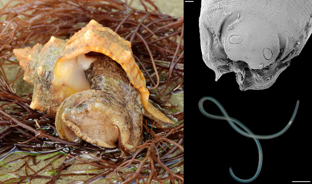
Left: Photo of a Purple-dye Murex by Holger Krisp, used under the Creative Commons (CC BY 3.0) license
Right: (top) SEM close-up photo of Sulcascaris larva's head, (bottom) a fourth-stage Sulcascaris larva  
(Photo of the nematode from Fig. 2 and Fig 4. of the paper)

Larvae of Sulcascaris have recently been reported from scallops and mussels - which raises some concerns since both are popular shellfish that are often eaten only lightly cooked or not at all. A recently published study adds another shellfish to that list - the purple dye murex, Bolinus brandaris. These large predatory snails are so-called because they used to be harvested to obtain a special type of purple dye. But in addition to their historic use in the textile industry, they are also commonly eaten in many parts of the Mediterranean.

A group of researchers in Italy obtained a haul of purple murex from fishermen on the coast of Baia Domizia, Italy, and brought the snails back to their laboratory to dissect them for parasites. Upon detailed examinations of the snails' organs, they found that 9 out of the 56 snails they obtained were infected with Sulcascaris larvae. However, infection intensity was very low, with most of the infected snails being parasitised by just a single nematode larva. These larval worms measured between one to five centimetres long, and were mostly lodged at the base of the snail's proboscis, with a few others found in the mantle cavity - the fleshy bag in a mollusc's body which houses its gills and other organs. 

Because of where those parasites are located in the snails, they can easily get overlooked during routine sanitary inspections, which only involve examining the outer appearance of the snail. The reason why those worms were mostly situated in those parts of the snail's anatomy might be due to their infection pathway. When the eggs of Sulcascaris are released from the turtle host, they settle onto the seafloor where they hatch into larval stages that lie in wait for an encounter with an unlucky murex. As the predatory sea snail moves across the sea floor, searching for prey with its proboscis, those larvae are sucked in via the inhalant current which transport them right into the snail's proboscis and mantle cavity.

Sea turtles with their strong beak and jaws can crack into these tasty snails which are off-limits to other animals, but it also means they end up with Sulcascaris in their gut. While this and previous studies on Sulcasacris have found that most shellfish carried only one or two individual nematodes, a turtle can eat a lot of shellfish, and over time may end up accumulating dozens or even hundreds of those worms in their stomach. When present in large numbers, these nematodes may cause ulcerous gastritis in sea turtles. But aside from that, not as much is known about this worm compared with its more famous, mammal-dwelling relatives, such as Anisakis.

So what does this mean for people who love eating shellfish? Based on prior experiments, it seems that Sulcasacaris can only infect sea turtles, so it is unlikely to become a zoonotic infection if it ends up being ingested by humans. Also, as mentioned above, when they are present, it's only one or two worms in each shellfish, and since purple murex are usually eaten after being cooked, this would kill the worm in the process. So the health risks presented by Sulcasacaris to any seafood consumers are relatively minimal.

However, like other anisakid worms, their tissue and secreted proteins may still potentially cause allergic reactions in some people, even after cooking. But not much is known about that possibility. The researchers suggested that at the very least, commercial fishermen should avoid harvesting snails from areas with sea turtles, since they are likely to be infected with Sulcascaris. This could be a win-win situation for both turtles and people - the turtles get to keep their feeding grounds to themselves, and seafood lovers can safely enjoy some worm-free sea snails. 

As the consumption of fish and other seafood increases around the world, there is a greater need for more studies on the wide variety of parasites that are found in seafood, along with people who have the skills and expertise to identify them - so we can continue to enjoy seafood without unintentionally barging into the life cycle of a parasite (and suffer its associating consequences).

Reference:
Santoro, M., Palomba, M., & Modica, M. V. (2022). Larvae of Sulcascaris sulcata (Nematoda: Anisakidae), a parasite of sea turtles, infect the edible purple dye murex Bolinus brandaris in the Tyrrhenian Sea. Food Control 132: 108547.

November 14, 2021

Fasciola nyanzae

Lake Kariba is the largest artificial lake in the world - Initially created in the late-1950s and early-1960s, it has since become inhabited by a wide variety of both endemic and introduced species. One of those introduced species is the water hyacinth (Eichhornia crassipes) which have made themselves quite at home in this giant artificial lake. These floating plants can proliferate at an alarming rate, clogging up the shorelines and sucking up vast quantity of water, nutrient and oxygen. They in turn are prime real estate for a variety of aquatic snails, and serves as breeding ground for many native and introduced snail species in the region.

Left: (a) Liver fluke Fasciola nyanzae, and (b and c) stomach flukes from hippos, from Fig. 1 of the paper.
Right: Photo of hippopotamus in Maasai Mara by Markrosenrosen, used under Creative Commons (CC BY-SA 4.0) license

So what does that have to do with parasites? For those who have been reading this blog for a while or know something about fluke biology would know the vital role snails play in the life cycles of digenean flukes - specifically the asexual part of their life cycles. Snails can get commandeered by flukes to act as parasite factories that churn out a stream of free-swimming clonal fluke larvae, and an infected snail can end up with 14-39% of their body mass being converted into parasite tissue. This means any place that is hosting an aquatic snail party would inadvertently become a fluke party too.

This study looked at how Lake Kariba has affected the transmission and infection of flukes in hippopotamus. Like any other large animals, hippos are host to a wide variety of parasites, but because they spend so much of their lives in water, this makes them an especially attractive host to a wide range of different flukes. In this study, researchers collected a variety of different aquatic snails from the northeastern shoreline of Lake Kariba, and examined them for fluke infections. They ended up finding six different species of snails that harboured hippo-infecting flukes.

Additionally, they were able to collect flukes directly from a male hippo which was culled near Kariba Town as part of the local wildlife management program. That hippo turned out to be home to a trove of flukes, ranging from long-bodied liver flukes dwelling in the bile ducts, to several hundred bright red stomach flukes that crowded the hippo's stomach. Using some good old-fashion morphological comparisons combined with DNA sequencing, they were able to identify those flukes and match them with the asexual stages which were found in some of the snails they collected.

The liver flukes, which were particularly large for flukes measuring at about 5 centimetres long, were identified as Fasciola nyanzae, a species known to occur in the region and are commonly recorded from hippos. But what's significant here is that whereas the usual snail host for F. nyanzae is Radix natalensis, a snail species which is native to the region, at Lake Kariba this liver fluke has also recruit two additional introduced snail species to serve as parasite factories to do its asexual reproductive biddings. This includes Radix plicatula from Asia, and Pseudosuccinea columella from North America. In fact, the invasive P. columella snail seems to be an even more receptive host to the hippo liver fluke than the native snails. What's more, F. nyanzae is not the only fluke that has taken a liking to the introduced snails. It seems that those bright red hippo stomach flukes also readily use both native and introduced snails for their asexual reproduction.

In addition, by examining the aquatic snails, the researchers were able to detect other flukes species which they didn't record from that male hippo they dissected, but are likely to be circulating in the local hippopotamus population. One of which was a species of hippo blood flukes (Schistosoma edwardiensis). Unlike the liver fluke or the stomach fluke, this blood fluke has only been found in native snails so far, but two of those snails were from the Bulinus genus, which has never recorded as hosts for S. edwardiensis before. This indicates that the fluke may be more prevalent and flexible about what snails it uses as hosts than previously thought.

While the human-infecting species of blood flukes such as Schistosoma mansoni and Schistosoma haematobium are extensively studied due to their public health significance, very little is known about blood flukes which infect wildlife, and information on species such as the blood flukes of hippos, including their ecology and life cycles, are very limited.

The conditions at Lake Kariba have created a haven for snails, which in turn makes it a hotspot for fluke infections, and the presence of introduced snail species exacerbates that. Because on top of contending with the native snails pumping out fluke larvae as they usually have, the hippos now have to deal with the flukes coming from the introduced snails as well. The introduced snails increase the overall parasite load in the environment - this phenomenon is known in disease ecology as "spillback", where an exotic host organism that was introduced to a new area might have started out largely free of its original parasites, but over time, they pick up some of the local parasite species and turn out to be even better hosts for them than their original hosts, thus amplifying the amount of parasite propagules in the environment.

Data and records on how environmental changes affect the epidemiology of wildlife diseases is severely lacking. And in order to obtain such information, it would require collaborations of researchers from many different fields including parasitology, veterinary science, ecology, and conservation biology. This study is but one small piece in a much larger story of how human activities impact the spread and transmission of infectious diseases on this planet.

Reference:

October 15, 2021

Caledoniella montrouzieri

Mantis shrimps (Stomatopoda) are some of the most formidable crustaceans in the sea; armed with trinocular colour vision, and a pair of powerful raptorial limbs that punch so hard, it generates supercavitating bubbles which collapse with such energy, the vapour within them briefly turns into white-hot plasma. Arguably, one of the most impressive animals in the sea. But to Caledoniella montrouzieri - a mantis shrimp is simply a big juicy host. 

Underside of a parasitised mantis shrimp, showing the male, female, and egg capsules of Caledoniella montrouzieri
Photo from Fig. 1 of the paper, taken by Ryutaro Goto.

Most parasitic snails belong to the Eulimidae or Pyramidellidae family, and both of them parasitise slow-moving or sedentary invertebrates such as echinoderms, molluscs, and polychaete worms. But Caledoniella has taken a different, independent route down to parasitism town. It doesn't belong to either of those families, and instead of a slow life feeding on some barely mobile hosts, it lives life in the fast lane, clinging to the belly of a nimble, predatory crustacean.

Such a lifestyle requires some specialised anatomy. In most snails, the foot is a flat muscular organ that is used for crawling over various surfaces. But in Caledoniella, the foot has been transformed into a big suction disc that allows it to cling firmly onto its very agile host. During the course of its evolution, it has also lost one of the key diagnostic characteristics of molluscs - the rasp-like radula in the mouth which snails use to scrape bits of food, be they algae or the flesh of other animals, into their mouth. Instead, it has a mouth that is more suited for suction feeding, and has highly developed salivary glands to facilitate its liquid diet. This snail is a vampire of mantis shrimp, sucking on their host's gill filament for that sweet, sweet hemolymph.

Caledoniella has some noticeable sexual dimorphism with the female snail being much larger than the males. When these snails mature, they pair up as a monogamous couple, living out their lives together on the underside of a mantis shrimp. But this happy couple likes to keep themselves to seperate parts of the mantis shrimp, with the female living near the tail of the shrimp, and the male living near the middle of the abdomen. Sitting between them are all the egg capsules they have been busily making together. This gastropod couple takes their toll on the mantis shrimp, which experience stunted growth, reduced moulting, and infertility.

So how did Caledoniella ended up with its unique way of life? The closest living relatives of Caledoniella are snails that live as roommates with mantis shrimps, hanging on the walls of the crustacean's burrow. While these snails are frequently in the presence of the burrow's main tenant, that's as far as their relationship with the mantis shrimp goes. They are strictly commensals that never lay their foot on the mantis shrimp, and it is likely that was the lifestyle of Caledoniella's ancestors. But at some stage, after living in such close quarters with mantis shrimps for so long, some of those meek wallflower snails just couldn't resist getting more intimate and started taking a bite of its crustacean roomie, thus giving rise to the clingy blood-sucking Caledoniella

But when you trace its evolutionary history even further back, it seems those mantis shrimp roommates have themselves evolved from snails that originally lived in the burrows of an entirely different animal - spoon worms! So the ancestors of Caledoniella switched from sharing quarters with spoon worms, to living with mantis shrimp, to living on mantis shrimps

Perhaps somewhat surprisingly, Caledoniella is not the only mollusc that spend their lives clinging to the mantis shrimp - there are also a few species of tiny clams from the Galeommatoidea family called yoyo clams that live attached to the mantis shrimp's belly.  But unlike Caledoniella, they don't go as far as to feed on their host's blood. These clams receive protection from living on the belly of this heavily-armed crustacean, and the host's agile movements provide it with plenty of water flow for all their respiratory and filter-feeding needs. While they aren't blood-suckers, they seem to have followed the same evolutionary pathway as Caledoniella, evolving from ancestors that originally lived as commensals in the burrows of mantis shrimps.

For molluscs, it seems that sharing room with a marine benthic invertebrate is a surefire gateway to becoming a clingy parasite.

Reference:
Goto, R., Takano, T., Eernisse, D. J., Kato, M., & Kano, Y. (2021). Snails riding mantis shrimps: Ectoparasites evolved from ancestors living as commensals on the host’s burrow wall. Molecular Phylogenetics and Evolution, 163:107122.

December 12, 2017

Megadenus atrae

A few months ago, I wrote about a snail that forms galls in the spines of sea urchins, and while most people might not think of snails as parasites - let alone parasites that live on animals like sea urchins, sea stars, and sea cucumbers - the parasite-host relationship of snails and echinoderms actually goes back hundreds of millions of years. There are fossils of snail boreholes and galls on ancient echinoderms. In fact, they are probably one of the few examples of parasitism that leaves a clear trace in the fossil record. If a sea cucumber is to write a parasitology textbook, most of it would be devoted to snails.

(1) A pair of Megadenus atrae - female on the left, male on the right; (2) Drawing of a M, atrae showing the proboscis (pr) and the pseudopallium (pp) cut away to show the shell (sh); (3) The shell of M. atrae - the larger ones are the female snail
Photos from Fig. 1 of the paper

Most of these parasites are from a family of snails call Eulimidae and the study that this blog post is covering was focused on a species call Megadenus atrae.  This parasitic snail has a few peculiar features when compared with the kind of snails that most people would be more familiar with. The shell is mostly wrapped up in a fleshy hood call the pseudopallium with only the tip visible, and it also has a giant sucker-like proboscis which it uses to cling to its host.

While other parasitic snails may simply attach to the skin or reside in the spines of their echinoderm hosts, this snails hangs out at a very specific spot - M. atrae lives in the cloaca of Holothuria atra - the black sea cucumber.

As strange as it may seem to us land-lubbers, the sea cucumber's butt is a popular hangout or gateway for many animals. There's the pearlfish which inserts its slim body into the sea cucumber through the echinoderm's cloaca and uses it as a kind of living shelter (some species also nibble on the sea cucumber's gonads while it is in there). There are also various crustaceans that are perfectly at home in a sea cucumber's butt. It is at this prime piece of real estate that M. atrae spends its adult life

In this study researchers collected black sea cucumbers from the chain of islands known as the Nansei Islands which stretches from the southern tip of Japan to the north eastern part of Taiwan, and recorded the presence of this parasitic snail. The snail is not particularly abundant, it was only found at two of the seven island sites they sampled from, and even on a reef flat at Kuroshima where they were most common, it was only found in one out of every ten sea cucumbers. Megadenus atrae has also been reported from other parts of the world including New Caledonia, India, and Australia. And in those other studies, the prevalence of this snail range from one in ten sea cucumbers to as few as one in a thousand.

Given that this parasitic snail is sparsely distributed in the sea cucumber population, this presents some challenges when it comes to reproduction - the likelihood of a larval snail encountering a host which is already occupied by another M. atrae is low enough, but the chance of that snail being of the compatible sex is even lower. Unlike other symbionts like pea crabs which can leave their host for a booty call, the only mobile stage of M. atrae is when it is a free-drifting immature larva. Once they are in a sea cucumber's butt, they are there for life

While it is possible that the snail can send out some kind of pheromone to recruit other M. atrae to settle in their host, how can they guarantee the new arrival would be of the suitable sex? After all there's no dating apps for snails living in a sea cucumber's butt.

Despite such obstacles, the researchers noticed that these snails were always found in pairs, and always as a female-male pair. They suggested that that this parasitic snail might have a sex determination system which is similar to that of the tongue-biter parasite and a range of other animals call protandry. With a protandric system, the larva starts out life as an immature male. If it settles down alone, it grows into a mature female snail. But if the snail larva happens to settle in a sea cucumber which is already occupied by a mature female, it will grow into a mature male. That way, M. atrae ensures that it will end up with a suitable reproductive partner no matter the circumstance.

So life finds a way, even for a parasitic snail trying to find a life partner amidst a sea of unlikely butts

Reference:
Takano, T., Warén, A., & Kano, Y. (2017). Megadenus atrae n. sp., an endoparasitic eulimid gastropod (Mollusca) from the black sea cucumber Holothuria atra Jaeger (Aspidochirotida: Holothuriidae) in the Indo-West Pacific. Systematic Parasitology 94: 699-709.

August 16, 2017

Sabinella troglodytes

Snails are host to a wide range of parasites, especially parasitic flukes that turn snails into clone factories to pump out streams of parasite larvae. But there are species of snails which are parasites themselves, and many of them are parasites of echinoderms - the phylum of animal which includes the likes of seastars, sea urchins, and sea cucumbers.

Left: live adult Sabinella troglodytes, Centre top: snail eggs inside a spine gall, Centre bottom: snail feeding on spine gall,
Right top: Female snail feeding on top of a spine gall, Right bottom: A spine gall showing feeding scar from snail
From Figure 1 and 2 of the paper
This post features Sabinella troglodytes - a parasitic snail found off the coast of Brazil which lives on the body of the slate pencil urchin Eucidaris tribuloides. The paper featured in this post presented a description of its life-cycle and other natural history observation of this gastropod. Parasites tend to be very specific about what part of their host's body they live on, and if there's one thing that sea urchins are known for, it is their spines, and that's what S. troglodytes feed and live on

Most molluscs have a rasping organ call a radula which they use while feeding to scrape away at their food. In the vampire snail this has been modified into something like a syringe which they can use to stab into a fish to drink their blood. In predatory whelks, the radula is used like a file to rasp away at the hard shell of their prey (usually another mollusc) to access the soft, gooey centre. But that is not how S. troglodytes feed on its sea urchin host. Unlike most of its gastropod relatives, S. troglodyte has lost its radula - so how can it bore into the spine of a sea urchin to reach its tasty core? Based on their observations, the researchers who conducted this study concluded S. troglodyte is secreting some kind of corrosive substance to eat through the tough walls of the spine in order to gain access to all that soft internal spine tissue.

But this parasitic snail is not content to simply just feed on the sea urchin, they also alter the urchin's spines to make it a more comfortable home. Sabinella troglodytes is one of many species of gall-forming snails that parasitise echinoderms. As their name indicates, the slate pencil urchin is covered in straight, pencil-shaped spines - but the spines housing S. troglodytes look almost  like fattened tubers. Much like how gall-wasps can induce bulbous growths on their host trees, these gall-forming snails can cause growth abnormalities in the sea urchin's tissue. This is also somewhat comparable to Accacoelium contortum, a parasitic fluke that lives on the gills of ocean sunfish while wrapped in a cosy little flesh bag made out of the host's tissue.

It is currently unknown how S. troglodytes alters the sea urchin's spines, but it could be due to some other components in the snail's saliva - in addition to corrosive agent to erode the sea urchin's spine, it might also be spitting out growth factors that alters the tissue of the spine. In addition to being a cosy place to feed and hide from threats, these galls seem to be a bit of a love nest for S. troglodytes during the summer months. The researchers noted that between December and February, almost all the galls were mostly occupied by snail couples (consisting of a female and her smaller male mate) which have settled down to raise a brood of eggs. But for the rest of the year, the galls were filled with juvenile snails which had probably inherited the gall from their parents.

While the spines of many sea urchins are straight and narrow, they are not immune to tampering by the right parasite. Sabinella troglodytes shows that with a little biological renovation, one can turn even something like a sea urchin's spine into a cosy home suitable for raising a healthy brood.

Reference:
The gall-former Sabinella troglodytes (caenogastropoda: Eulimidae) and its association with Eucidaris tribuloides (Echinodermata: Echinoidea). Journal of Conchology 42: 371-377.

April 15, 2017

Amphiorchis sp.

Sea turtles have a lot of different parasites infecting them - in a previous post I wrote about a recently published study on a parasitic copepod that eats sea turtle skin. But as well as external parasites, turtles are also infected by a range of internal parasites, many of which are digenean flukes, but the ones that cause the most harm are the blood flukes. While most parasitic flukes that infect turtles live in the intestine and cause relatively little harm unless they occur in large numbers, blood flukes, as their name indicates, live in the circulatory system.

Top: shell of the worm snail Thylaeodus rugulosus,
Bottom: cercaria of Amphiorchis sp.
Photo from Fig. 1. of the paper
Infection by these blood flukes can cause a range of disease symptoms, but by far the main source of grief to their reptilian host comes from the eggs they lay in the hundreds and thousands. These microscopic eggs get circulated in the turtle's blood vessels and many of them become lodged in various parts of the turtle's body where they can cause damage to the surrounding tissue as they triggered the body's immune response. Infected turtles often have internal lesions throughout their tissue and various organs.

But how these flukes get into the turtles in the first place has long been a mystery. Like other digenean trematode flukes, blood flukes require some kind of invertebrate host - usually a snail - in which they undergo asexual/clonal reproduction to produce free-swimming larval stages call cercariae (which is the stage that infects the turtle). But there are many different species of snails in the sea, which species is/are the one(s) pumping out those turtle parasites? It is like looking for a needle in a haystack in a bigger haystack which is the size of an iceberg.

Recently, a group of very sick loggerhead turtles presented an opportunity to find out more about the life-cycle of these blood flukes. At the Sea Turtle Rescue Centre (ARCA del Mar) (which was where the study described in the previous post took place). Some juvenile turtles were exhibiting symptoms that matched those caused by blood fluke infections and it seems that they were infected by a species of fluke from the Amphiorchis genus. So how were they getting infected? The water supply at the facility is semi-closed and pre-treated to remove any contaminants - so the turtles must be getting infected by cercariae which were coming from inside the facility.

The silver lining to all this was that it was a great opportunity to work out what Amphiorchis is using as a first host to produce clonal larvae. As mentioned above, for most species of flukes, this is usually a snail, and there is only one species of snail living in the facility - worm snails that were encrusting on pipes that delivered water to the facility. Dissection of some specimens confirmed that those snails were filled with the asexual stages of Amphiorchis and thus the source of infection.

The worm snail is a peculiar family of snails call Vermetidae. Unlike other snails, this family of tube-shaped molluscs have evolved to live like tube worms or barnacles by cementing themselves to a hard surface, and casting out a sticky mucus net to haul in microalga, zooplankton, or anything else that gets caught in its snot web (see this video here). This might explain why some sea turtles end up getting such a heavy infections out in the wild. Worm snails are abundant on reefs, or form part of reefs themselves, and sea turtles often hang out around such habitats.

Furthermore, the turtle's shell also happens to be a good surfaces for these snail to stick to - while few encrusting snails in themselves usually wouldn't cause much problem to a sea turtle, if they are infected with Amphiorchis or other blood flukes, these snails get converted into little parasite factories that pumps out a stream of turtle-infecting larvae - and what better host for those tiny, short-lived cercariae to infect than the turtle that the host snail is already encrusted on?

Reference:
Cribb, T. H., Crespo-Picazo, J. L., Cutmore, S. C., Stacy, B. A., Chapman, P. A., & García-Párraga, D. (2016). Elucidation of the first definitively identified life cycle for a marine turtle blood fluke (Trematoda: Spirorchiidae) enables informed control. International Journal for Parasitology 47: 61-67.

December 11, 2016

Leucochloridium paradoxum (revisited)

Parasites manipulating their hosts' appearance and behaviour is one aspect of parasitology which seems to have captured the public's imagination. The idea of body-snatching parasitic horrors taking over a host in both body and mind is one that evokes (and exceeds) the scenarios of many horror movies. Among the more well-known example of such parasitic body-snatchers is Leucochloridium paradoxum - the infamous zombie snail parasite, also referred to as the "green brood sacs".

But while L. paradoxum is the most well-known among its kind, it is just one of about a dozen different species in the Leucochloridium genus, all of which infect small land snails (mostly amber snails) and produce the pulsating brood sacs that people recognise. Traditionally, scientists have used the different colours and shapes of the brood sacs to tell apart different Leucochloridium species. More recently, this has been supplemented with genetic analysis, which has confirmed the validity of using brood sac colour and shape for species identification.

Left: A snail infected with two different Leucochloridium Right: Broodsacs of L. paradoxum and L. perturbatum from a double-infected snail
Photos from Fig. 1 of this paper 
In this study, researchers from Russia apply both techniques to examine cases of multiple Leucochloridium infections. Yes, as if being host to a single species of mind-manipulating parasite isn't bad enough, an amber snail can get infected with two (or more)! The researchers examined snails collected from the town of Lyuban in Russia, and upon dissecting them, found that while most of the infected snails were parasitised by the infamous L. paradoxum, a few snails had both L. paradoxum (green brood sacs) and another species call L. perturbatum (brown brood sacs). While simultaneous infections of different flukes species in snails are not uncommon, they also came across the first recorded case of a snail that was infected with three Leucochloridium species - L. paradoxum, L perturbatum, and the third species L. vogtianum which aren't as colourful, but was covered in warty projections

In other trematodes, competition between fluke asexual stages within the snails usually end up with one species overwhelming the other and gaining monopoly on the host. So it is possible that those snails that harboured multiple infection were merely be in the middle of a transitional state before one of the parasite colony is eliminated by the other. Had the snail been examined much later on, it might have revealed only a single parasite colony without any traces of a prior cohabitation with another species.

What most people might not know about Leucochloridium is that the prominent brood sacs are merely a part of an asexually-produced parasite colony inside the host snail. Unlike the asexual stages of many other trematodes which exist as genetically-identical but physically discrete stages call sporocysts or rediae, the asexual stages of Leucochloridium are stitched together into a writhing mass. This living colony is differentiated into different parts in a way that is comparable to the colonies of siphonophores such as the Portuguese Man'O'War. At centre of the parasitic mass, deep inside the snail's body, is where embryonic parasites are produced. As the embryos develop, they move through the colony's branches and into the extremities that form the colourful brood sacs, each packed full of mature parasite larvae that are ready to infect a bird.

This study also revealed another observation which provides insight into how these parasites reach the bird final host. The usual story is that infected snail are manipulated by the parasite into crawling to an exposed location where they can be easily spotted by hungry birds. The bird then mistaken the snail's pulsating, brood sac-engorged eyestalks for caterpillars, and peck them off. This is a classic story of parasite manipulation, told many times in multiple books and documentaries. While the validity of this story was partly demonstrated in 2013 when a study was published showing snails infected with Leucochlordium are indeed attracted to exposed and well-lit locations, it has yet to be demonstrated whether this actually enhance the likelihood of them (or at least their parasite) being eaten by birds
Broodsacs of L. paradoxum leaving the host snail. From Fig. 1 of this paper

But the researchers in this study observed that those pulsating brood sacs are not limited to expressing themselves in the snail's eyestalks. These sacs of parasite larvae can in fact leave the snail - possibly by rupturing through the snail's body wall. If the brood sacs of these parasites can exit the snail on their own and remain viable while still pulsating in the outside world for a brief period of time, then that significantly alter the above oft-repeated narrative of how this parasite is transmitted to its final host.

The parasitised snails might not need to have its eyes pecked out by the bird for Leucochloridium to reach its final host after all. Instead of treating the snail as a sacrificial lamb, the parasite could be using it as a unwitting courier that brings itself to an exposed location, drop off a few brood sacs, then those twitching brood sacs would attract the attention of a hungry bird on their own. It is still not a pleasant life for the infected snail - it is still stuck with a constantly regenerating parasite colony which is taking up almost a quarter of its body mass, but at least Leucochloridium would not be adding further injuries to insult by soliciting a avian attack.

Reference:
Ataev, G. L., Zhukova, A. A., Tokmakova, А. S., & Prokhorova, Е. E. (2016). Multiple infection of amber Succinea putris snails with sporocysts of Leucochloridium spp.(Trematoda). Parasitology Research 115:3203–3208.

P.S. Leucochloridium is a very striking-looking parasite and has been subjected to numerous artistic interpretations, so here's one of my own in the form of a Parasite Monster Girl version of a Leucochloridium-infected snail.

November 26, 2016

Cardiocephaloides longicollis

Human activities are having very significant impacts on the ecosystems of this planet and it is affecting every organisms. Parasites are not exempted from that - indeed parasites with complex life-cycles which involve many different host animals are in prime position to have their usual way of life altered by human intervention. The study being featured here today is on a parasitic fluke - Cardiocephaloides longicollis - which has a life-cycle that involves a carnivorous scavenging whelks, a variety of fish, and gulls. The researchers behind this study set out to investigate how commercial fisheries is affect the transmission dynamics of this parasite.

Left: Stained specimens of C. longicollis under light microscopy from here
Right: SEM of a closely related species Cardiocephaloides physalis from here
The asexual stages of C. longicollis reside in the body of whelks which acts as a kind of clone factory for the parasite, producing a stream of swimming larvae call cercariae. These larvae then go in the water to infect a variety of different fish. While C. longicollis has previously been recorded in 19 fish species, in this study the researchers found a further 12 species which are also viable hosts for C. longicollis, making for a grand total of 31 species of fish. The final host for this parasite are gulls, which acquire the fluke when they eat parasitised fish.

When it comes to C. longicollis infections, fish that hang around near the sea floor or the coast are the most loaded, most likely because they are in close proximity to the whelks which are sources of infection. Furthermore practically all the fish above a certain size (about 14 cm in length) are infected.  Fish in those size range have on average 73 C. longicollis larvae in their brain, with one unlucky fish recorded to have 220. Ironically, while these larger fish are the motherlode when it comes to parasites as they have been accumulating parasites for longer, since they live in deeper waters they are out of the gulls' reach. So regardless of their heavy larval fluke burden, because gulls can't get to them, all those parasites are at a dead end, destined to die or end up in the stomach of another predator which is not a gull - at least not without human intervention.

Many of the 31 species of fish which C. longicollis infects are either targeted by commercial fishing operations, or end up as by-catch. Many of those by-catch fishes - some of which are loaded with parasites - are discarded at the port. This pile of of parasite-laden fish present opportunistic gulls with a rich and accessible feast. It is a similar situation at fish farms, where the researchers found over half the fish there are infected with C. longicollis. At these facilities, organic matter from left-over feedstock, fish poop, and dead fish would also attract hungry gulls. But they're not the only ones who are attending the seafood party - being opportunistic carnivores, the whelks also come along to scavenge - so you end up with a situation where two of the host for C. longicollis are hanging out at the same location.

As the gulls feed on the discarded fish, they also are also getting infected with C. longicollis. Meanwhile, the flukes which have already reached maturity in the gulls' gut from previous feeding bouts are laying eggs which get pooped out into the water, right next to the whelks which have come for the scraps. And as mentioned above, the whelks are next host in the parasite's life-cycle, and some of those attending the feast will end up serving as parasite factories for C. longicollis in the future. For these parasites, this entire arrangement is a blessing - whereas without the activities of commercial fishing many C. longicollis larvae would have been consigned to a dead end in a large, benthic-dwelling fish, never to reach their final host. Indeed, the researchers found the fluke to be more abundant in areas with intensive fishing activity and aquaculture.

Cardiocephaloides longicollis is not the only parasite benefiting from commercial fishing activities, a study published a few years ago showed that overfishing can also benefits the tongue-biter parasite. A more recent study shows that clams living at commercially harvested sites are more heavily infected with parasitic flukes. While this does not apply for all parasites, as many would actually be negatively affected by commercial harvesting as their host population dwindles, for some species like C. longicollis human activities provide them with a rich opportunity for expansion.

Reference:
Born-Torrijos, A., Poulin, R., Pérez-del-Olmo, A., Culurgioni, J., Raga, J. A., & Holzer, A. S. (2016). An optimised multi-host trematode life cycle: fishery discards enhance trophic parasite transmission to scavenging birds. International Journal for Parasitology 46: 745-753.

November 12, 2015

Colubraria reticulata

Vampires have undergone a lot of image change over the centuries and they are a common part of many culture's mythology. But vampires are also a common part of nature. Blood sucking is a life style found in over 14000 known living species. Even those vampires themselves have blood suckers that feed on them. But living as a blood-sucker require special adaptations, and one particularly unlikely vampire is Colubraria (formerly known as Cumia) reticulata, the vampire snail. It is a marine snail that feed on fish blood and it belongs to a family of vampire snails called the Colubrariidae - at least six species are known to feed on blood and it is quite likely that it is a trait shared by the entire family.
Image modified from Figure 2 of the paper

So just how does a snail feed on a comparatively agile animal like a fish? First of all, they feed at night when fish are asleep, a survival tactic shared by other blood-feeders like vampire bats. They also have modified mouthpart can can slice flesh like a tiny scalpel, which is mounted at the end of a long proboscis that can stretch to three times its body length. This enables it to bypass even a parrotfish's mucus sleeping bag which normally protects it against other nocturnal blood-suckers.

But those behavioural and anatomical adaptations are just the start, most of the tools C. reticulata brings to this blood feast exist on a molecular level. The vampire snail is able to secrete a range of specialised proteins, most of which have multiple effects on the host and overlap in their functions.

First of all when the snail is about to cut into the fish's flesh, it spits out an anaesthetic similar to compounds secreted by other blood suckers like mosquitoes, to numb the area of incision. Once C. reticulata gets access under the fish's skin, other types of compounds come into play. A major problem for any would-be vampire is the natural tendency for blood to clot. Imagine drinking a smoothie and suddenly it turns into a big block of solid curd. So during feeding, C. reticulata secretes a chemical cocktail that disrupts the process of blood clotting and wound healing. Furthermore, the anti-coagulant action needs to be active until the blood is fully digested, so the snail also have secondary glands in its oesophagus that secrete other types of proteins to keep the blood liquefied as it sits in the snail's gut.

In addition to anti-coagulants, C. reticulata also spits out vasopressive compounds that increases the fish's blood pressure. This is very important to the vampire snail's feeding style because its long proboscis is actually not very muscular - so it is not that good at sucking blood. Instead, the snail injects compounds that increase the fish's blood pressure so that it will actually be pumping blood into the snail's gut. When scientists looked into the vampire snail's molecular arsenal in more details, they found that many of the proteins secrete by the vampire snail can be considered as pretty standard fare for a vampire and are similar to those found in terrestrial blood-feeders like ticks and mosquitoes.

However, C. reticulata also has a few tricks up its shell which are unique compared with other vampires, in particular the complex of protein which it secretes to temporarily suppress the fish's coagulation and healing mechanism. This is actually quite a feat because comparing with other vertebrate animals, fish are very good at repairing vascular injuries, especially in delicate blood-rich organs like the gills which are exposed to the external environment.

Another substance unique to the vampire snail is turritoxin - which is also produced by the coneshell. At this point, scientists are unsure how vampire snail (or the cone shell) uses turritoxin in their hunting behaviour, though it is possible they release it as a way of lulling the fish into a compliant state. Scientists have observed that fish which are approached by the coneshell enters a kind of "hypnotic" state before they get stung with the coneshell's highly lethal neurotoxin. Perhaps the vampire snail also release turritoxin to coax its victim into a deeper state of sleep.

By investigating the molecular arsenal of the vampire snail, scientists can gain insight into how the vampire snail evolved to be a blood-feeder. In addition, some of compounds secreted by C. reticulata can finely manipulate the physiology of their host, and examining them in detail may lead to the development of compounds with useful medical and pharmaceutical applications.

Reference:
Modica, M. V., Lombardo, F., Franchini, P., & Oliverio, M. (2015). The venomous cocktail of the vampire snail Colubraria reticulata (Mollusca, Gastropoda). BMC Genomics,16: 441.

October 9, 2014

Calyptraeotheres garthi

There are many parasites that castrate their hosts - the parasitic barnacle that feed on the velvet belly lantern shark, the nightmarish Sacculina that takes over the body of a crab and turns it into a baby-sitting zombie, or the nematode that sterilise queen hornets and turn them into mobile nurseries.

Limpet without (left) and with (right) C. garthi
Image from Fig. 2 of the paper
There are two main ways that a parasite might stop its host from having babies. It can manipulate host physiology and suck up resources that would have otherwise gone into growing and maintaining the host's reproductive organs, which then simply shrivel up from being starved of nutrients. Alternatively, a parasite might actively occupying the space where the reproductive organs or resulting broods would normally be, displacing any would-be eggs and/or offspring.

Pea crabs are tiny crabs that specialise in making a living inside the body of marine invertebrates like various molluscs and echinoderms. The species featured in the study we are looking at today is Calyptraeotheres garthi, which lives inside limpets on the coast of Argentina. The researchers that conducted this study started noticing that most limpets that have crabs tend not to produce any egg sacs during the reproductive seasons, so they tried to find why that is by examining limpets from the field and by raising both crab-free and crab-infected limpets in the lab to compare their reproductive output.

Out in the wild, about a third of the crab-free limpets carried eggs during the breeding season, and a few of the limpets infected with smaller male or juvenile crabs managed to produce at least some eggs. But the limpets that were harbouring fully-mature female crabs had no eggs at all. This was similar to what they observed in their captive limpet population - while half of the uninfected limpets could spawn and produce a brood, none of the crab-laden limpets managed to do so.

Even though the crab-infected limpets did not produce any eggs, they had intact ovaries which were filled with oocytes (egg cells) ready to go. And when the researchers they remove crabs from infected limpets, they quickly recovered. Within a week or two after crab removal, those limpets started producing eggs again. So how was C. garthi stopping the limpet from producing a brood? Is the crab hogging all the nutrients and leave none to the developing eggs?

These limpets feed by collecting phytoplankton (floating, single-celled algae) into a mucus string around the gill fringe, and that is what C. garthi feeds on - pilfered strings of algae-loaded slime from its host. While you might think this free-loading would be taking a major toll on the limpets, they do not seem to be too affected by this. Crab-infected limpets carried on feeding and digesting at the same rate as crab-free ones, so the little crustaceans was not affecting the limpet's usual energy intake - at least not to a level that the host cannot compensate.

Calyptraeotheres garthi with a stomach full of phytoplankton
Image from Fig. 4 of the paper
So C. garthi is not stealing much nutrient from the hosts, but is it actually adding caviar to its green salad and treating itself to the limpet's eggs? After all, it is in the perfect position to snack on a serving of freshly-produced eggs. But when the researchers examined field-collected limpets that harboured crabs but still managed to squeeze out a few eggs, none of their egg sacs showed no signs of damage by the crab, which means C. garthi were only interested in one thing - sweet green slime strings.

Despite not being a severe physiological drain, their physical presence occupy the spot where the limpet would carry its brood of eggs. So while the limpet can still carry on feeding and digesting as normal, it gets brood-blocked by the crab.

The relationship between the limpet and the crab is made even more complicated by seasonal changes. During summer, the larger limpets that are infected with C. garthi are healthier than crab-free limpets, but in winter the situation is reversed. However, on top of that, during winter, only the larger limpets with crabs suffer a decline in health, while those below a certain size threshold gets away with carrying around a food-stealing crab without any severe consequences.

From our perspective, under certain circumstances, it might actually seem beneficial to have a pea crab, seeing as crab-harbouring limpets seems to be healthier during certain times of year. But from an evolutionary perspective, this pea crab is extremely harmful - by preventing its host from reproducing, it is effectively terminating that individual limpet's genetic lineage - all just for a mouthful of green slime.

Reference:
Ocampo, E. H., Nuñez, J. D., Cledón, M., & Baeza, J. A. (2014). Parasitic castration in slipper limpets infested by the symbiotic crab Calyptraeotheres garthi. Marine Biology, 161: 2107-2120.