"So, naturalists observe, a flea has smaller fleas that on him prey; and these have smaller still to bite ’em; and so proceed ad infinitum."
- Jonathan Swift

November 22, 2012

Pseudanisakis sp.

As has been discussed in a number of previous posts, most parasites don't get the whole host to themselves and often have to compete with other parasites for resources. In the case of gastrointestinal parasites, this can mean jockeying for the best real estate along the highway of pre-digested food that is the intestine. In some cases, the ideal position might already be occupied and the parasite needs to shift elsewhere to what is known in ecology as the "realised niche width". How this pans out depends on both what host they happen to be in and what other parasites happens to be around.

A researcher from University of Otago investigated how competition affects intestinal worms in different species of skates and how they are distributed within the gut. In the lower intestinal tract of elasmobranchs (sharks, skates, and rays) is the spiral valve - a series of folds and whorls that increases the surface area (and thus nutrient absorbent surface) of the intestinal wall. Different species have different number of whorls and this is where most intestinal worms of elasmobranchs live.
image modified from here

The most common types of tapeworms found in elasmobranchs are the tetraphyllideans (last year we featured a species which lives in the Great White shark) - this name translates roughly into "four leaves", so-called because their scolices (plural for scolex - the attachment organ of tapeworms) consists of four intricate lobes that fold out almost like a flower (you can see some of them here). These elasmobranch tapeworms are very specialised, and the shape of their scolex fits perfectly into the intestinal folds of their host and no other species (see this for example).

But the parasite we are focusing upon today is actually a nematode (roundworm) - Pseudanisakis sp. (photo on the right) - it infects three species of skates and shares them with a number of other parasites. When Pseudanisakis shares the spiral valve of the little skate (Leucoraja erinacea) with two species of tetraphyllidean tapeworms, its presence causes one of the tapeworms - Pseudanthobothrium purtoni - to shift from its usual position in the spiral valve and move more towards the anterior whorls. Contrast this with what happens in the smooth skate (Malacoraja senta) where Pseudaniskis simply lives alongside two other species of parasites (both also tetraphyllidean tapeworms) without anyone pushing anyone else out of place. But when Pseudanisakis is confronted with a different type of tapeworm, as is the case in the gut of the thorny skate (Amblyraja radiate), the nematode becomes the one that is forced to compromise, and the worm that causes Pseudanisakis to submit is Grillotia sp.

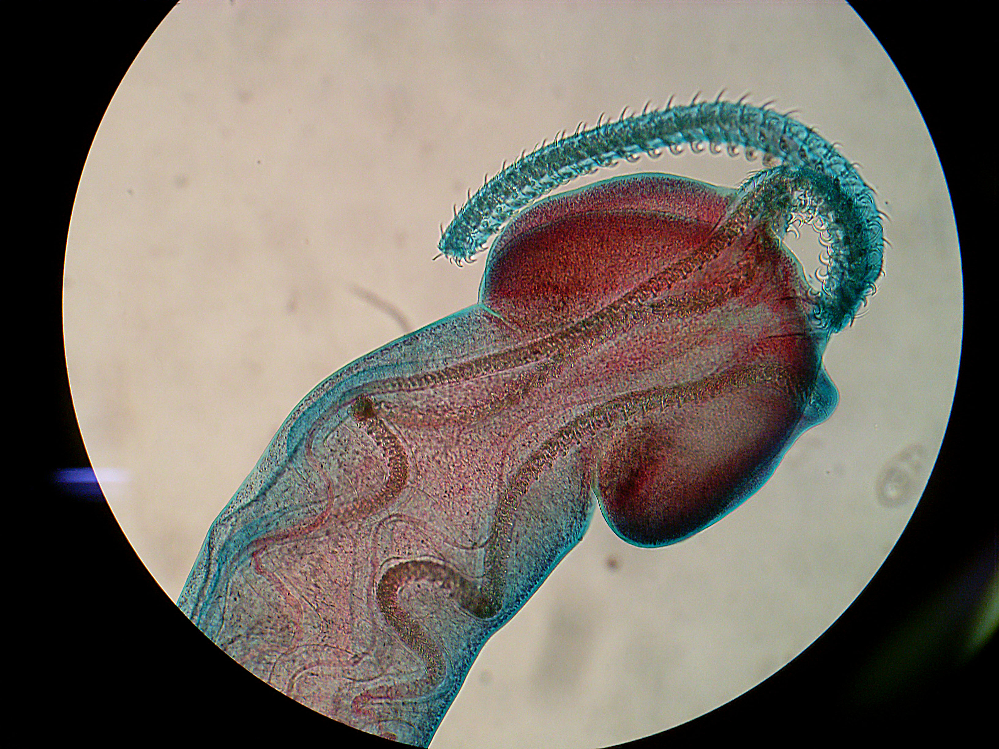
Grillotia belongs to a different group of tapeworms called the trypanorhynchs. Instead of four intricate lobes that fit snugly into the folds of the intestinal wall, it has four tentacles lined with hooked barbs that upon contact with the intestinal wall of its host, shoot out and embed themselves in the host's tissue (the photo on the left shows the scolex of a larval trypanorhynch with the tentacle just slightly protruding, see also this photo of a worm with one of its tentacles more fully extended). For whatever reason, in the presence of Grillotia, Pseudanisakis is compelled to move.

There appears to be a pecking order amongst the intestinal worms of skates, with trypanorhynchan tapeworms on top, followed by nematodes, then tetraphyllidean tapeworms trailing behind. Note that this kind of competition between these species only seems to occurs between worms that live in the spiral valve of skates. Similar worms living in the spiral valves of sharks seems to just leave each other alone. At this point, it remains uncertain why that is the case.

Reference:
Randhawa, H.S. (2012) Numerical and functional responses of intestinal helminths in three rajid skates: evidence for competition between parasites? Parasitology 139: 1784-1793

November 13, 2012

Amblyomma nodosum

The parasite being featured today is Amblyomma nodosum (image on the right - male top, female bottom) - a species of specialised tick that happens to be one of only three species of parasite that were found while examining three roadkilled giant anteaters from Minas Gerais, Brazil. There are 100 species of Amblyomma from around the world (33 of which are from Brazil) and they have been described from a variety of hosts from amphibians and reptiles to birds and mammals, but A. nodosum is a specialist that lives exclusively on the giant anteater (Mymercophaga tridactyla) and the collared anteater (Tamandua tetradactyla). It was also the most abundant of all the parasites found on the anteaters in the study we are featuring today, occurring in moderately high numbers (average of 58 ticks per anteater).

The second parasite that was found is the chigoe flea Tunga penetrans. Unlike A. nodosum, this ectoparasite infects a wide range of hosts, and while most fleas simply hop onto a host, drink up some blood and jump away, T. penetrans females burrow into the skin and *stay* there, feeding on blood and laying eggs. They only occurred in low numbers on giant anteaters and were found burrowing into the footpad and nowhere else on the body (see image below).

The third parasite found was also the sole internal parasite in the anteaters, the tapeworm Oochoristica tetragonocephala. Tapeworms from this genus are known to infect a range of hosts including lizards, snakes, and a variety of mammals. The larva needs to infect an invertebrate host, specifically an arthropod, before reaching the gut of a reptilian or mammalian host (by the said reptile or mammal eating the infected arthropod) and maturing into an adult worm. In the case of this species infecting the anteater, ants and termites are the most likely candidates for where the larval stages reside, given the host's specialised diet.

Relatively speaking, the giant anteater has very a sparse parasite fauna. Usually, a mammal of its size would be infected with a dozen or more different species of parasites. But because of its specialised diet and solitary life style, there are very few opportunity for most parasites (except specialists or very abundant generalists) to infect the giant anteater (as reflected by its paltry parasite fauna). Such an example shows how the ecology of the host organism can often shape what parasites it is infected with.

Photos from figures in the paper.

Reference:
Frank R, Melaun C, Martins MM, Santos AL, Heukelbach J, Klimpel S. (2012) Tunga penetrans and further parasites in the giant anteater (Myrmecophaga tridactyla) from Minas Gerais, Brazil. Parasitology Research 111:1907-1912

October 28, 2012

Hyperia curticephala

C. plocamia photo by
Rubén Arturo Guzmán Pittman
Generally speaking, jellyfish are not very appetising as food. They are composed mostly of water and armed with batteries of nasty stinging cells. Both of those characteristics together they make an unfulfilling and potentially painful meal. Nevertheless, they are fed upon by large pelagic fishes, and there are even some marine animals such as sea turtles that can live on a diet composed entirely of sea jellies. For those lacking the stomach for such squishy and venomous prey, there is still a way for them to obtain nutritional benefits from jellyfishes - and the parasite we are featuring today provides one such pathway.

The study we are looking at today focuses on a little parasitic crustacean that belongs to a group known as the Hyperiidea. They are amphipods that have evolved to live inside gelatinous animals of the open ocean. In the case of Hyperia curticephala, it dwells within the bell of the medusa Chrysaora plocamia - a rather large jellyfish that can grow up to a metre (a bit over 3 feet) in diameter.

H. curticephala image from here
Like other hyperiids, individuals of H. curticephala feed on the jellies that they live in. In turn, they can also provide food for those that feed on them. An avid consumer of these little crustaceans is the palm ruff (Seriolella violacea) - a fish that can grow to about 65 cm (about 2 feet) long. The palm ruff is one of a number of fish that are known to be "medusafish" as they are often found in close association with medusa jellyfishes.

When scientists examined the stomach contents of small (about 6-10 cm / 2-4 inches long) palm ruffs, they found them to be packed full of H. curticephala and nothing else. As they grew larger, the fish started having a more varied diet, but hyperiids still make up for over 97% of their prey. The amount of H. curticephala in the stomach of palm ruffs reaches a peak in February, just as the parasite also reaches very high abundances in the medusa when some individual C. plocamia can be infected with over a thousand amphipods (which in turn provides a floating banquet for any hungry palm ruff). The abundance of H. curticephala also reaches a high during November, but this was not reflected in the stomach content of the fish - so why is that? The scientists suggested that during this season, most of the medusae available are still quite small and while collectively they might be harbouring a high abundance of H. curticephala, because of their smaller bell size they are inaccessible to the palm ruff (which needs to get in or under the medusa's bell to reach the hyperiids). But by February, the medusae have grown to sufficient size that the fish are able to swim inside the jellyfish's bell to peck at the hyperiids.

Smaller fish can easily swim inside the jellyfish to feed on the parasites and are often found loitering within the host medusa (which also provides them with protection). Larger juveniles cannot enter the bell and have to settle for pecking off parasites, which happens to be in more accessible positions. In this manner, the palm ruffs act as cleaners for C. plocamia, protecting the jellyfish from the parasitic H. curticephala rather like cleaner wrasses that eat ectoparasites off coral reef fishes.

Reference:
Riascos, J.M., Vergara, M., Fajardo, J., Villegas, V., Pacheco, A.S. (2012) The role of hyperiid parasites as a trophic link between jellyfish and fishes. Journal of Fish Biology 81:1686–1695

October 15, 2012

Marshallagia marshalli

Photo by Billy Lindblom
A host can be infected by many different species of parasites (see this post for example). While in some cases, co-infecting parasites can get along just fine, in others, co-infecting parasites end up competing with each other because they both use the same resources from the host. When it comes to such conflict of interest, the stronger competitors can often push other species aside, or even bar their entry altogether. So what can a parasite do in such a situation? Well, it can try and catch their competitors off guard by getting in during the off seasons.

During their life-cycle, many parasites go through a free-living stage where they spend some time in the outside environment; either as an egg or a spore, or as a larva that has just hatched or while they are moving from one host to the next. Outside the cozy interior of they host, they can be exposed to some pretty harsh conditions. The parasite we are looking at today is found in the gut of Svalbard Reindeer (Rangifer tarandus platyrhynchus), which live on the Svalbard archipelago in the high arctic. During winter, Svalbard reindeer do not migrate, but instead move around the local area in search of any forage that is still accessible, which is not easy as the ground becomes completely covered by snow during winter. So not exactly the most cozy environment, especially not for the microscopic larval worms which infect these reindeer.

Marshallagia marshalli
egg from here
There are (non-parasitic) nematodes in Antarctica that can survive extreme cold, but it is not known if the free-living stages of some of their parasitic relatives can do the same. The two most abundant species of nematode worms in Svalbard reindeer are Marshallagia marshalli and Ostertagia gruehneri. For today's post, we will be focusing on a study which looked at the transmission dynamics of M. marshalli. Previous studies suggest that while other worms simply overwinter in the host and only lay eggs during summer, M. marshalli does not care for seasons; it just keeps laying eggs and infecting reindeer all year round, even through winter when their eggs and larvae will be resting on cold, snow-covered grounds.

At Spitsbergen, Norway, a group of researchers conducted an experiment to find out if reindeer did indeed pick up any additional worms during winter. To do so, they first fed some reindeer with anti-parasite drugs just before winter to purge them of any worms they already had. The drug wears off after a month, so the deer can start picking up worms again during winter if there are any infectious parasites around. What they found was that in the treated reindeer, after the purge there was no increase in O. gruehneri throughout winter, but the number of adult M. marshalli steadily increased, indicating the reindeer were picking up M. marshalli larvae throughout this period.

Marshallagia marshalli is a generalist parasite which also infects a wide range of hoofed animals ranging from sheep in Saudi Arabia, to saiga antelopes in Kazakhstan, to bighorn sheep in Montana, and reindeers in the Arctic - unlike O. gruehneri which is a reindeer specialist. While you'd expect that the reindeer specialist would have evolved such cold-resistant larvae, instead it simply refrains from laying eggs during winter so that transmission only occurs during summer. Because M. marshalli is a parasite of ruminants in dry deserts, their ability to survive such cold conditions might simply come with being able to infect hosts in generally arid and inhospitable environments. The caveat here is that M. marshalli might be a species complex (a group of closely-related lineages which have been classified as a single species due to their similarities), and the species/sub-species that infects Svalbard reindeer might have evolved to withstand the cold as a specialised adaptation for the conditions found in the high arctic.

So why has M. marshalli evolved such cold-resistant larvae instead of doing what O. gruehneri does and simply lay their eggs during summer when the larvae will be exposed to more favourable conditions? As mentioned above, Ostertagia gruehneri is a reindeer specialist, so perhaps in order for M. marshalli to have a fighting chance while sharing a host a well-adapted specialist like O. gruehneri, it needs to come in from the cold. Given how the infection dynamics of these two parasites are so seasonally-dependent, it is unknown how future climate change will affect their respective abundance in their hosts, and what consequences this will have on the reindeer population.

Reference:
Carlsson, A.M., Justin Irvine, R., Wilson, K., Piertney, S.B., Halvorsen, O., Coulson, S.J., Stien, A., Albon, S.D. (2012) Disease transmission in an extreme environment: nematode parasites infect reindeer during the Arctic winter. International Journal for Parasitology 42:789-795

September 30, 2012

Gyliauchen volubilis

Fish image taken by Richard Field, found at FishBase
Today's parasite is Gyliauchen volubilis - an intestinal fluke from a family of parasites that exclusively inhabit the gut of herbivorous fishes, in this case, the rabbitfish Siganus rivulatus, (see photo) which feed mostly on seaweed. The larvae of G. volubilis infect the rabbitfish by sticking to aquatic vegetation and wrapping themselves up into little cysts, which are then swallowed by their herbivorous host alongside their food (a strategy reminiscent of Philophthalmus sp. which we featured back in May).

The number of G. volubilis that infect each individual fish varies considerably, with some host to only a dozen G. volubilis, while others may have over a hundred flukes in their gut. Today's post is based on a study that looked at how infection level (or population size from the parasite's perspective) can affect the adult life of a fluke inside the rabbitfish's gut. While you may study this simply by looking inside the intestine of naturally infected fish, the problem with this approach is that you cannot know if there were other key events in the fish's life that might have affected the parasite population that you find. What you really need in order to get a more accurate picture is to start off with a blank slate.

Gyliauchen volubilis image
modified from original by
M.O. Al-Jahdali in this paper
That was exactly what a researcher in Saudi Arabia did to find out. For this study he took 70 pre-marked rabbitfish from an area where fish were found to be free of intestinal parasites, and moved them to a netpen in a lagoon where rabbitfish are known to be infected with G. volubilis. Ten weeks later, he recaptured the marked fish and noted how many G. volubilis they picked up while they were in the lagoon, and recorded the developmental stages of the flukes he found.

He found that when a fish's gut is occupied by fewer than about 60 G. volubilis individuals, flukes that newly arrived had a good chance of settling into a nice spot within the intestine. But, as the gut gets more crowded, he started finding more and more dead flukes - most of them were young flukes which had just arrived in the fishes with a mouthful of algae and have barely exited the cyst they came in. When the population of already established G. volubilis reached above 100, these new arrivals starts dying in droves, and the number of "dead on arrival" increased almost exponentially. At high population density, the gut is littered with dozens of dead worms - most of them young, and in some cases none of the newly excysted worms survived.

Crowding also alters the mating behaviour of these flukes. Like most flukes, G. volubilis are hermaphrodites with simultaneously functional male and female sex organs. When the gut is sparsely populated, they kept mostly to themselves - being hermaphrodites they simply reproduce by mixing their own sperm and eggs together - a process also known as "selfing" (other hermaphroditic animals and some flowers do this too). But when the neighbourhood gets more crowded, they get a bit more "social" and G. volubilis become embroiled in "mating groups". For those that produce eggs via selfing, they lay many small eggs. Because it doesn't get more incestuous than mating with yourself, it pays to hedge your bets and lay a lot of eggs in case some of them turn out to be defective. In contrast, flukes that had an opportunity to mate with others tended to lay fewer eggs, but they were comparatively larger - when your eggs are likely to turn out okay and defect-free, you might as well invest more into them to give them the best start in life.

As the flukes grow in size, they also adopt different mating habits, and as hermaphrodites, they also alter how many resources are allocated to the different sex organs to suit their habits. Smaller flukes that have just recently reached sexual maturity usually assume the role of sperm acceptors, receiving them into an organ called a seminal receptacle. This organ becomes very swollen with sperm in these smaller flukes. Medium-size flukes tend to pair up with a single mating partner with which they mutually exchange both sperm and eggs. Large flukes tend have shrivelled-up seminal receptacles and assume the role of sperm donors, inseminating multiple smaller flukes and rarely if ever pair with a worm of equal size.

In short, while starting out life in a crowded fish gut could be a dead end for many, for flukes that do survive that initial gauntlet, they also end up with more mating opportunities.

Reference:
Al-Jahdali, M.O. (2012) Infrapopulations of Gyliauchen volubilis Nagaty, 1956 (Trematoda: Gyliauchenidae) in the rabbitfish Siganus rivulatus (Teleostei: Siganidae) from the Saudi coast of the Red Sea. Parasite 19:227-238.

September 16, 2012

Bolbosoma balaenae

Image from Figure 1 of the paper
Today's parasite is an acanthocephalan (also known as a thorny-headed worm) and its name should be a clue to what it infects - baleen whales. And what do most baleen whales eat? Krill - lots and LOTS of it. The authors of the study I am writing about in this post found Bolbosoma balaenae larvae infecting krill that were caught during a plankton trawl off the coast of Ría de Vigo, Spain in the NW Iberian Peninsula.

The krill serve as hosts for larval B. balanae and from there, they proceed to infect the next host of their life-cycle, which as mentioned above, are baleen whales where they develop into adult worms. Acanthocephalans as a whole generally only have two hosts in their life-cycle - a small arthropod intermediate host where the larval worm resides, and the vertebrate definitive host where the adult lives and reproduces. But many of the thorny-headed worms that infect marine mammals add another host into the life-cycle between the crustacean host and the vertebrate host - this extra host is known as a paratenic host. The paratenic host is different from the intermediate host, and here's why.

For parasites with complex, multi-host life-cycles, the intermediate host is an obligate component for successful completion of the cycle. It is where the larval parasites gather resources to undergo development into the next stage, and at the same time, the intermediate host also serves as a mean of transporting the larvae into the definitive host (usually by getting itself eaten by the said host). It is in the definitive host where the parasite reaches sexual maturity. In contrast, a paratenic host serves only as a transport, and while the parasite has to infect an intermediate host to complete its life-cycle, infecting the paratenic host is optional. Seeing how the parasite can technically go through its life without ever hopping inside the paratenic host, why do it at all?

Image from Figure 1 of the paper
In the case of other acanthocephalans that infect marine mammals (such as Corynosoma cetaceum), if they are accidentally ingested by their marine mammal hosts while still inside the tiny crustacean intermediate hosts, they will still reach adulthood. But because the chances of that happening is negligibly slim compared to the likelihood of the crustacean host being eaten by a fish, which itself is then eaten by the said marine mammal, incorporating a paratenic host greatly enhances its chances of completing its life-cycle.

However, all this is unnecessary for B. balaenae, as their next host - fin whales and minke whales - do in fact feed on those tiny crustaceans. The authors of this study found that the infection prevalence of B. balaenae in krill is very low - only one in every thousand krill was infected with B. balaenae. But considering that a fin whale gulps down about 10 kg (22 lb) worth of krill with every mouthful and eats about 1800 kg (4000 lb) of those little crustaceans each day,  they can easily pick a few hundred worms very quickly even though the infection level is relatively low in krill.

Just like another acanthocephalan we have previously featured on this blog, Acanthocephalus dirus, instead of simply shedding eggs that are released into the environment with the host's faeces, the female worm actually leaves the gut once she is filled with fertilised eggs (see this paper). So even though the whale is constantly being infected with new worms with every mouthful, there is also a constant turnover in the population in the form of mature female worms exiting the host.

Reference:
Gregori, M., Aznar, F.J., Abollo, E., Roura, Á., González, Á.F. and Pascual, S. (2012) Nyctiphanes couchii as intermediate host for the acanthocephalan Bolbosoma balaenae in temperate waters of the NE Atlantic. Diseases of Aquatic Organisms 99: 37-47.

September 7, 2012

Antricola marginatus

People usually associate bats with the image of vampires and blood feeding, even though most bats are not blood drinkers. However, bats are themselves host to all manners of blood-feeding parasites. Today, we are looking one such blood sucker - Antricola marginatus - a tick with a caring, maternal side that people don't usually associate with the word "parasite" (though we have featured a few parasites on this blog which go out of their ways to give their offspring with the best possible start to life).

Image from Figure 1 of the paper
While collecting ticks in a cave which is home to nine species of bats (if you are wondering, none of those bats are vampires) in the Yucatan, Mexico, a trio of researchers came across eight female A. marginatus that were covered in massive broods of little baby tick. Each of the female ticks carried between a hundred to four hundred nymphs on their backs. Those little nymphs are very attached to their mother - when the researchers tried to brush some nymphs off, they quickly scramble back onto mother's back at their own volition.

Over the course of its evolution, A. marginatus has almost completely given up its vampiric life-style of drinking bat blood in favour of... something less glamourous - eating bat droppings. However, they still go through a stage in their life as nymphs when they retain their taste for blood. So how are the nymphs suppose to disperse to a suitable host when their mothers are scrambling around and munching on bat poop? The researchers suggested that A. marginatus facilitates her babies by making regular visits to roosting bats, where the nymphs can disembark and drink all they want.

Indeed, when they brush a nymph-ladened mother tick onto a rabbit's ear, the nymphs quickly jump off and started gorging themselves on blood. However, after three days of chugging down rabbit blood, they died - this is not surprising because as I have discussed in a previous post, blood-feeding parasite can be remarkably picky about their hosts, and for some parasites even a slight host species difference can result in deterioration in survival, let alone the large difference between bats and rabbits. So it was no surprises that those nymphs dropped dead after a few days of imbibing rabbit blood. Back in their natural environment of the bat cave, the next warm-bodied mammal A. marginatus would have off-loaded her nymphs on to would have been roosting bat.

Maternal care has been reported for other arachnids like spiders and scorpions, but not ticks. It is unknown just how unique A. marginatus is among ticks with its maternal behaviours, or if there are many other caring, motherly ticks out there which are just waiting to be discovered.

Reference:
Labruna MB, Nava S, Guzmán-Cornejo C, Venzal JM. (2012) Maternal Care in the Soft Tick Antricola marginatus. Journal of Parasitology 98: 876-877

August 27, 2012

Metschnikowia bicuspidata

If you are a regular reader of this blog, at some point you would have read about the concept of coevolutionary arms races between hosts and parasites (see this for example). Previously, we have featured Pasteuria ramosa - a bacterial parasite of the waterflea Daphnia. Pasteuria ramosa is very picky about its host - specific strains are compatible only with specific host genetic lines, and as we have talked about in that previous post, this parasite is very harmful. Because of how virulent P. ramosa is to waterfleas and because the resistance by the host is dependent upon being the lucky genotype that is not compatible with whatever strain of the parasite which is most common at the time, this sets up an ideal situation for a Red Queen-style evolutionary arms race (and it is one that has been going on for long time).

Uninfected (top right) and
infected waterflea (lower left)
Photo by Meghan Duffy
But in some areas where P. ramosa is found, it also co-occurs with a different parasite - the one that we are featuring today: Metschnikowia bicuspidata. It is a yeast that also infects Daphnia (other fungal parasites also named Metschnikowia biscuspidata have been reported to cause disease in shrimps, crabs, even fish - but it is more likely that they are similar-looking fungi that have been lumped together). The study we are looking at today was conducted by a collaborative group of three researchers who wanted to find out what happens when waterfleas are confronted by both parasites.

Under such circumstances, will the presence of M. bicuspidata exacerbate the existing arms race between Daphnia and P. ramosa, or will it simply get in the way? If resistance for P. ramosa is also associated with resistance to M. bicuspidata, then it means Daphnia has a general mechanism for resisting both parasites. This scenario will simply select for general parasite resistance in the Daphnia population, reducing the level of genetic variation in the population (the raw material for ongoing Red Queen-style evolutionary arms race). On another hand, if Daphnia resistant to P. ramosa are negatively associated with resistance to M. bicuspidata, then it means resistance for one parasite will come at the cost to another - this trade-off in defending against two different parasites sets up an additional selective pressure that can potentially accelerate the arms race.

There are a number of key differences between the two parasites. While P. ramosa reduces the reproductive capacity of the host more than M. bicuspidata, the latter kills the host quicker. Metschnikowia bicuspidata is extremely lethal, killing infected waterfleas within 2-3 weeks of infection (whereas waterfleas can live up to 5-7 weeks after being infected by P. ramosa). The fungus releases its spores after the waterflea dies, and those infective spores can even survive passage through a fish's gut if their host Daphnia is eaten.

And unlike P. ramosa, infection success of M. bicuspidata depends not so much on encountering a host with the right genes, but through sheer persistence - the more often a waterflea is exposed to M. bicuspidata spores, the more likely that they become infected. This difference also manifests in the nature of outbreaks caused by the two parasites. Outbreaks of P. ramosa tend to be rarer and more limited, especially in genetically diverse populations, whereas M. biscuspidata is more prone to massive outbreaks that spread widely across the whole population. Even though it is not as discriminate about host genotype as P. ramosa, it is not as if M. bicuspidata does not influence the evolution of its host. But the way it affects host evolution is different to that of P. ramosa - instead of selecting for specific genotypes, it influences how much the waterfleas allocate their resources into either reproduction or parasite resistance.

In this study, the researchers found that different genetic lines of waterfleas varied considerably in their resistance to M. bicuspidata, but a waterflea's resistance to the fungal parasite did not in turn predict how well it also resisted P. ramosa. Instead, as found in previous studies, infection success of different P. ramosa strains depended upon the specific combination of host genotype and parasite genotype. This indicates that waterfleas have very different ways of resisting the two parasites, and that resistance to one does not lend protection to the other, but at the same time, nor does protection against one parasite increases a waterflea's vulnerability to the other.

Therefore, as far as the Red Queen arms race between waterfleas and P.ramosa is concerned, even though M. bicuspidata looms as a significant threat to the waterflea population, it is unlikely to significantly alter the coevolutionary dynamics between Daphnia and P. ramosa.

Reference:
Auld SKJR, Hall SR, Duffy MA (2012) Epidemiology of a Daphnia-Multiparasite System and Its Implications for the Red Queen. PLoS ONE 7(6): e39564. doi:10.1371/journal.pone.0039564

August 16, 2012

Eimeria echidnae

We have previously featured a number of coccidian parasites on this blog from birds (here and here), alligators, and groundhogs. Today's coccidian parasite lives in a strange ant-eating, egg-laying mammal from Australia - the short-beaked echidna Tachyglossus aculeatus.

photo from Figure 1 of the paper
The parasite we are featuring today is found in the gut of the echidna where it resides alongside another species of Eimeria - E. tachyglossi. Both these coccidians are found exclusively in echidna guts (generally coccidians are highly host-specific), and both are known to cause mild to severe inflammation of the small intestine, and in some cases, associated with fatality in systemic infections where the parasites have spread to the echidna's other organs. However, the exact role they might play in disease is still unclear. The study we are featuring today was conducted to establish the baseline, background level of Eimeria infection found in healthy echidnas.

The researchers of this study collected fecal sample from echidnas from various zoos and wildlife parks, and examined them for oocysts (see accompanying photo) - the infective stage of coccidia that are shed by infected animals. They found that most echidna shed between a few thousand to tens of thousands of oocysts in each gram of feces. While that may sound a like lot, all the echidnas involved in the study were clinically healthy, and the oocyst numbers were comparable to those from wild marsupials. Furthermore, infection intensity did not change over the different seasons, though oocysts (the parasite's infective stage) were more commonly shed by animals that were housed in outdoor enclosures

Additionally, they also found that while wild and short-term captive echidna shed oocyst of both E. echidnae and E. tachyglossi, echidna that have been held in captivity for an extended period of time only shed E. echidnae, indicating that captive conditions are unfavourable for E. tachyglossi transmission . Because coccidian oocysts are commonly found in the soil, presumably the echidnas become infected while feeding on ants; as they poke their snout in the dirt and use their long sticky tongue to lick up ants, they also end up ingesting a lot of soil (see this video of a hungry echidna on the prowl)

Most newborn mammals become infected with coccidia within their first week or month of life. In contrast, juvenile echidnas that have not been weaned were found to be free of coccidia. Given that echidnas become infected with E. echidnae through exposure to oocysts while feeding on ants, and young echidnas do not start feeding on ants until they are weaned at 6 months old, this age-dependent diet shift most likely explains the absence of E. echidnae infection in juvenile echidnas.

Reference:
Debenham JJ, Johnson R, Vogelnest L, Phalen DN, Whittington R, Slapeta J. (2012) Year-long presence of Eimeria echidnae and absence of Eimeria tachyglossi in captive short-beaked echidnas (Tachyglossus aculeatus). Journal of Parasitology 98:543-549

August 7, 2012

Mysidobdella californiensis

Photo taken from Figure 3 of the paper
Marine leeches are commonly known to feed on various vertebrate hosts - mainly fish and sea turtles. However, today's parasite stands out from the pack by associating itself with an arthropod. Instead of fish or turtles, Mysidobdella californiensis sticks its sucker onto mysid shrimps. Mysids are also known as opossum shrimps because the females have a little brood pouch (called a marsupium) in which they carry developing young.

The discovery of Mysidobdella californiensis actually occurred rather serendipitously. Back in the summer and fall of 2010, an unprecedentedly huge swarm of mysid shrimp appeared off the central Californian coast. Some of those shrimps got sucked into the water clarification system at the Bodega Marine Laboratory. With all this shrimp in the system, the lab staff began collecting them opportunistically for fish food. But then, they started noticing these little leeches attached to the shrimps, so they made a concerted effort to collect the shrimps directly from the water clarifier, and examine them under the microscope.

What they found were tiny leeches about 1.5 cm (a bit above half an inch) long. Approximately one in every six shrimp were found to have leeches on them, and each infected shrimp was carrying between one to three leeches. Seeing as this is a new species, at this stage very little is known about its biology except what can be inferred based on what we know of a related species - M. borealis - which has been studied in slightly more details. It is unclear whether M. californiensis (and related species) merely hitch-hike on the shrimp and use it to carry them to potential hosts, or if they in fact feed on the shrimp. In laboratory trials on M. borealis, the leeches refused to feed on any of the fishes that they were presented with, and none of the leeches were found to have fish blood cells in their gut. It is possible that Mysidobdella as a genus specialise in feeding on mysid shrimps. If that is indeed the case, then Mysidobdella would be the only marine leech known to feed on the blood of invertebrates rather than vertebrates. However, mysid blood has yet to be found in the gut of these leeches, so at least at this point, the diet of M. californiensis remains a mystery.

Reference:
Burreson, E.M., Kim, B. and Passarelli, J.K. (2012) A New Species of Mysidobdella (Hirudinida: Piscicolidae) from Mysids along the California Coast. Journal of Parasitology 98: 341-343.