"So, naturalists observe, a flea has smaller fleas that on him prey; and these have smaller still to bite ’em; and so proceed ad infinitum."
- Jonathan Swift

November 14, 2024

Saccularina sp.

The bay scallop (Argopecten irradians) is a highly prized shellfish, but it has suffered through a rough history from overharvesting, habitat loss, and natural enemies. This culminated in a massive population decline in the 1980s that led to the closure of all its fisheries across certain regions along North Carolina. With the depletion of its wild populations, efforts are being made to raise the bay scallops in aquaculture to meet demands. But now a new woe has fallen upon this besieged bivalve with the appearance of a never-before-seen parasite.

Left: Bay scallop infected with Saccularina, the red arrows indicating the parasite's sporocysts in the gills. Right: the cercaria stage of Saccularina which is a "cystophorous"-type cercariae.
From Fig. 1 and 2 of the paper

In 2012, a researcher started noticing a gill-dwelling parasite in both caged and wild bay scallops along the coast of North Carolina, and later at the Gulf Coast of Florida. These parasites are readily visible in the shellfish’s gills as they become swollen with the parasite's presence. Examination under a microscope revealed the parasites to be a species of parasitic fluke which is using the scallop for the asexual stage of its life cycle. Essentially, these trematode flukes are converting the shellfish into a parasite clone factory that pumps out a stream of free-swimming larvae to infect the next host in the life cycle.

Given such an operation consumes a lot of the host’s resources, this can interfere with the scallop’s growth and survival, and thus it is a major concern to the scallop fisheries in North Carolina. The key to managing any parasitic infection is an understanding of its natural history and life cycle, and unfortunately, given its relatively recent discovery, the life cycle of this parasite is almost entirely unknown. However, related fluke species can give us some clues, and as it turns out, this bay scallop parasite is no ordinary fluke.

First of all, DNA analysis showed that one of this fluke's closest relatives is Saccularina magnacetabula, a trematode species found on the other side of the world in Australia. While it is genetically similar enough to the bay scallop parasite for them both to be in the same genus (Saccularina), there is enough geographical and genetic distance between them that they are clearly different species. Furthermore, instead of scallops, S. magnacetabula infects the Sydney cockle (Anadara trapezia) as the chosen bivalve for its asexual stage. As for the parasite's next stops, it's a multi-part journey involving tiny crustaceans, followed by a type of smallish Australian fish called whiting (Sillago sp.), and finally the adult fluke completes its life cycle nestled in the fin membranes of the giant herring (Elops hawaiensis).

Saccularina magnacetabula, and by extension, the bay scallop parasite, belongs to a family of flukes called Didymozoidae - a flukey group of flukes with some very unusual anatomy and habits. While the adult stages of most trematodes are generally leaf-shaped, didymozoids come in all kinds of shapes and sizes. And they are found in a wide range of different bony fishes all over the world, mostly marine species. Additionally, instead of living in the final host’s gut like most flukes do, didymozoids cram themselves into all kinds of nooks and crannies such as the muscles, the gills, or even the fin membranes as is the case for S. magnacetabula.

While there are many known species of didymozoids, the life cycles for most of them are a mystery, with the hosts for the asexual stage known only for a few species. But those handful of species alone showed didymozoids to have quite the eclectic range. Aside from bivalves like cockles and scallops, the asexual stage of other didymozoids infect snails, but not just any regular snails - one species is known to use pelagic sea snails (which are also called “sea elephants”), while another species infects worm snails which are peculiar sea snails with twirly shells that encrust on rocks and other hard surfaces.

So, based on the information above, we can make some inference about the likely source of the bay scallop parasite. It’ll have to be some kind of predatory sea-dwelling fish harbouring the adult stage of the fluke, and given S. magnacetabula completes its life cycle in the giant herring, the bay scallop parasite is most likely completing its life cycle in some kind of predatory herring-type fish in the region, which means ladyfish or Atlantic tarpon

While we may have some clues about the bay scallop parasite’s life cycle, how they might have gotten there is more of a mystery though. This parasite was first seen in bay scallops in 2012, but if the disseminator of this parasite is really a local fish species such as the tarpon, why has it only been noticed now? Whatever its origin turns out to be, it seems the bay scallop parasite is not ready to give up its (many) secrets.

Reference:
Boggess, H. F., Varney, R. L., Freshwater, D. W., Ben-Horin, T., Preister, C., McCurry, H., Wilbur, A. E. & Buck, J. C. (2024). A newly discovered trematode parasite infecting the bay scallop, Argopecten irradians. Aquaculture 589: 740960.

October 7, 2024

Nectonema sp

Sometimes a new scientific discovery comes about while one is doing the most mundane things, and it might not even be a scientist who happens to be doing it. Last year, an unusually high number of tanner crabs started showing up in the waters off the southern coast of Hokkaido. While these crabs are a bane for flounder fishermen as they have a habit of tearing up their nets, tanner crabs are easy to catch and they taste good, so numerous crabs have ended up in markets all over Japan, being sold for a relatively low price.

Top right: a cooked tanner crab with a coiled-up Nectonema worm inside of it. Top left: another cooked crab with a smaller Nectonema worm inside of it. Bottom: A Nectonema worm extracted and unraveled from the first tanner crab (scale bar = 2 cm). Photos from Figure 1 of the paper, taken by Rieko Yamamoto 

So what does this have to do with parasitology? Well, earlier this year a woman named Rieko Yamamoto had bought and boiled up some tanner crabs for a meal, but upon opening her would-be crab dinner, she discovered that one of the crabs came with an extra helping of worm, all coiled up like a bundle of cables. This worm was about 82 centimetres long and took up a lot of space in the crab's body. But instead of doing what some people might do, which is to toss the crab out the window in disgust, she calmly placed the parasitised crab in the freezer and contacted Dr. Keiichi Kakui, an invertebrate zoologist at Hokkaido university, who was able to identify the worm as Nectonema.

Nectonema is a genus of horsehair worm, and while horsehair worms are more commonly known from land-dwelling arthropods such as crickets and praying mantis, there is one offshoot lineage of horsehair worms that have taken up life within the denizens of the seas. Nectonema has previously been reported in many types of crustaceans, including rock crabsshrimpslobsterssquat lobsters, and even marine isopods, but this is the first time that it has been found in a tanner crab.

A week after this discovery, Ms Yamamoto bought another eight crabs and found one of those crabs also came with a worm, which means this parasite might not be all that uncommon among tanner crabs. Fortunately, Nectonema doesn't cause any harm to humans, so there are no public health issues here, though it might be an alarming sight to those who are unfamiliar with these worms.

The life cycle of these marine horsehair worms is a mystery, though if their more well-studied relatives is anything to go by, it might involve the larva infecting a smaller invertebrate first, before being eaten by the final host where it can grow to its full adult size. While horsehair worms in land-dwelling hosts are known for altering the behaviour of their hosts, such behavioural manipulation is due to the worm needing to move its terrestrial host into a water body to complete its life cycle. This is not necessary for Nectonema since it is already surrounded by water in the sea.

Nature is full of surprises, but if you are prepared and observant, you might come across a scientific discovery while having your next meal. So if you ever find a worm in your dinner - don't panic! It might turn out to be an important scientific discovery.

Reference:
Kakui, K. (2024). Nectonema horsehair worms (Nematomorpha) parasitic in the Tanner crab Chionoecetes bairdi, with a note on the relationship between host and parasite phylogeny. Diseases of Aquatic Organisms 159: 153-157.

September 14, 2024

Epifagus virginiana

Epifagus virginiana is a parasitic plant that grows on the roots of American beech trees. Also known as "beechdrops", clumps of their brown stems can be found protruding from the forest floor, reaching up to 30 cm tall and lined with purple-white flowers. To most people, they look like just another ordinary plant amidst the undergrowth. But Epifagus lacks a key component which is usually a defining characteristic of plants - chlorophyll, the pigment which allows plants to harness solar power. Instead, the way this plant obtains its nutrients is via an underground tuber attached to the roots of its host. The beechdrop is a very discerning parasite - as its name indicates, it usually goes after beech trees, but sometimes they switch up their target and end up engaging in botanical cannibalism.

Left: The stem and flowers of beechdrops, Epifagus virginiana, Centre: a beechdrops tuber, with adventitious root (ar) indicated, Right: two beechdrops tubers linked via a parasitic connection.
Photos from Fig. 1 and Fig. 2 of the paper.

A botanist named Dr. Luiza Teixeira-Costa was conducting a study on these parasites in the mixed forest at Powdermill Nature Reserve in Pennsylvania, where there is a dense population of beechdrops. After carefully excavating a dozen of those parasitic plants, she noticed one of the specimens was composed of two Epifagus plants locked in a peculiar pairing. When she looked at that specimen more closely, she found that the pair consisted of two beechdrops tubers clinging tightly to each other, as if one of the plant was parasitising its fellow parasite.

To see if there's an actual parasitic connection between the tubers and that this was not two plants that had wrapped around each other via happenstance, Dr. Teixeira-Costa tested whether fluid could be passed between the two plants by injecting one of them with a special tracking solution, and examining it with micro CT scan to create a 3D image of the plants' internal structure. The scanning revealed that there is continuity in the vascular tissue that connects the two plants. It would be like if you find a pair of animals that have joined together and are connected via their circulatory systems. But that was one specimen - perhaps this was just a freakish one-off occurrence?

In order to get a better picture of this phenomenon, Dr. Teixeira-Costa examined the collections of Epifagus specimens at the Meise Botanic Garden and the Harvard University Herbarium, to see if there are more of such pairings. Out of 150 Epifagus that were in those collections, four of them were composed of a pair of E. virginiana plants attached to each other, in a similar way to how these parasites would usually attach to their host. 

In addition she also searched through the online digital archive of herbarium specimen images from 233 herbaria via the SERNEC portal. Out of the 3097 Epifagus specimens in those collections, she found 52 specimens that showed potential signs of parasitising a fellow beechdrops. All those specimens had been collected from across the plant's distribution range in the United States. What this exhaustive search revealed is that while such cases of beechdrops-on-beechdrops is not a frequent occurrence, it is not negligibly rare either. Furthermore, it is widespread and not restricted to just one particular region.

Orobanchaceae plants such as Epifagus are usually able to recognise its own kind and avoid this kind of friendly misfire - so what made some beechdrops turn on one of their own? One possible scenario might involve an Epifagus plant attaching itself to a beech tree root, and was followed by the seed of another Epifagus which responded to the stimuli given off by the tree root. But instead of latching onto the beech tree root, it ended up parasitising one of its fellow Epifagus that was already there.

These examples of intraspecific parasitism have been described as "botanical version of cannibalism" and have been documented in a range of other parasitic plants. If anything, compared with the beechdrops, other parasitic plants seem to be far less discriminating about parasitising their own kind, particularly among mistletoes and dodders. In the world of botanical parasites, sometimes a parasitic plant's worst enemy is a fellow parasite.

Reference:

August 13, 2024

Selenidium elongatum

A passing glance at the parasite in today's post might lead you to think that it is a worm, perhaps a nematode. But a closer look would reveal that not only is it much, much smaller than most nematodes, it also has a visible nucleus - like what you'd find with say, a cell. Despite how it looks, this parasite is not a worm, but a gregarine - which is a group of single-celled parasites that infect all kinds of invertebrates, including insects, crustaceanssea cucumbers, and even sea squirts.

Left: Selenidium elongatum from Myxicola sp. Quadra. Right: Selenidium elongatum from M. aesthetica
scale bar = 20 μm. m = mucron; n = nucleus. Photomicrographs from Figure 1 of the paper 

Gregarine belongs to the phylum Apicomplexa - which includes Toxoplasma gondii, Plasmodium (the malaria parasite), and Cryptosporidium among its ranks. But the cells of gregarines grow much larger than those human parasites, with some species reaching half a millimetre in length (and one species, Porospora gigantea, exceeding 10 mm in length). They also come in all kinds of different shapes, including one species which is shaped like a microscopic rubber chicken, and they cling to the host's tissue using their mucron, an organelle which functions like the suckers of a fluke or a tapeworm.

The study featured in this post looked at gregarines and other symbionts living in two species of feather duster worms from Harriot Bay in British Columbia. As their names indicate, those worms are shaped like feather dusters, they live in tubes and use their long feathery appendages to filter food particles and plankton from the surrounding waters. The two species that the researchers examined were Myxicola sp. Quadra, which lives in tubes on muddy seafloors, and Myxicola aesthetica, a shallower water dweller that attaches to firmer substrates like rocks or shells.

The researchers examined about 50 of those worms and found that nearly all of them were infected with gregarine parasites, consisting of two species in the Selenidium genus - S. mesnili and S. elongatum. Those gregarines lived in the gut of the marine worms in a comparable way to how parasitic worms inhabit the gut of vertebrate animals. Though they belong to the same genus, the two Selenidium species are different to each other in many ways. The cells of S. mesnili are shaped kind of like skinny lemons, while S. elongatum, as its name indicates, has a long cell that makes it look like a single-celled version of a roundworm.

Aside from their size and shape, they also differ in other ways. Selenidium elongatum lives in the intestine of its worm host, and is found in both species of feather duster worms that the researchers sampled. Meanwhile, S. mesnili was only found in Myxicola sp. Quadra, and it lives exclusively in the host's pharynx and oesophagus. These differences might have arisen from the way these gregarines obtain nutrients from the host's digestive tract, or it might have something to do with the life cycles and transmission routes of these parasites. But Selenidium were not alone in the guts of those feather duster worms, living inside the gut of Myxicola aesthetica next to S. elongatum was a species of ciliate called Pennarella elegantia that swam freely in the worm's gut content.

Gregarines are poorly known but they seem ubiquitous in invertebrates, and their relationship with the host isn't always parasitic - there are evidence to indicate they can sometimes be beneficial to their host. And there are many more of them out there which are waiting to be discovered. What these gregarines show is that if you know where to look and what to look for, you will find a rich vibrant world even within the guts of a mud-dwelling worm.

Reference:

July 11, 2024

Parvatrema spp.

Parasites are known for their complex life cycles, especially among parasitic flatworms such as flukes. And the flukes that are being featured in today's post have life cycles that make them a fluke among flukes. This blog post is about an extensive study that culminated from 25 years of work, where a group of researchers were able to identify and describe five different fluke species with a peculiar life cycle adaptation.

Parvatrema sp. “quadriramis” cercaria stage (left), young metacercaria (centre), parthenogenetic metacercaria containing fully-formed metacercariae (right). Insert: Parvtrema parthenogenetic metacercariae in the hepatopancreas of a limpet.
Photos from Fig. 4 and Fig. 11 of the paper

In the cold waters of the northern European seas and the Sea of Okhotsk, there is a group of parasites with life cycles that defy the conventions of its class. They are five closely related species of flukes in the genus Parvatrema, and they spend parts of their lives lurking quietly in the bodies of clams and snails. In many ways, they're just like other digenean flukes, with multi-host life cycles that involve turning their first mollusc host into a clone factory, producing clonal larvae which go off to infect a second host, and culminating in sexually mature adults living in the gut of vertebrate animals. But these Parvatrema flukes have evolved to do some things differently once they reach their second host.

These flukes infect sea snails such as limpets and periwinkles as their second host. Some species sit in the extrapallial space - the fluid-filled gap between the snail's fleshy mantle and the shell, others get into the gonads and digestive organs. Usually, this is a relatively dormant stage of the fluke life cycle, where they literally sit and wait to be swallowed by the appropriate final host. But with these Parvatrema flukes, instead of simply sitting around waiting to be eaten by a bird like other flukes would, they have another round of asexual reproduction, as a treat.

Each of those immature flukes become filled with numerous miniature clones of itself until it is stuffed to the point of exploding. Some of them take it further, with unlimited consecutive generations of parthenogenetic clones, each fluke exploding into multiple clones and then each of those clones explodes into even more clones and so on, like a never ending series of Matryoshka dolls. On top of that, some of those Parvatrema doing unlimited fluke works are also able to produce cercariae - the free-swimming larval stages - which then go off to infect more sea snails to start the asexual cycle again. 

So why did they evolve this unique developmental stage? Parvatrema are tiny flukes, the adult stage only live for a few days in the gut of a bird, and they produce less than a hundred eggs - a relatively low number compared with other flukes which may produce thousands or even millions of eggs over their lifetime. To make things worse, the likelihood of any one fluke successfully infecting the right hosts at each consecutive stages of its life cycle is astronomically low, so they need to multiply their numbers at every chance they get.

Furthermore, the final hosts for these flukes are migratory birds which only come once a year - so they need to make the most of their brief stay by making sure that if they only eat one infected snail, instead of just getting a single or a dozen flukes in each snail, they're getting the whole gaggle of fluke clones arriving en masse into the bird's gut in their hundreds or thousands, ready to get on with the business of producing the next generation.

Essentially, these Parvatrema flukes recapitulate the process that most other digenean flukes only undergo in the first host. Asexual reproduction in the first host is arguably one of the key evolutionary innovation of digenean flukes, allowing them to offset the losses associated with the process of being transmitted from one host to the next. Since Parvatrema seems to do asexual reproduction at every possible opportunity, they can provide us with insights into how flukes evolved their one weird asexual trick that gave them an edge in the transmission game.

Reference:
Galaktionov, K. V., Gonchar, A., Postanogova, D., Miroliubov, A., & Bodrov, S. Y. (2024). Parvatrema spp.(Digenea, Gymnophallidae) with parthenogenetic metacercariae: diversity, distribution and host specificity in the Palaearctic. International Journal for Parasitology. 54: 333-355

June 10, 2024

Forficuloecus pezopori

Parasites are a major part of biodiversity, but they spend most of their time hidden in plain sight. Even with some animals that have been known to science for centuries, their parasite fauna remains completely unknown. This can either be due to the lack of research interest into their parasites, or the host animal is just really rare, so there has been very little to no opportunities to study the parasites that live on or in them. In some cases, those rare animals are at risk of going extinct, which means their parasites and symbionts may also disappear before we even know they exist. This post is a story about a ground parrot and its hidden louse.

Left: Forficuloecus pezopori louse viewed under light microscope, Right: a western ground parrot (Pezoporus flaviventris).
Both photos from graphical abstract of the paper

The parasite being featured in this post is Forficuloecus pezopori, and it is the first known parasite from the western ground parrot (Pezoporus flaviventris), also known as Kyloring. Kyloring is one of the rarest parrots in the world and is considered critically endangered, which is bad news for F. pezopori, because we have barely gotten to know this little insect, and it may already be at risk of disappearing along with its feathered host. Lice are particularly vulnerable to co-extinction as they are completely helpless off the host's body and are often specifically adapted to living on just one particular host species.

Forficuloecus pezopori was found on some captive ground parrots at Perth Zoo, the lice were hanging around the feathers at the back of the birds' head and nape. As far we know, the western ground parrot is the only host for this parasite. There is a slim chance that it might also be found on the western ground parrot's closest relative, the eastern ground parrot (Pezoporus wallicus), but we can't verify that at this point, because we know so little about the parasites of ground parrots in Australia.

For example, a subspecies of eastern ground parrot in Tasmania was found to support a type of feather mite called Dubininia pezopori but it is uncertain whether that mite is unique to just that particular subspecies, or if it is also found on the mainland eastern ground parrots, since nothing is actually known about the parasites and symbionts of those birds. Given the eastern and western ground parrot have been separated since the Pleistocene about 2 million years ago, there would have been enough separation in time and space for the two species to develop their own distinct collection of parasites. So as far as we can tell, the Kyloring is the only host for F. pezopori.

Parasites on endangered hosts such as the Kyloring are in a precarious position, because not only are they at risk of dying out alongside their hosts, historically, there have been cases of parasites being wiped out in the process of people trying to conserve their hosts. For example, during the California condor breeding program, a unique species of condor louse was wiped out due to the pesticide-based delousing that the birds received when they were taken into captivity. And the California condor louse is not the only victim of extinction via conservation efforts. The Iberian lynx louse also suffered the same fate

Even more tragic is the case of Rallicola extinctus - a louse of the Huia, which was a species of New Zealand bird that became extinct early in the 20th century, with its last confirmed sighting in 1907. But the louse that it hosted was not even formally described until 1990 - many decades after the host had already gone extinct, hence the species name R. extinctus. Forficuloecus pezopori and many other lice species are at risk of such entangled fates, or become victims of well-meaning conservation efforts.

While a lot of people may not mourn the loss of parasites, it might be their hosts that end up missing them the most. The presence of parasites may help them develop a properly functioning immune system, and their absence could leave the host with a range of physiological disorders. And these parasites might be better off together with their hosts as they can tell us a lot about how the host animals live, and the ecosystem they exist in.

To rectify the mistakes of the past, the researchers suggested that any future studies on wild populations of ground parrots should incorporate a routine louse check to see how common F. pezopori are among those birds, but not to remove any of the lice that are found, and just let those little insects be. Especially since they don't seem to harm healthy, wild parrots. At the same time, lice infection on captive birds can serve as an opportunity to learn more about F. pezopori - saving the host along with its parasites at the same time

Living organisms are intertwined within a network of ecological interactions, if you pull on one loose thread you might trigger a series of co-extinctions and unravel the entire tapestry. Because of those connections, we could be losing more species than we realise.  Though they are often hidden out of sight, and thus out of minds, losing parasites and symbionts would leave us with emptier ecosystems and a lesser world.

Reference:

May 10, 2024

Dracunculus insignis

The Guinea Worm, Dracunculus medinensis, is an agonising parasite for those who have to endure its wrath. The female worm can grow up to 80 cm long and when it comes time for it to release its offspring, it does so by poking its body partially out of the host's arms or legs, all while causing a fiery pain that forces the host to immerse their limbs into the water, allowing the worm to release its larvae. This parasite has afflicted humans since antiquity, with description of pathologies and treatment associated with the worm dating from ancient Egypt, and depiction of the parasite in a 15th century painting

In the modern era, the Guinea worm has been the subject of an eradication effort by the World Health Organization (WHO) since the 1980s. An obituary was even written about this parasite in 2013. But while this campaign has been largely successful, the effort to completely eradicate the Guinea worm has hit an obstacle in some regions as the worm has taken to using dogs as alternative hosts in place of humans.

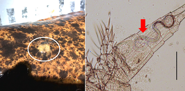
Left: Large bundle of Dracunculus insignis in the paws of a river otter (Lontra canadensis), Right: A Dracunculus worm being removed from a river otter (Lontra canadensis)

But aside from the infamous Guinea worm, there are many other species of Dracunculus out there which are found in a wide range of animals, many of which are actually reptiles. Of those, Dracunculus insignis is considered the most important because in addition to parasitising many species of wildlife,
it can also parasitise cats and dogs. The female worm can grow to 30 cm long, and about 300 days after the initial infection, the mature worm - now loaded with larvae - will migrate to the extremities and exit through a lesion, to explosively release a load of baby worms to begin the cycle anew.

This study looked at Dracunculus worms in river otters from North America. The worms the researchers examined came from various sources, including wildlife parasite surveys, as well as dead otters which were obtained from trappers. In addition, they also collected some worms from an otter in Florida that was recovering in a rehabilitation centre after being struck by a car. During its stay in rehab, worms started emerging on their own out of the otter's body. It was just one thing after another for that unlucky otter.

The worms dwelled in swollen abscesses under the skin on the otter's back, and examination of dead otters obtained from trappers revealed that some of the worms were also located in swellings deep in the limb joints or in the otter's paws, particularly D. insignis. In total, the researchers found four different Dracunculus species in the otters - alongside D. insignis, there was also D. lutrae, as well as two other unique lineages of Dracunculus, one of which was first discovered in a Virginia opossum. It seems that otters are just a cornucopia of different Dracunculus species, some of which are currently undiscovered. Just last year, another newly found species of Dracunculus - D. jaguape - was described from neotropical otters (Lontra longicaudis).

Like other Dracunculus, those worms have larvae that stowaway in tiny crustaceans called copepods where they moult and grow. But unlike the Guinea worm which usually infect people when they drink from stagnant water that contains parasitised copepods, in order to get inside otters, the larval Dracunculus would need to take a detour up the food chain into larger aquatic animals such as a fish or amphibians which are on these otters' menu. Incidentally, that is also the suspected route through which the Guinea worm is infecting dogs in places like Chad, because in contrast to how humans drink water, the way that dogs lap water with their tongue means they are unlikely to end up swallowing infected copepods.

While most of the research on Dracunculus have focused specifically on the Guinea worm and its "classical" route of transmission through infected copepods, this has blinded us to the other potential ways that these parasites can circulate in the environment. Understanding how D. insignis and other wildlife-borne Dracunculus complete their life cycles can provide insight into the different ways that these parasites reach their hosts, which in turn can help us better understand how to control the Guinea worm in affected communities.

Reference:

April 11, 2024

Anoplocephala gorillae

Tapeworms are found in all kinds of vertebrate animals, and while their life cycles and transmission usually rely upon parasitised prey being eaten by predatory final hosts, some tapeworms have evolved ways to infect herbivorous animals as well. Anoplocephala is a genus of tapeworms that parasitise a wide range of herbivorous mammals including elephants, rhinos, hyrax, zebras, and more. The most well-studied species is Anoplocephala perfoliata because it happens to be a parasite of horses, and heavy infection with that tapeworm can cause gastrointestinal diseases. But the species featured in this post are found in a close relative of humans, specifically the Mountain Gorilla (Gorilla beringei beringei), and its name is Anoplocephala gorillae.

Left: Anterior of four Anoplocephala gorillae with their scolices (attachment organ) visible. Right: Proglottids (reproductive segments) of Anoplocephala gorillae collected from faecal samples.
Photos of the parasite from Figure 2 and Figure 4 of the paper

This post is about a study which took place at the Volcanoes National Park (VoNP), in the Rwandan part of the Virunga Massif - a complex of protected areas spanning the borders of Rwanda, Uganda and the Democratic Republic of the Congo (DRC). The aim of the study was to examine the epidemiology of tapeworms in mountain gorillas, and to improve the diagnostic tools for detecting such parasites. To do so, researchers examined faecal samples which were collected by park personnel and Gorilla Doctors veterinarians from groups of habituated gorillas in the VoNP. Whenever possible, each of those samples were identified to specific gorilla individuals, allowing veterinarians to keep track of each gorilla's health and parasite status.

Researchers estimated the abundance of tapeworms in the gorillas by counting the number of tapeworm eggs in each gram of faeces. Generally speaking, more eggs means more worms, but egg production varies between individual worms at different times, so multiple samples needed to be taken to ensure a more accurate count. Out of the 1500 samples they examined, about seven percent had egg counts of over a thousand eggs per gram of faeces, though the average was much lower at 384 eggs per gram. While A. gorillae seems to dominate the tapeworm fauna of these gorillas, the faeces of one gorilla, an infant male named Inkingi, also had another tapeworm species in the genus Bertiella. It is relatively easy to distinguish the eggs from those two different tapeworms - Anoplocephala has quadrangular or triangular-shaped eggs with flat sides and thick shells, whereas Bertiella has spherical eggs with thin shells

In addition to those faecal samples, any gorillas that had died were retrieved from the wild and necropsied as a part of the local veterinary surveillance program. For the purpose of this study, five deceased gorillas that were recovered between 2015–2018 were necropsied and examined for tapeworms. In total, 53 A. gorillae tapeworms were collected, and they varied in size from 1.5 to 13 cm long. Most of them were found in the small intestine, but there were also some in the caecum and colon.

So how do the gorillas end up with all those tapeworms in the first place? While the eggs are released into the environment packaged in the gorilla's faeces, they cannot infect the gorillas directly. Like other tapeworms, they have to go through an intermediate host, which as mentioned earlier, is usually a prey animal. But since gorillas are herbivores, how can tapeworms gain entry into their guts? 

Based on what is known for other Anoplocephalidae tapeworms, gorillas become infected by swallowing mites that are parasitised by the tapeworm's larvae. These mites are tiny, barely pinhead-size, thus can be easily swallowed among a mouthful of foliage. While the prevalence of Anoplocephala among mites might be extremely low, like other herbivorous mammals, gorillas go through a lot of plant matter, eating 18-45 kilograms of vegetation a day. So just a few infected mite sprinkled in would be enough to ensure that the gorillas get infected,

While the deceased gorillas that were necropsied in this study had large numbers of tapeworms dwelling in their gut, they were all in good condition, and had died from other sources of trauma rather than disease. So in contrast to A. perfoliata which can cause major pathologies in horses, A. gorillae is content with a more peaceful existence, just living quietly as a part of the gorilla's regular gut symbiont fauna.

Reference:

March 9, 2024

Veneriserva pygoclava

There are many ways to become a parasite, and there are parasites with vastly different ancestries that end up joining the same path on the road of parasitism. In some cases, sharing the same path can also mean adopting a certain shape. This post is about Veneriserva pygoclava, a worm that lives inside a worm, more specifically it is a polychaete worm that has evolved to parasitise another type of polychaete worm which are commonly called "sea mice".

Top left: Ventral view of an infected Aphrodita longipalpa with a Veneriserva pygoclava parasite inside. Bottom Left: MicroCT scan image of an Aphrodita longipalpa with Veneriserva pygoclava female highlighted in yellow and juvenile highlighted in blue. Right: A female Veneriserva pygoclava (top) and a male (bottom).
Photos from Fig. 1 and Fig. 3 of the paper

The genus name of this parasitic polychaete translates into "Venus' servant" though this worm is certainly a servant for nobody but itself. You'd think that living inside the body of another animal would restrict how big it can get, but the female Veneriserva grows to about seven centimetres long, which is twice as long as its host. Surprisingly enough, being longer than the host is not unusual among these kinds of parasitic polychaete worms. Despite its size and the amount of space it occupies within the host, it does not seem to cause any injuries or damage to the host's internal organs.

Living this endoparasitic lifestyle requires some specialised adaptations, and over the course of its evolution, Veneriserva has ended up with a body plan which is very similar to that of tapeworms. Despite both being called "worms", tapeworm and polychaete worms are from entirely separate animal phyla and their path to this "tapeworm body plan" (for the lack of a better term) were very different.

Tapeworms evolved from free-living flatworms, which are fairly simple animals, at least in terms of their body plan. A flatworm has no body cavity, its gut is more or less a blind-end sac (with some branches in larger flatworms), and it doesn't even have a circulatory system. If anything, in order to adapt to a parasitic lifestyle, tapeworms have evolved to become more complex than their free-living ancestors. Over the course of the tapeworm's evolution, they have gained a new attachment organ - the scolex - which is a heavily modified head, while the rest of the body has become an efficient conveyor belt of reproductive organs. These parasitic flatworms have even evolved a brand new type of "skin" called a tegument which allows it to absorb nutrients as well as protect itself against the host's enzymes, and some tapeworms even have the most complex central nervous system among all the flatworms, enabling them to navigate and maneuver in the dark, fleshy tunnels that are their host's intestinal tract.

In contrast, polychaetes are segmented worms, and are actually more similar to us in their body plan, equipped with a full body cavity, muscular gastrointestinal tract, and a closed circulatory system with blood vessels. But Veneriserva has abandoned much of that, because when you're living inside another animal, being built like a tapeworm seems to be the way to go.

Veneriserva does have a mouth of sorts, but it is not connected to any digestive tract to speak of. In fact, the digestive tract has been reduced down to a throat with a blind-end. Instead, the mouth of Veneriserva serves as a grabber to hold the parasite in place, functioning much like a tapeworm's scolex. Additionally, Veneriserva has also evolved its own version of the tapeworm's tegument, which is covered in fine microscopic finger-like projects (rather like the lining of your small intestine, just inside out) allowing it to absorb nutrients through its skin. There are also patches of cilia on the skin which may serve to stir the host's bodily fluids in order to bring more nutrients into contact with the parasite's skin.

However, when it comes to sex, there is one key difference between Veneriserva and tapeworms. Tapeworms are hermaphroditic - any tapeworm can mate with any other individual of the same species, or even with itself if it is desperate and alone. In contrast Veneriserva have separate female and male sexes which are clearly distinguishable - male worms are tiny compared to their much larger partners (see accompanying photo).

This "attachment organ + loads of gonads" type of body plan that tapeworms and Veneriserva have both independently evolved is also found in other internal parasites. For example, acanthocephalans - thorny-headed worms - are parasitic worms which live in the gastrointestinal tract of vertebrate animals, and are somewhat related to rotifers. Despite being in a different phylum, they share some key anatomical similarities to tapeworms, with their own version of the tegument, a body dominated by gonads, and a prickly anchor at its "head" to stay attached to the host's intestinal wall. Another example is Thyonicola, the parasitic snail which uses a thin stalk to attach itself to the intestines of its sea cucumber host, while the rest of the body is simply a long tube of reproductive organs and developing eggs. There are even some parasitic dinoflagellates that have evolved to resemble tapeworms. 

Judging from how common this "tapeworm-style" anatomy is across different parasite groups, it seems that when you are an internal parasite, you have to get into shape - and that shape happens to be that of a tapeworm.

Reference:

February 12, 2024

Ascarophis globuligera

To land-dwelling humans, deep sea hydrothermal vents would seem like a vision of hell, amidst the crushing darkness you have plumes of superheated water, mixed with noxious sulfides, erupting from fissures on the seafloor. But for many deep sea animals, this "hell" is in fact a vibrant oasis in the middle of the abyss. This lively habitat is made possible thanks to bacteria that are able to extract energy from the sulphurous waters billowing from those vents. In the absence of sunlight, these chemoautotrophs form the foundation of the food chain. Some tube worms have been able to co-opt the power of these bacteria by housing the microbes in their gills, enabling them to grow to enormous sizes. Their tubes form dense, forest-like habitats for many other animals including other polychaete worms, fishes, crustaceans, and molluscs. This sets the stage for all kinds of complex ecological interactions, and that includes parasitism.

Left: Anterior of Ascarophis globuligera from Fig. 6 of the paper, Right: Photo of Thermarces cerberus (pink vent fish) by Dr Lauren Dykman, used with permission 

This post is about a paper reporting on three newly described species of Ascarophis nematodes that have been found in the guts of some deep sea hydrothermal vent fishes. Some of those worms were collected as a part of a larger study which focused on looking for parasites from hydrothermal vent animals, and along with freshly caught specimens, the researchers also examined preserved fishes collected by past expeditions. While they only managed to recover a few specimens of Ascarophis nematodes, some of which were fragmentary, those were enough to provide a scientific description for three different species - A. justinei, A. globuligera, and A. monofilamentosa.

The three species differed slightly in which fish species they infect - A. justinei is found in both the pink vent fish and a species of viviparous brotula, whereas A. globuligera has only been found in the pink vent fish, and A. monofilamentosa lives in a species of zoarchid fish named Pyrolycus manusanus. While it is not possible to conduct experimental infections to work out exactly how these nematodes transmit between hosts, their life cycles can be inferred based on what is known about other Ascarophis species which are found in shallower waters. This usually involves a crustacean, often amphipods, serving as the intermediate host for the parasite's larvae. Amphipods are plentiful around hydrothermal vents, and these crustaceans are eaten by a range of animals including deep sea fishes such as the pink vent fish, making them the ideal vehicle for Ascarophis to complete its life cycle.

The need for Ascarophis to reach an amphipod host may explain why each of those Ascarophis species has differently shaped eggs. For example, A. justinei has eggs which are regular, ovoid shape rather similar to other known species of Ascarophis, but the eggs of A. globuligera have a bulge on their side (which gave the species its name), and A. monofilamentosa eggs have a long filament dangling from them which is about fifteen times the length of the egg itself. These differently shaped eggs could mean slightly different transmission strategies. The extra ornament on the eggs of A. globuligera might serve to entice a hungry amphipod by resembling something edible (as with some tapeworm eggs that infect crustaceans by mimicking diatoms), or in the case of A. monofilamentosa, its long filament may prevent the eggs from drifting away into the empty abyss by wrapping them around a structure, or entangle them around something which might get eaten by an amphipod.

Some Ascarophis species are actually known to take a shortcut with their life-cycles -  instead of waiting for a fish host to come along, they become sexually mature and start laying eggs inside the amphipod, bypassing the need to enter a fish host to complete their life cycles. It is unknown whether any of the newly described deep sea species are capable of doing this, but in an ephemeral habitat like hydrothermal vents, it would be useful to have such an option as insurance.

There are many biomes on this planet which are completely inhospitable to humans. But that does not stop them from being as rich and vibrant as those that we are more familiar with, and wherever there is a thriving ecosystem, you will find parasites taking part in its web of interactions.

Reference:
Moravec, F., Dykman, L. N., & Davis, D. B. (2024). Three new species of Ascarophis van Beneden, 1871 (Nematoda: Cystidicolidae) from deep-sea hydrothermal vent fishes of the Pacific Ocean. Systematic Parasitology 101: 2.