"So, naturalists observe, a flea has smaller fleas that on him prey; and these have smaller still to bite ’em; and so proceed ad infinitum."
- Jonathan Swift
Showing posts with label bivalve. Show all posts

November 14, 2024

Saccularina sp.

The bay scallop (Argopecten irradians) is a highly prized shellfish, but it has suffered through a rough history from overharvesting, habitat loss, and natural enemies. This culminated in a massive population decline in the 1980s that led to the closure of all its fisheries across certain regions along North Carolina. With the depletion of its wild populations, efforts are being made to raise the bay scallops in aquaculture to meet demands. But now a new woe has fallen upon this besieged bivalve with the appearance of a never-before-seen parasite.

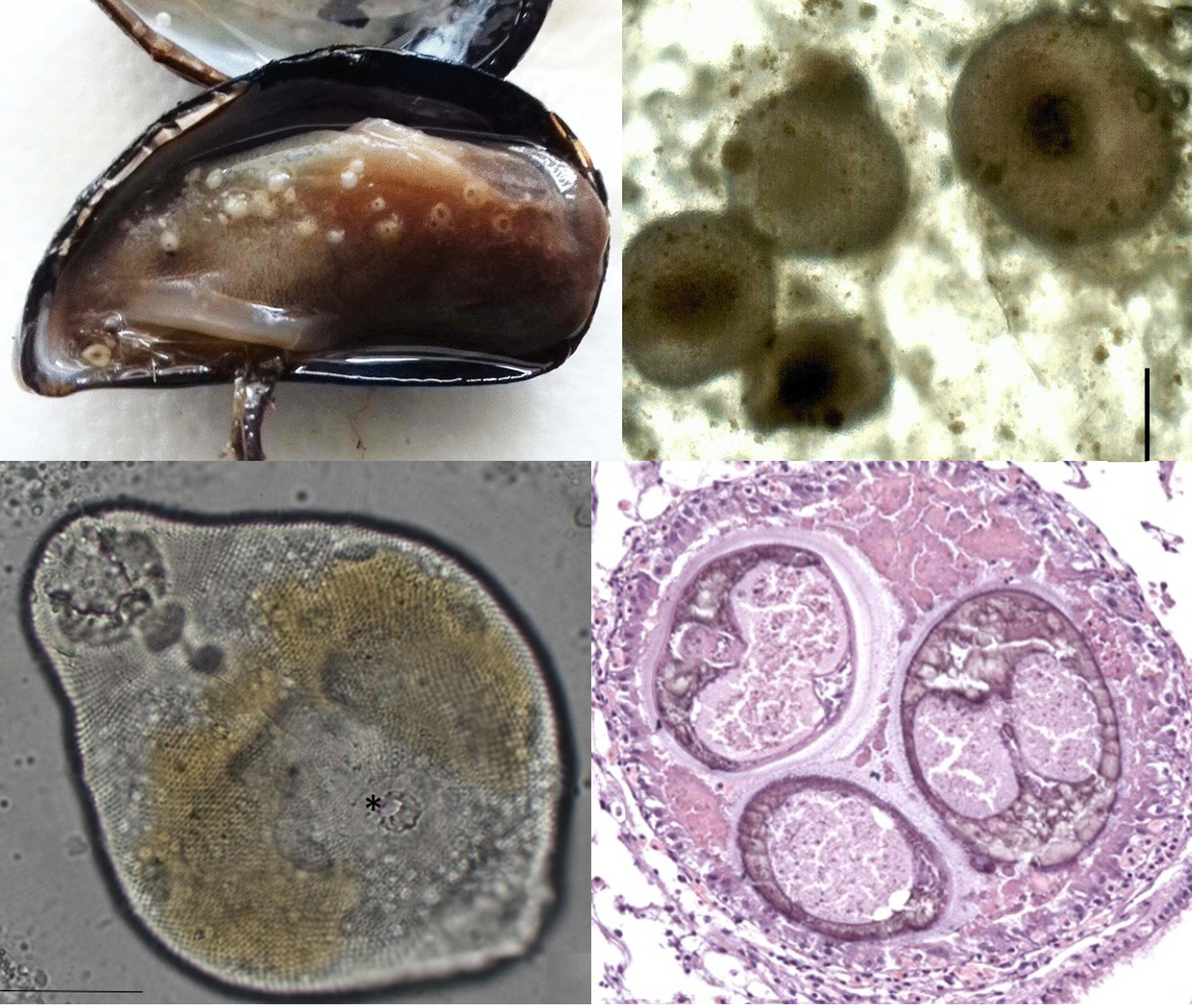
Left: Bay scallop infected with Saccularina, the red arrows indicating the parasite's sporocysts in the gills. Right: the cercaria stage of Saccularina which is a "cystophorous"-type cercariae.
From Fig. 1 and 2 of the paper

In 2012, a researcher started noticing a gill-dwelling parasite in both caged and wild bay scallops along the coast of North Carolina, and later at the Gulf Coast of Florida. These parasites are readily visible in the shellfish’s gills as they become swollen with the parasite's presence. Examination under a microscope revealed the parasites to be a species of parasitic fluke which is using the scallop for the asexual stage of its life cycle. Essentially, these trematode flukes are converting the shellfish into a parasite clone factory that pumps out a stream of free-swimming larvae to infect the next host in the life cycle.

Given such an operation consumes a lot of the host’s resources, this can interfere with the scallop’s growth and survival, and thus it is a major concern to the scallop fisheries in North Carolina. The key to managing any parasitic infection is an understanding of its natural history and life cycle, and unfortunately, given its relatively recent discovery, the life cycle of this parasite is almost entirely unknown. However, related fluke species can give us some clues, and as it turns out, this bay scallop parasite is no ordinary fluke.

First of all, DNA analysis showed that one of this fluke's closest relatives is Saccularina magnacetabula, a trematode species found on the other side of the world in Australia. While it is genetically similar enough to the bay scallop parasite for them both to be in the same genus (Saccularina), there is enough geographical and genetic distance between them that they are clearly different species. Furthermore, instead of scallops, S. magnacetabula infects the Sydney cockle (Anadara trapezia) as the chosen bivalve for its asexual stage. As for the parasite's next stops, it's a multi-part journey involving tiny crustaceans, followed by a type of smallish Australian fish called whiting (Sillago sp.), and finally the adult fluke completes its life cycle nestled in the fin membranes of the giant herring (Elops hawaiensis).

Saccularina magnacetabula, and by extension, the bay scallop parasite, belongs to a family of flukes called Didymozoidae - a flukey group of flukes with some very unusual anatomy and habits. While the adult stages of most trematodes are generally leaf-shaped, didymozoids come in all kinds of shapes and sizes. And they are found in a wide range of different bony fishes all over the world, mostly marine species. Additionally, instead of living in the final host’s gut like most flukes do, didymozoids cram themselves into all kinds of nooks and crannies such as the muscles, the gills, or even the fin membranes as is the case for S. magnacetabula.

While there are many known species of didymozoids, the life cycles for most of them are a mystery, with the hosts for the asexual stage known only for a few species. But those handful of species alone showed didymozoids to have quite the eclectic range. Aside from bivalves like cockles and scallops, the asexual stage of other didymozoids infect snails, but not just any regular snails - one species is known to use pelagic sea snails (which are also called “sea elephants”), while another species infects worm snails which are peculiar sea snails with twirly shells that encrust on rocks and other hard surfaces.

So, based on the information above, we can make some inference about the likely source of the bay scallop parasite. It’ll have to be some kind of predatory sea-dwelling fish harbouring the adult stage of the fluke, and given S. magnacetabula completes its life cycle in the giant herring, the bay scallop parasite is most likely completing its life cycle in some kind of predatory herring-type fish in the region, which means ladyfish or Atlantic tarpon

While we may have some clues about the bay scallop parasite’s life cycle, how they might have gotten there is more of a mystery though. This parasite was first seen in bay scallops in 2012, but if the disseminator of this parasite is really a local fish species such as the tarpon, why has it only been noticed now? Whatever its origin turns out to be, it seems the bay scallop parasite is not ready to give up its (many) secrets.

Reference:
Boggess, H. F., Varney, R. L., Freshwater, D. W., Ben-Horin, T., Preister, C., McCurry, H., Wilbur, A. E. & Buck, J. C. (2024). A newly discovered trematode parasite infecting the bay scallop, Argopecten irradians. Aquaculture 589: 740960.

July 11, 2024

Parvatrema spp.

Parasites are known for their complex life cycles, especially among parasitic flatworms such as flukes. And the flukes that are being featured in today's post have life cycles that make them a fluke among flukes. This blog post is about an extensive study that culminated from 25 years of work, where a group of researchers were able to identify and describe five different fluke species with a peculiar life cycle adaptation.

Parvatrema sp. “quadriramis” cercaria stage (left), young metacercaria (centre), parthenogenetic metacercaria containing fully-formed metacercariae (right). Insert: Parvtrema parthenogenetic metacercariae in the hepatopancreas of a limpet.
Photos from Fig. 4 and Fig. 11 of the paper

In the cold waters of the northern European seas and the Sea of Okhotsk, there is a group of parasites with life cycles that defy the conventions of its class. They are five closely related species of flukes in the genus Parvatrema, and they spend parts of their lives lurking quietly in the bodies of clams and snails. In many ways, they're just like other digenean flukes, with multi-host life cycles that involve turning their first mollusc host into a clone factory, producing clonal larvae which go off to infect a second host, and culminating in sexually mature adults living in the gut of vertebrate animals. But these Parvatrema flukes have evolved to do some things differently once they reach their second host.

These flukes infect sea snails such as limpets and periwinkles as their second host. Some species sit in the extrapallial space - the fluid-filled gap between the snail's fleshy mantle and the shell, others get into the gonads and digestive organs. Usually, this is a relatively dormant stage of the fluke life cycle, where they literally sit and wait to be swallowed by the appropriate final host. But with these Parvatrema flukes, instead of simply sitting around waiting to be eaten by a bird like other flukes would, they have another round of asexual reproduction, as a treat.

Each of those immature flukes become filled with numerous miniature clones of itself until it is stuffed to the point of exploding. Some of them take it further, with unlimited consecutive generations of parthenogenetic clones, each fluke exploding into multiple clones and then each of those clones explodes into even more clones and so on, like a never ending series of Matryoshka dolls. On top of that, some of those Parvatrema doing unlimited fluke works are also able to produce cercariae - the free-swimming larval stages - which then go off to infect more sea snails to start the asexual cycle again. 

So why did they evolve this unique developmental stage? Parvatrema are tiny flukes, the adult stage only live for a few days in the gut of a bird, and they produce less than a hundred eggs - a relatively low number compared with other flukes which may produce thousands or even millions of eggs over their lifetime. To make things worse, the likelihood of any one fluke successfully infecting the right hosts at each consecutive stages of its life cycle is astronomically low, so they need to multiply their numbers at every chance they get.

Furthermore, the final hosts for these flukes are migratory birds which only come once a year - so they need to make the most of their brief stay by making sure that if they only eat one infected snail, instead of just getting a single or a dozen flukes in each snail, they're getting the whole gaggle of fluke clones arriving en masse into the bird's gut in their hundreds or thousands, ready to get on with the business of producing the next generation.

Essentially, these Parvatrema flukes recapitulate the process that most other digenean flukes only undergo in the first host. Asexual reproduction in the first host is arguably one of the key evolutionary innovation of digenean flukes, allowing them to offset the losses associated with the process of being transmitted from one host to the next. Since Parvatrema seems to do asexual reproduction at every possible opportunity, they can provide us with insights into how flukes evolved their one weird asexual trick that gave them an edge in the transmission game.

Reference:
Galaktionov, K. V., Gonchar, A., Postanogova, D., Miroliubov, A., & Bodrov, S. Y. (2024). Parvatrema spp.(Digenea, Gymnophallidae) with parthenogenetic metacercariae: diversity, distribution and host specificity in the Palaearctic. International Journal for Parasitology. 54: 333-355

February 13, 2023

Parvatrema sp.

Pearls may look beautiful to us, but for some parasites, they represent a slow and claustrophobic death. Pearls are secreted by the soft and fleshy mantle, the part of a mollusc's body that also produces the shell. Indeed, pearls and shells are made from the same material - calcium carbonate. For the shellfish that produce them, pearls are battle scars of their fight against parasites.

Top left: Mussel infected with Parvatrema, Top right: Pearls from a mussel Bottom left: Parvatrema metacercaria stage from a mussel, Bottom right: Cross-section of a pearl showing three flukes trapped within.  
Top row of photos from Fig 1 of this paper. Bottom row of photos from Fig. 2 of the paper.

Bivalves are host to a wide range of different parasites that use them as a home, a site of propagation, or even as a convenient vehicle to their next host. One of the most common types of parasites that infect bivalves are trematode flukes. Some species embed themselves stubbornly in the mollsuc's tissue, others impair their ability to use parts of their body, and there are even some that end up castrating their shellfish host. Sometimes, these seemingly passive molluscs put up a fight against these tiny intruders, especially when they get into the mantle fold. And they do so by secreting calcium carbonate around the invading parasite, smothering these flukes alive - and the result of that gruesome interaction is a pearl.

The study being featured in this post looked at the frequency of pearls and parasites in mussels on the northwestern Adriatic coast. The flukes that are most commonly associated with pearls there are those from the Gymnophallidae family, and this study focus on one particular genus - Parvatrema. These flukes use mussels as their intermediate host, where the larvae temporarily reside and develop until they are eaten by shorebirds - this parasite's final host.

Out of the 158 mussels that the researchers examined, about two-thirds of them were infected, and most of the mussels had a mix of both live flukes and pearls.Their parasite load varied quite a lot, from some mussels with a few flukes, to one with over 3700 flukes. But on average, each mussel harboured about 200 flukes. The flukes were scattered throughout the mussel's body, but most were concentrated near the gonads, and some were found at the base of the gills. A few were squeezed in between the mantle and the shell - and it is those that are at the most risk of being turned into pearls. 

Speaking of which, about half the mussels that the researchers examined had pearls of some sort in them. But there were far fewer pearls than there were flukes. Each mussel had 35 pearls on average, but they were nowhere near the size of pearls most people associate with jewellery. These pearls were about the same size as fine sand grains, but they were pearls nevertheless - complete with entombed fluke(s) in each of them.

The high prevalence of Parvatrema in mussels from this area means that it could be risky to set up mussel farms there, at least near the coast where the parasite's bird hosts like to hang out. No one wants to buy mussels riddled with parasites, and while pearls are considered as valuable, the type of pearls found in these mussels only decrease their market value. That is one of the reasons why some mussel farming operation are located offshore where they won't be exposed to Parvatrema and other parasitic flukes. 

Based on the results of the study, pearl formation seems a bit hit-or-miss as a defensive mechanism. The majority of flukes get away with living rent-free in the mussels without setting off the pearly deathtraps, and it's not entirely clear why some of them trigger pearl formation, while most flukes are left alone. Despite this, some recent studies indicate that bivalves are not the only molluscs that can entomb their parasites that way. Some land snails are also capable of sealing away various parasites such as flukes and roundworms into their shell. 

So it seems the molluscs have evolved a general two-in-one defensive package that can potentially protect them against both predators and parasites. While neither shell nor pearls offer guaranteed protection against predators and parasites respectively, it's still better than having nothing at all.

Reference:
Marchiori, E., Quaglio, F., Franzo, G., Brocca, G., Aleksi, S., Cerchier, P., Cassini, R. & Marcer, F. (2023). Pearl formation associated with gymnophallid metacercariae in Mytilus galloprovincialis from the Northwestern Adriatic coast: Preliminary observations. Journal of Invertebrate Pathology 196: 107854.

November 12, 2019

Electrovermis zappum

Fish blood flukes are common parasites in the aquatic environment and many species have been described from all kinds of fish all over the world. However the full life cycle is only known for relatively few of such flukes, because while the adult parasite can be fairly common in the fish host population, the asexual stage living in the invertebrate host can be quite rare and difficult to find. The study featured in this blog post described the life cycle of Electrovermis zappum - a blood fluke that lives in the heart of the lesser electric ray, but spends part of its life cycle in a beach clam.

Left: An adult Electrovermis zappum, Right: the life cycle of E. zappum. From the Graphical Abstract of the paper
When it comes metamorphosis and transformation, most people usually think of caterpillars turning into butterflies, but such level of change pales in comparison to the different forms that digenean flukes take on at each stage of their life cycles. The adult E. zappum fluke is a long skinny worm about 1.5 mm long, living in the heart of an electric ray. Over half of its length is composed of reproductive organs, devoted to producing a steady stream of eggs. The eggs that manage to make their way out of the ray's body hatch into cilia-covered larvae called miracidia. This microscopic ciliated mote then infects a coquina clam.

It then undergoes another set of transformation as it enters the asexual stage of the life cycle. The lone miracidium turns itself into a clone army of self-propagating units call sporocysts which take over the clam's body. These sporocysts look like microscopic marbles, each measuring about one-tenth of a millimetre across, and packed within those translucent spheres are the next stage of the fluke's life cycle. Within each sporocyst are half a dozen skinny, tadpole-shaped larvae called cercariae - these develop and grow within the nurturing wall of the sporocysts until they are ready to be released into the water column, at which point the sporocyst will start growing the next batch of cercariae from its reserve of undifferentiated germinal cell balls.

A single infected clam can be filled with several hundred of those sporocysts, which occupy the space where the clam's gonads would have been, with some also spilling over into the digestive system. This process essentially turns the clam into a parasite factory that churns out thousands upon thousands of infective fluke larvae, saturating the surrounding waters. Both the bottom-dwelling electric ray and the coquina clam are found right next to each other in the swash zone of beach, so the cercariae are released right where the rays are likely to be.

Most of these short-lived, microscopic larvae will perish - eaten by other marine creatures or simply exhausting their energy reserves before encountering an electric ray. But enough of them will come into contact with an electric ray to continue the life cycle. When a cercaria comes into contact with a ray, it will discard its paddle-like tail, and burrow though the skin and into the blood vessels. It will then traverse the vast network of the fish's circulatory system until it finally settle within the heart's pulsating lumen, and start the cycle anew.

Because the asexual stage in the coquina clams allows E. zappum to continuously spam the water with waves of tiny baby flukes, this means it only takes a relatively small number infected clams for E. zappum to saturate the water with enough infective stages to maintain a viable population of the parasite in the ray hosts. Indeed, this was reflected in what the researchers found in this study - while the adult fluke was fairly common in the electric rays (fourteen of the fifty four rays the researchers examined were infected with adult E. zappum), infected beach clams were extremely rare - only SIX of 1174 clams that they examined at were infected.

On the beaches where these coquina clams and electric rays are found, each square metre of beach are densely packed with thousands of coquina clams. So looking for an infected clam amidst all that is like panning for gold - time-consuming and labour-intensive work which involves spending hours upon hours in front of a microscope with a bucket of shellfish. This is one of the reason why the full life cycle of so few of these flukes have been described.

Furthermore unlike most other digenean flukes that tend to infect mollusc (mostly snails) at their asexual stage - which narrows down the list of potential animals to examine, some fish blood flukes are known to infect some unusual invertebrates. While E. zappum is relatively conventional in that it still uses a mollusc for the asexual stage of its life cycle, there are some species which have really gone off the beaten evolutionary path and have evolved to infect polychaete worms.

Blood flukes have been reported from other species of rays in other parts of the world. Based on their DNA, the blood flukes that infect cartilaginous fish all belong to their own special evolutionary branch among the fish blood flukes, and that the common ancestor of all the living blood fluke lineages, including those that infect mammals and birds today, might have originated over 400 million years ago.

So long before there were dinosaurs, long before there were mammals, even before a lineage of fish began crawling onto land, and at around the same time as when the earliest iterations of sharks and ratfish were prowling the Silurian seas, the ancestors of these flukes were already going through their life cycles, and well-acquainted with the hearts of vertebrate animals.

Reference:
Warren, M. B., & Bullard, S. A. (2019). First elucidation of a blood fluke (Electrovermis zappum n. gen., n. sp.) life cycle including a chondrichthyan or bivalve. International Journal for Parasitology: Parasites and Wildlife 10: 170-183.

August 13, 2019

Caulobothrium sp.

Scallops are highly prized as seafood because of their tasty adductor muscle and roe, but humans are not the only ones with a taste for scallops. These bivalves are on the menu for a wide range of marine animals including various crabs, snails, seastars, marine mammals, and fishes. And many parasites make use of these predator-prey interactions to complete their life cycles.

Scallops are an important part of the Peruvian aquaculture, but little is known about their parasites there. In the study we're looking at today, researchers collected samples of scallops from a scallop ranch in Sechura Bay over the course of three years between 2013 to 2015, to examine them for parasites. They ended up looking through a total of 890 scallops, and the parasite that they encountered most frequently were whitish cysts that turned out to be tapeworm larvae belonging to the genus Caulobothrium.
SEM and light microscopy photos of tapeworm larvae. The lower left photo shows the tapeworm's scolex
Photos from Fig. 1 and 2 of the paper
Those tapeworm larvae were embedded in the scallops' gonads, and their numbers ranged from just twenty to over two hundred per scallop. While the number of infected scallops varied each year, they were nevertheless consistently high, with about eighty to ninety percent of scallops harbouring tapeworms. While this level of prevalence may seem unusually high, this is actually comparable to previous studies on tapeworms in scallops from other regions, so this is nothing too out of the ordinary.

Ultimately, those tapeworms are waiting for a rendezvous with the final host which, based on what is known about other species of Caulobothrium around the world, is the most likely a ray of some sort. Tapeworm species in the Caulobothrium genus have been reported from eagle rays in the waters of United States and Chile, as well as stingrays on the coast of Australia. On the coast of Peru, the adult stages of Caulobothrium have been found in the gut of both eagle rays and cownose rays, and given the circumstances, it is likely that the tapeworms found in the scallop gonads represented the larval stage of those worms.

Rays have specialised jaws armed with heavy, rounded teeth that allow them to crunch through the shell of bivalves such as scallops, and this tapeworm make use of their taste for shellfish to complete their life cycle.

Tapeworm larvae are not the only parasites with an affinity for scallop roe. Flukes in the Bucephalidae family also infect the gonads of scallops and turn them into parasite factories that churn out streams of parasite larvae. Much like those flukes, the presence of so many tapeworm larvae in the scallop gonads can impair the scallop's reproductive capacity, which as you can imagine, would be a concern for scallop aquaculture since they can potentially reduce the number of scallop larvae produced during spawning season.

In terms of infected scallops' edibility, Caulobothrium is known for being host specialists which can only infect rays, so there is no real risk of these tapeworms infecting humans, but on an aesthetic level to most would-be consumers, scallops with tapeworm-filled roe simply look too gross to eat.

The life cycles of most marine tapeworms are not well understood, and of the over one thousands species of tapeworms which have been described from sharks and rays, the full life cycle is only known for a measly FOUR species. Finding and documenting the larval stage of such tapeworms in marine animals such as scallops can help us put together the biological puzzles that are their complicated life cycles, and work out the roles these parasite play in marine ecosystems.

Reference:
Castro, T., Mateo, D. R., Greenwood, S. J., & Mateo, E. C. (2019). First report of the metacestode Caulobothrium sp. in the Peruvian scallop Argopecten purpuratus from Sechura Bay, Piura, Peru. Parasitology Research 118: 2369–237.

April 26, 2015

Nepinnotheres novaezelandiae

Life as a pea crab seems pretty sweet, you spend most of your time sitting snug and protected within the armoured shelter of a shellfish, while your host's filtration current bring you a constant stream of oxygen and food - everything that a pea crab needs for a good life. Well, almost everything - because there's more to life than just being protected and fed. Much like other organisms pea crabs need to reproduce - that's how evolution works, and unlike many other living things, a pea crab cannot just clone itself.
Male Nepinnotheres novaezelandiae squeezing in between
the valves of a mussel. From video here.
So when it comes to reproduction, the balance of living the pea crab life tips from "pretty sweet" to "absolutely terrible" - especially if you are a male pea crab. For them, trying to find a mate is a harrowing challenge than none of us can possibly imagine. First of all, to reach a potential mate, you have to leave your host, which means you have to pass the gates that are the valves of the host mussel, without being caught in between them. At that stage, those valves that had offer such formidable protection for the pea crabs then become death traps, with about 13% of male crabs meeting their end at this molluscan gate - their bodies litter the mussel bed.


Once outside, the male pea crab faces even more challenges. These tiny crustaceans, which are more accustom to a cosy life inside a shellfish, have to cross the treacherous, open areas of the mussel bed, filled with horrible monsters (in the form of predators like fish, octopus, and larger crustaceans) for which an exposed pea crab is just a convenient snack. Furthermore, male crabs only make up 20% of the population despite the more or less equal sex ratio of immature pea crabs. The length that they have to go to just to find a mate probably has something to do with that...

Despite the odds, almost 90% of all female crabs in the population carry fertilised eggs, so some male crabs must be having successes - but how?

The researchers who conducted this study noticed that the male pea crabs always set out under the cover of darkness when they will be less likely to be spotted by predators, and also because mussels are more relaxed at night. From the researchers' perspective, this also means that all the experiments and observation of pea crab behaviour had to be done in the dark. So in addition to sea water tanks, they set up some infra-red cameras to capture footages of all this activity - like some kind of voyeuristic shellfish reality TV show.

So what would coax a male crab out of his cosy home? To find out, the researchers constructed a flow-through observation chamber lined with PVC tubes in which they placed pea crab-infected mussels. When they placed a mussel with a female crab upstream of one with a male pea crab, the male crab would exit their host 60% of the time, roused into action by something which seem to secreted by the mussel (or the female crab in the mussel) upstream.

Male Nepinnotheres novaezelandiae tickling the mantle edge
of a mussel. From videos here.
The crab then makes its way to the mussel where the female crab resides. Once there, the pea crab patiently tickles the mussel's mantle fringe with its legs to try and convince the bivalve to let it enter. This is also the reason why the male crab only do this at night, because a mussel's response to such tickling can be very different in daylight. Try the same trick during the day and the bivalves would slam shut, crushing the amorous crab between its valves. On average, the crab will spend over three hours fiddling away at the mussel to coax the shellfish into opening up.

Additionally, in a different flow-through seawater tank where the crabs were given more freedom to roam from one host to another, the researchers recorded how long it took for the male pea crab to leave its host and reach a mussel containing a female crab. The entire journey from exiting the original host mussel to reaching their final destination took seven hours on average, though this varies from a quick hour-and-a-half jolt, to an eighteen-and-a-half hour-long trek for one particularly unfortunate individual.

So when love (or at least lust) is in the water, the pea crab will give up the easy life, and risk life and limbs for an evening rendezvous.

Reference:
Trottier, O., & Jeffs, A. G. (2015). Mate locating and access behaviour of the parasitic pea crab, Nepinnotheres novaezelandiae, an important parasite of the mussel Perna canaliculus. Parasite, 22: 13.

June 16, 2013

Himasthla elongata

Photo taken by and used
with permission from Kirill V. Galaktionov
Today's post is bit of a trip down nostalgia lane for me, as the experimental model used in the study we are featuring today is a host-parasite combination similar to one I worked on for somes years during my PhD and postdoc - bivalves and flukes (specifically flukes from a family called the Echinostomatidae - identifiable by their fetching array of collar spines). Much like a parasite that I worked on (Curtuteria australis), Himasthla elongata encysts in the foot muscle of its host and transforms into a stage called the metacercaria (see left photo). But whereas C. australis infects cockles on the mudflats of New Zealand, H. elongata infects mussels on the rock shores of the White Sea.

By embedding itself in the mussel's foot, this parasite hinders the mollusc's ability to move and produce the all-important byssus threads that anchor them to rocks or other substrates. If it becomes infected with too many H. elongata, the mussel loses its ability to use its foot and its survival becomes compromised. Thus this parasite selects for the evolution of mussels that are resistant against it, resulting in a coevolution arms race between the mussels and H. elongata.

To find out how parasites and mussels fare against each other and the role that genetic variants in both the parasite and host population play in coevolution, a group of Russian researchers conducted a series of parasite survival studies and experimental infections. First of all, they did an in vitro experiment where they exposed the infective larval stage of H. elongata (called cercariae) to the blood of different mussels. This was followed by an experimental infection study where they exposed some of those same "blood donor" mussels to H. elongata larvae and measured how well they were they at resisting the parasite.
Photo taken by and
used with permission
from Kirill V. Galaktionov

The researchers obtained parasite-free mussels from an experimental aquaculture farm to act both as blood donors and infection targets for H. elongata cercariae, while the parasites themselves came from infected periwinkles that the researchers collected from an intertidal inlet. These periwinkles harboured the asexual proliferative stages of H. elongata which produce cercariae (see photo on the right). Because H. elongata undergoes asexual multiplication in the periwinkle host, the researchers were able to obtain multiple genetically-identical (clones, essentially) cercariae from each infected snail and test them against a group of genetically-varied mussels.

The researchers paired up 51 different H. elongata clonal lines to blood samples from 161 randomly selected mussels for a total 764 parasite versus host blood combinations* (!). They found that a handful of mussels had blood that killed every single cercaria that came in contact with it and another handful had blood where all the cercariae survived and successfully turn into metacercariae. It seem that H. elongata is adapted specifically to surviving contact with mussel blood (just that it seems that some are better adapted than others), because when they tried to incubate H. elongata cercariae in the blood of the soft-shell clam (Mya arenaria), all the cercariae died within an hour or two.

In a follow-up experiment, they selected 39 of those mussels that had previously served as "blood donors"and exposed each to one of twelve H. elongata clones that were used in the in vitro experiment and found that the results of the in vitro experiment were pretty good indicators of the outcome of those experimental exposures - mussels with blood that killed all the H. elongata they came in contact with were also better than most at fighting off infection by the parasitic fluke. The rest of the mussels were fairly vulnerable to H. elongata and a small handful offered almost no resistance. The larger mussels were generally better at fighting off the parasites with just a little over a quarter of the H. elongata cercariae getting through, while more than half of the cercariae successfully established in the smaller mussels, regardless of the host or parasite genotype.

The parasites themselves also varied in their effectiveness at infecting mussels. Most of the H. elongata clones were fairly good at it, there were a few "superstars" that were especially effective at becoming metacercariae in mussels, while there were also a few "duds" that were hopeless, regardless of which particular mussel they were up against.

Other host-parasite coevolution arms races operate under so-called "gene-for-gene"-type interaction. Examples of which include the bacterial parasite Pasteuria ramosa in waterfleas where a specific parasite strain is most successful at infecting a specific host strain, or the arms race between parasitoid wasps and aphids' protective symbionts where you have wasp lines that can overcome most of aphid protective symbiont strains out there, but remain vulnerable to one specific strain of the symbiont.

What those Russian scientists found with the mussel-Himasthla elongata system does not seem as absolute. Instead, we see variation in overall performance in the population of both host and parasite: there are parasites that ranged from being super effective at what they do, all the way down to complete duds and everything in between. They in turn are going up against mussels with varied level of resistance against them, and how much of a fight those bivalves put up can also be affected by the age and/or body size of the host. However, what it does have in common with those "gene-for-gene"-type coevolutionary systems is that there is a genetic component to either infectivity or resistance, and none of the host are completely resistant to all parasites, just as not all the parasites are completely effective at infecting the available hosts.

Reference:
Levakin, I. A., Losev, E. A., Nikolaev, K. E., & Galaktionov, K. V. (2013). In vitro encystment of Himasthla elongata cercariae (Digenea, Echinostomatidae) in the haemolymph of blue mussels Mytilus edulis as a tool for assessing cercarial infectivity and molluscan susceptibility. Journal of Helminthology, 87: 180-188.

*because there was simply not enough blood and cercariae to go around, not every H. elongata clone was exposed to the blood from every mussel

March 19, 2013

Duplicibothrium minutum

In June 2010 at Folly Beach, South Carolina, the local community was shocked and dismayed by the sight of millions of dead dwarf surf clams (Mulinia lateralis) that carpeted the beach with rotting shellfish. During the course of an investigation into what might have caused this die-off, researchers discovered some tapeworm larvae in the clams which had not been previously reported from that area.

image of Duplicibothrium minutum from figures in the paper
Because the larval stages of tapeworms have few of the morphological features that usually serve as diagnostic markers to identify different species, the researchers looked to their DNA for clues on their identity. They sequenced a section of the tapeworms' DNA and compared it with known DNA sequences of tapeworms (we have previously featured a study which used the same technique, know as DNA barcoding, to figure out the life cycle of a Great White shark tapeworm.) They were able to determine that the most common species of parasite in those clams was the tapeworm we are featuring today; Duplicibothrium minutum. Out of the 200 clams that the researchers dissected, 150 of them were infected with D. minutum, while four of clams were infected with another species of tapeworm - Rhodobothrium paucitesticulare (three of which were also infected with D. minutum).

photo of Rhodobothrium paucitesticulare 
from figures in the paper
The larvae of these two tapeworms occupied different part of the clam's body - whereas D. minutum were often found in pairs in the bivalve's digestive glands, R. paucitesticulare larvae tucked themselves away at the gap just beneath the clam's fleshy mantle. Both tapeworms are gut parasites of the Cownose ray, (Rhinoptera bonasus) which commonly feed on mollusks and other invertebrates that they suck up from sandflats and crush with their hard dental plates. Rhodobothrium paucitesticulare only infects the dwarf surf clam and one other species of clam (Donax variabilis), while D. minutum has a much wider host range and has also been found in two other species of clams (Donax variabilis and Angulus versicolor) as well as the Florida crown conch (Melongena corona).

Unlike some parasitic flukes that can alter the burrowing behaviour of clams and other bivalves, neither tapeworm caused much noticeable harm to the host clams. The presence of D. minutum caused some minor enlargement at the opening of the digestive glands, but there were no signs of inflammation, and the R. paucitesticulare larvae seem to be completely benign and did not affect the clam's health at all. So while those tapeworms seems to be very common in the clam population, they were not causing nearly enough harm to be considered responsible for the mass die-off.

Reference:
de Buron I, Roth PB, Bergquist DC, Knott DM. (2013) Mulinia lateralis (Mollusca: Bivalvia) die-off in South Carolina: discovery of a vector for two elasmobranch cestode species. Journal of Parasitology 99: 51-55

October 20, 2010

October 20 - Urastoma cyprinae

Yesterday you met a copepod that lives in the mantle of bivalves. Today, meet a turbellarian (a platyhelminth) that could be cozying up to those crustaceans. Urastoma cyprinae spends the majority of its life in the gills of bivalves, specifically mussels such as the gastronomically and thus economically important species Mytilus edulis, Mytilus galloprovincialis, and Crassostrea virginica. They live and feed and grow in the mussels, but their complete life cycle stumped researchers because eggs were never observed in these hosts. Researchers in Spain recently discovered that when U. cyprinae is ready to lay eggs, it leaves the bivalve, secretes a protective cocoon around itself and produces an egg sac. About 24 days later, the young hatch out (shown in the photo), make their way out of the coocoon and swim off to find new mussel hosts. The adults sometimes appear to just die in the cocoons, but sometimes also were observed to escape from it themselves and head off to lay more eggs.

Read more about these parasites here and here.

Photo by Celia Crespo González.

October 19, 2010

October 19 - Pseudomyicola spinosus

Pseudomyicola spinosus is a parasitic copepod that is found in more than 50 species of bivalves around the world, ranging from clams to mussels to scallops. It dwells in the mantle cavity of the bivalve, where it grazes on mucus produced by the host. This copepod has a pair of hook-like attachment appendages that allow it to cling to the host tissue and avoid being swept away by the constant water flow that passes through the mantle cavity. In large numbers, they can cause considerable tissue damage to the host - the constant attachment and reattachment of the copepod (which can be highly mobile within the host's body cavity) aggravate host tissue, causing epithelial erosion and induce over-production of mucus. At lower infection levels, the tissue damage caused by the copepod is almost negligible, but it does have a more subtle effect on its host. It has been found that infection with just a few P. spinosus is associated with higher levels of infections by metcercarial cysts of echinostomatid trematodes such as Curtuteria australis and Acanthoparyphium. Once again, this is possibly due to the effects of the copepod's attachment appendages, which damage the epidermis in such a way that facilitates subsequent invasion by trematode cercariae.

References:

Cáceres-Martínez, J. and Vásquez-Yeomans, R. (1997). Presence and histopathological effects of the copepod Pseudomyicola spinosus in Mytilus galloprovincialis and Mytilus californianus. Journal of Invertebrate Pathology 70, 150–155.

Leung, T. L. F. and Poulin, R. (2007). Interactions between parasites of the cockle Austrovenus stutchburyi: Hitch-hikers, resident-cleaners, and habitat-facilitators. Parasitology 134, 247–255.

Post and image by Tommy Leung.

August 17, 2010

August 17 - Acanthoparyphium sp. B

The purpose of this blog is to celebrate parasite biodiversity, but what some people might not realise is that much of parasite biodiversity might simply be hidden from sight, in more ways than one. It isn't just that parasites are hidden within the body of their free-living host, sometimes, previously unrecognised species could be right under your nose and you wouldn't know it even if you see it.

For example, take today's parasite. For quite a few years, it was thought that the Acanthoparyphium found at Otago Harbour, South Island, New Zealand consisted of a single species. It infects the marine snail Zeacumantus subcarinatus as the first host, within which it undergoes asexual replication to produce free-living larvae call cercariae which then infect the New Zealand cockle Austrovenus stutchburyi (which also happens to be the second host for another trematode, Curtuteria australis - featured on January 15). However, during some routine investigation with molecular markers, it was serendipitously discovered that what was initially thought to be a single species of trematode actually turned out to be composed of at least four different species that are morphologically indistinguishable from each other (or at least very similar), but genetically distinct. At the moment, they are provisionally known as Acanthoparyphium species A, B, C, and D. While they all utilise the same species of snail as the first host in their life-cycles, since the initial discovery, it has been found that they differ in term of the next host they infect. Whereas the encysted stages of species A were commonly found in cockles, those of species B were found to infect ragworms, and the second host of species C and D remains unknown.

Cases like this goes to show that there's more than meets to the eye with most parasites!

For more details see:
Leung, T.L.F., Keeney, D.B. and Poulin, R. 2009. Cryptic species complexes in manipulative echinostomatid trematodes: when two become six. Parasitology 136: 241-252

Photo credit: Haseeb Randhawa and Matthew Downes.
Contributed by Tommy Leung (with B for Birthday - Happy Birthday, Tommy!)

June 27, 2010

June 27 - Stegotricha enterikos


Stegotricha enterikos is a parasite of oysters and has been found infecting them on both coasts of North America. They are quite prevalent and though they live within the body of the oyster and ingest the epithelial cells of the oyster, they don't appear to induce a great deal of pathology in the shellfish.

The image comes from this site.

June 24, 2010

June 24 - Zaops ostreum

How many kind of food can you name comes with its own side dish? Well, the eastern oyster (Crassostrea virginica) should be on that list. This week. we've already seen how a trematode infection can improve the taste of oysters, but it seems that oyster also comes with another gastronomic treat in the form of the pea crab Zaops ostreum. Pea crabs (family Pinnotheridae) are small soft bodied crabs which live inside a variety of marine invertebrates, with most species living in bivalves. Zaops ostreum infect the oyster as a tiny first stage larvae, and grow to maturity within the bivalve's mantle cavity, feeding upon food-laden mucus strings produced by its host's filtering action. It is a true parasite in that it causes harm to its host. Not only does it steal food from the oyster, it also forms an obstruction within the body cavity and erode the gill tissue. From a culinary perspective, there are many serving suggestions available for pea crabs - they can be served raw, deep fried, or sautéed, and can be eaten either as a side dish to oysters, or even on their own (if you can get enough of them to make a meal!).

Photo and contribution by Tommy Leung.

June 23, 2010

June 23 - Bucephalus haimeanus

Do you enjoy seafood? Especially shellfish? Especially raw oysters? Whether you know it or not, the next time you tuck into some raw oysters, there might be a parasite which is responsible for enhancing your gastronomic experience. Usually, having parasites in your shellfish can be seriously bad news. However, apart from the squeamishness factor, the harm caused by your seafood being infected with the asexual stage of a trematode is minimal to neglible.

As we have seen in prior blog entries, the asexual stages of trematode parasites castrate their first intermediate host, utilising it to produce free-living larval stages which are released into the environment. Now if this first host happens to be oysters - which is the case for Bucephalus haimeanus, this has interesting culinary implications. In uninfected oysters, after they spawn they exhaust the all the nutrients and resources they've stored up and become almost tasteless after spawning season is over. However, because infected oysters are castrated, they stay fat and retain their flavor through out the year.

So from a gastronomic point of view, parasitised oysters are a class above their unafflicted brethen!

Contributed by Tommy Leung.

May 27, 2010

May 27 - Pinnotheres pisum


Some crabs, like hermit crabs, live in the discarded shells of mollusks, but some, like the pea crab Pinnotheres pisum, just can't wait for the resident to move out first and move in as a roommate -- but the kind of bad roommate that steals your food and damages your house. P. pisum, which is at most about half and inch wide, lives in the mantle cavities of bivalves such as mussels and clams where it picks the food off their gills and can severely damage them in the process (kind of like you might imagine if something with 10 legs lived in your fridge full-time.) I chose this photo from wikipedia (click on it for a blown-up view), which shows a pea crab that has fallen out of the clam that an otter is eating. Better go find another landlord, little crab!

February 5, 2010

February 5 - Lampsilis fasciola


Mussels may seem like unassuming creatures, hanging out on the riverbed until they’re caught and served with a delicious garlic butter sauce, but many species have parasitic larvae that are crafty and downright aggressive when it comes to attracting their hosts. The larvae, known as glochida, usually can’t move around to seek out their hosts, so they employ a wide variety of lures to bring those hosts to them. Meet Lampsilis fasciola, commonly known as a "pocketbook mussel". The glochidia of this species develop inside their mother for approximately one year, and then the mother mussel moves them to the outside of her shell into a pouch on her mantle. Here’s where it gets interesting. The mantle looks remarkably like a small fish, complete with eyespots, and the movement of the mantle looks like a small fish swimming – just the right meal for a larger fish passing by. When a hungry fish bites the mantle, the glochidia-filled pouch ruptures and the larvae emerge, latching onto the inside of the fish’s gills with adhesive threads and teeth on their shells’ valves. Once in their host, the glochidia encyst and develop into juvenile mussels and then drop down to the riverbed to mature, mate, and get ready to lure the next generation of host fish.

Contributed by Kate Bowell.

January 15, 2010

January 15 - Curtuteria australis


Curtuteria australis Allison 1979 (Platyhelimthes: Digenea: Echinostomatidae) infects bivalves (in this case, a cockle, a type of clam) on the mudflats and sandflats of New Zealand. This parasite lodges itself in the foot of the bivalves where it forms a hard cyst. As more and more parasites accumulate, the cockle loses its ability to dig itself into the underlying sediment, leaving it stranded on the surface of the mudflat/sandflat. There, it is exposed to predation by shorebirds such as oystercatchers, which are the parasites' next hosts. These parasites also have a cascading effect on the rest of the ecosystem - as the mudflat is filled with stranded bivalves, the nature of the substrate changes from one consisting largely of mud and sand, to one littered with the hard shells of bivalves. This, in turn, alters the biotic community which inhabits the rest of the ecosystem.

The photo shows a cockle's foot with encysted metacercariae of Curtuteria australis tagged with a fluorescent dye.

Contributed by Tommy Leung.
Read the full paper here.