"So, naturalists observe, a flea has smaller fleas that on him prey; and these have smaller still to bite ’em; and so proceed ad infinitum."
- Jonathan Swift
Showing posts with label nematode. Show all posts

October 14, 2026

Anisakis spp. (et al.)

In the ocean, squids occupy a sort of middle position in the food web. While they are voracious predators that attack anything that moves, they themselves are prey for a variety of animals like larger predatory fish and marine mammals. Even the mighty giant squid is food to whales and fish at various stages of their lives. This makes them the perfect vehicle for parasites looking to climb their way up the food chain, starting from infecting small plankton to end up in the bowels of a large fish or a whale.

Left: An Antarctic flying squid (Todarodes filippovae). Top right: Anisakis larvae encysted on squid stomach (indicated by red arrows), Bottom right: Anisakis physeteris larva from a squid.
Photo of flying squid and infected squid stomach provided by Dr Chenhua Li, photo of larval Anisakis from this paper

To investigate the role played by squid in parasite transmission, scientists from the University of Otago dissected a whole bunch of squids and what they found was a scientific treasure trove of parasites. They had on their hands a mixed platter of large squids,  including the likes of arrow squid (Notodarus sloanii), flying squids (Todarodes spp.), glass squid (Teuthowenia pellucida), cock-eyed squid (Histoteuthis sp.), and many more - include a giant squid (Architeuthis dux)! In total, they were able to obtain 11 different species of squid, fit for a giant cephalopod banquet. But this would have been a banquet with parasite fillings, because they also found six types of roundworms, three types of tapeworms, and a mystery fluke among those cephalopods.

Of the six different roundworm species, three of them were in the Anisakis genus - a notorious public health hazard. Anisakis worms complete their life cycles in whales, but they can cause problems when they end up in humans and their occurrence in people is highly dependent on eating habits. For example in Japan, due to the popularity of raw seafood, Anisakis causes over 19000 cases of illness per year, whereas it is far less common in other regions with different seafood consumption preferences.

The scientists also found the larvae of Crassicauda, a genus of very long (>3 m) whale parasite that lives in all kinds of awkward nooks and crannies in the whale's body, such as under their skin, in their kidneys, in their genitalia, or even occupying multiple parts of the body simultaneously. The complete life cycles of those worms are unknown, though evidently squids are involved for at least a part of it. Similarly mysterious is Lappetascaris, a nematode which has previously been featured on this blog. The full life cycle of these worms are also shrouded in mystery, but based on what little we know, they would have been on their way to a swordfish or other large predatory bony fishes. 

Meanwhile, the tapeworms are ridesharing these squid in order to reach a different destination - sharks. Two of the tapeworms found were trypanorhynchans, which also use various fish as vehicles to reach their final hosts. They are armed with four gnarly hook-lined tentacles that anchor them to their hosts' intestinal wall once they finally reach a shark's bowel. The other tapeworm species was a genetic match with a tapeworm which has previously been reported to dwell in the belly of porbeagle sharks, which is perhaps to be expected as these sharks have been documented to frequently dine on squid.

Amidst all the worms riding these squid to various different destinations was a fluke lurking in the mysterious googly-eyed glass squid. It was not readily identifiable to a known species but based on its DNA, it seems to be genetically similar to a species of fluke which lives in the throat of ocean sunfish, from the family Accacoeliidae. Very little is known about the life cycle of this family of flukes, and what little we know indicates they use various oceanic invertebrates such as pelagic snails and man o'war as intermediate hosts. Given the habits and diet of the glass squid, it might have inadvertently picked up these flukes while chowing down its usual prey.

So simply by looking under the hood of some squids have given us some remarkable insights into the ecological interactions in the ocean, along with parasitic worms are able to zip between hosts via such interactions. For those parasites, cephalopods are like well-connected travel hubs through which they can reach a myriad array of the ocean's inhabitants, all linked via a network of transmissions.

Reference:

July 14, 2026

Echinomermella matsi

Nematodes (roundworms) just can't get enough of being parasites. Parasitism has independently evolved at least fifteen times among nematodes, with each of them realising their best lives inside the body of another animal (and for some of them, inside plants). But how did they end up like this? Well, the worm being featured in this post may offer some answers (while raising many others).

Left: A bundle of Echinomermella matsi inside of an opened sea urchin. Right: An E. matsi juvenile viewed under a microscope. Photos from Fig. 2 of the paper.

Echinomermella matsi is a species of roundworm that parasitises sea urchins. It can grow fairly large and is very noticeable, opening up a heavily infected sea urchin is like looking at a bowl of udon noodles. In some urchin populations, these worms can be rather common, and they exert a demanding toll on their hosts, so much so that they can end up castrating the sea urchin as they divert resources away from their host's gonads. But sometimes, mere castration is not enough, and this worm can also shorten the urchin's lifespan.

All this affects the population dynamics of sea urchins, which has flow on effects on the rest of the intertidal ecosystem as sea urchins are often major grazers of kelp and seaweed in such environments. Given the impact that this nematode can have on urchin populations, there is some interest in figure out what makes this worm ticks, and this study took a genomic approach to that question.

It has previously been thought that Echinomermella and similar parasitic nematodes might be marine relatives of mermithid nematodes, which are a family of roundworms that infect insects on land. But subsequent studies have found that their resemblance is merely superficial. While like the mermithids, they can take up a lot of room in their host's body cavity, that is hardly a unique characteristic. Turns out, Echinomermella and related worms seem to belong in a group of nematodes that have all independently become parasites of marine invertebrates.

So how does our urchin-dwelling friend fit into all this? The researchers' analyses found that Echinomermella belong to a larger group of nematode worms called Plectida which contains both parasitic and free-living species, with some species found in the sea and others in freshwater. The parasitic species within that group infect all kinds of marine organisms, even single-celled foraminiferans.  The closest living relative of Echinomermella is a nematode called Trophomera, a parasitoid worn that lives in a range of different deep sea invertebrates including crustaceans and bivalves.

Since Echinomermella is evolutionarily close to both free-living and parasitic worms, that can provide us with some clues as to how it evolved to be parasitic. One of the hypothesis is that the ancestor of Echinomermella and other parasitic plectids started out as marine nematodes. Their long slender bodies would allow them to squeeze into all kinds of tight spaces, including the insides of other animals. And because the body cavity of marine invertebrates contains fluid which is almost at the same solute concentration as seawater, it seems natural for those worms to squeeze in there to escape from the bustle of the outside world. Once they're in, they're protected from potential predators and other sources of danger, all while bathing in delicious nutrient-rich fluids.

There are multiple paths to parasitism, many of which have been explored and mastered by nematodes. In a dangerous and uncertain world, perhaps shacking up in the cozy wet insides of other creatures is the best way to live when you are just a worm.

Reference:

July 10, 2025

Kamegainema cingula

Dracunculidae is a family of worms with a nasty reputation. Most of this is due to Dracunculus medinensis, also known as the Guinea Worm which causes its human hosts such painful suffering that the World Health Organisation has set out to eradicate it. As of now, Guinea Worm has mostly been eliminated from the human population, though it persists in dogs. But there are also many other species of dracunculid worms out there, infecting various animals including other mammals, as well as reptiles. In fact the majority of known dracunculid worms are parasites of snakes.

Left: Lesion caused by Kamegainema cingula on the skin of a Japanese Giant Salamander (from graphical abstract of the paper). Right: Larval K. cingula  (indicated by the red arrow) in a copepod (from Fig. 4 of this paper)

Most species in the Dracunculidae family are in the Dracunculus genus, but Kamegainema cingula is one which stands out from the crowd. Aside from belonging to its own distinct genus, it also lives in a very distinct host - the Japanese Giant Salamander. Usually, K. cingula dwells underneath the salamander's skin, hidden out of sight. But when it comes time for the parasite to release its larvae, it produces a lesion on the salamander's skin from which it unloads its offspring.

Researchers in Japan studying these parasites examined salamanders from rivers systems in the Hyōgo and Kyoto prefectures. They did so by walking through the streams at night, examining the salamanders they came across for skin lesions associated with K. cingula, and extracting the parasite whenever it was possible to do so without injuring the salamander. The worms were only visible during April to June, between spring and early summer, when they would poke out of those skin lesions to release their larvae. 

Like the infamous Guinea Worm, the larval K. cingula uses tiny copepods as its intermediate host - which scientists have previously confirmed via experimental infection. From there, it is possible that the juvenile worms might infect fish as paratenic (transport) hosts first before reaching the salamander, since adult giant salamanders won't usually eat speck-size copepods, but they would definitely eat a fish.

Genetic analyses of those worms revealed that evolutionary speaking, Kamegainema occupies a key position in the Guinea worm family tree, having split off early during its evolution from the Guinea worms and the rest of its relatives. In a way, K. cingula and its life cycle could possibly represent the ancestral condition for dracunculid worms, where the parasite is able to release its larvae freely into the surrounding waters whenever it wants.

There are two other families of roundworms which are related to the dracunculids - Micropleuridae which includes worms that infect crocodiles, turtles, and sharks, and the Philometridae, a family of fish parasites that squeezes themselves into awkward places in a fish's body including their muscles and gonads. It is notable that all those worms mostly infect aquatic hosts or at least animals that spend extended periods of time in the water. In this way, by infecting an amphibian, Kamegainema is sort of at an evolutionary crossroad between worms like the fish-dwelling philometrids and those that infect land-dwelling animals like the Guinea worm.

So the ancestor of the Guinea worm might have started out infecting aquatic animals, but when the dracunculid worms started infecting land-dwellers, it needed some drastic adaptations to ensure that it can still reach the water to complete its life cycle. And that adaptation ended up being the ability to coax their hosts into the water by inflicting fiery pain upon them. 

The idea that a successful parasite is one that lives in harmony with its host and inflicts minimal harm has no basis in nature - sometimes, the solution to completing a life cycle includes causing the host agony and suffering.

Reference:

May 10, 2024

Dracunculus insignis

The Guinea Worm, Dracunculus medinensis, is an agonising parasite for those who have to endure its wrath. The female worm can grow up to 80 cm long and when it comes time for it to release its offspring, it does so by poking its body partially out of the host's arms or legs, all while causing a fiery pain that forces the host to immerse their limbs into the water, allowing the worm to release its larvae. This parasite has afflicted humans since antiquity, with description of pathologies and treatment associated with the worm dating from ancient Egypt, and depiction of the parasite in a 15th century painting. 

In the modern era, the Guinea worm has been the subject of an eradication effort by the World Health Organization (WHO) since the 1980s. An obituary was even written about this parasite in 2013. But while this campaign has been largely successful, the effort to completely eradicate the Guinea worm has hit an obstacle in some regions as the worm has taken to using dogs as alternative hosts in place of humans.

Left: Large bundle of Dracunculus insignis in the paws of a river otter (Lontra canadensis), Right: A Dracunculus worm being removed from a river otter (Lontra canadensis)

But aside from the infamous Guinea worm, there are many other species of Dracunculus out there which are found in a wide range of animals, many of which are actually reptiles. Of those, Dracunculus insignis is considered the most important because in addition to parasitising many species of wildlife,
it can also parasitise cats and dogs. The female worm can grow to 30 cm long, and about 300 days after the initial infection, the mature worm - now loaded with larvae - will migrate to the extremities and exit through a lesion, to explosively release a load of baby worms to begin the cycle anew.

This study looked at Dracunculus worms in river otters from North America. The worms the researchers examined came from various sources, including wildlife parasite surveys, as well as dead otters which were obtained from trappers. In addition, they also collected some worms from an otter in Florida that was recovering in a rehabilitation centre after being struck by a car. During its stay in rehab, worms started emerging on their own out of the otter's body. It was just one thing after another for that unlucky otter.

The worms dwelled in swollen abscesses under the skin on the otter's back, and examination of dead otters obtained from trappers revealed that some of the worms were also located in swellings deep in the limb joints or in the otter's paws, particularly D. insignis. In total, the researchers found four different Dracunculus species in the otters - alongside D. insignis, there was also D. lutrae, as well as two other unique lineages of Dracunculus, one of which was first discovered in a Virginia opossum. It seems that otters are just a cornucopia of different Dracunculus species, some of which are currently undiscovered. Just last year, another newly found species of Dracunculus - D. jaguape - was described from neotropical otters (Lontra longicaudis).

Like other Dracunculus, those worms have larvae that stowaway in tiny crustaceans called copepods where they moult and grow. But unlike the Guinea worm which usually infect people when they drink from stagnant water that contains parasitised copepods, in order to get inside otters, the larval Dracunculus would need to take a detour up the food chain into larger aquatic animals such as a fish or amphibians which are on these otters' menu. Incidentally, that is also the suspected route through which the Guinea worm is infecting dogs in places like Chad, because in contrast to how humans drink water, the way that dogs lap water with their tongue means they are unlikely to end up swallowing infected copepods.

While most of the research on Dracunculus have focused specifically on the Guinea worm and its "classical" route of transmission through infected copepods, this has blinded us to the other potential ways that these parasites can circulate in the environment. Understanding how D. insignis and other wildlife-borne Dracunculus complete their life cycles can provide insight into the different ways that these parasites reach their hosts, which in turn can help us better understand how to control the Guinea worm in affected communities.

Reference:

February 12, 2024

Ascarophis globuligera

To land-dwelling humans, deep sea hydrothermal vents would seem like a vision of hell, amidst the crushing darkness you have plumes of superheated water, mixed with noxious sulfides, erupting from fissures on the seafloor. But for many deep sea animals, this "hell" is in fact a vibrant oasis in the middle of the abyss. This lively habitat is made possible thanks to bacteria that are able to extract energy from the sulphurous waters billowing from those vents. In the absence of sunlight, these chemoautotrophs form the foundation of the food chain. Some tube worms have been able to co-opt the power of these bacteria by housing the microbes in their gills, enabling them to grow to enormous sizes. Their tubes form dense, forest-like habitats for many other animals including other polychaete worms, fishes, crustaceans, and molluscs. This sets the stage for all kinds of complex ecological interactions, and that includes parasitism.

Left: Anterior of Ascarophis globuligera from Fig. 6 of the paper, Right: Photo of Thermarces cerberus (pink vent fish) by Dr Lauren Dykman, used with permission 

This post is about a paper reporting on three newly described species of Ascarophis nematodes that have been found in the guts of some deep sea hydrothermal vent fishes. Some of those worms were collected as a part of a larger study which focused on looking for parasites from hydrothermal vent animals, and along with freshly caught specimens, the researchers also examined preserved fishes collected by past expeditions. While they only managed to recover a few specimens of Ascarophis nematodes, some of which were fragmentary, those were enough to provide a scientific description for three different species - A. justinei, A. globuligera, and A. monofilamentosa.

The three species differed slightly in which fish species they infect - A. justinei is found in both the pink vent fish and a species of viviparous brotula, whereas A. globuligera has only been found in the pink vent fish, and A. monofilamentosa lives in a species of zoarchid fish named Pyrolycus manusanus. While it is not possible to conduct experimental infections to work out exactly how these nematodes transmit between hosts, their life cycles can be inferred based on what is known about other Ascarophis species which are found in shallower waters. This usually involves a crustacean, often amphipods, serving as the intermediate host for the parasite's larvae. Amphipods are plentiful around hydrothermal vents, and these crustaceans are eaten by a range of animals including deep sea fishes such as the pink vent fish, making them the ideal vehicle for Ascarophis to complete its life cycle.

The need for Ascarophis to reach an amphipod host may explain why each of those Ascarophis species has differently shaped eggs. For example, A. justinei has eggs which are regular, ovoid shape rather similar to other known species of Ascarophis, but the eggs of A. globuligera have a bulge on their side (which gave the species its name), and A. monofilamentosa eggs have a long filament dangling from them which is about fifteen times the length of the egg itself. These differently shaped eggs could mean slightly different transmission strategies. The extra ornament on the eggs of A. globuligera might serve to entice a hungry amphipod by resembling something edible (as with some tapeworm eggs that infect crustaceans by mimicking diatoms), or in the case of A. monofilamentosa, its long filament may prevent the eggs from drifting away into the empty abyss by wrapping them around a structure, or entangle them around something which might get eaten by an amphipod.

Some Ascarophis species are actually known to take a shortcut with their life-cycles -  instead of waiting for a fish host to come along, they become sexually mature and start laying eggs inside the amphipod, bypassing the need to enter a fish host to complete their life cycles. It is unknown whether any of the newly described deep sea species are capable of doing this, but in an ephemeral habitat like hydrothermal vents, it would be useful to have such an option as insurance.

There are many biomes on this planet which are completely inhospitable to humans. But that does not stop them from being as rich and vibrant as those that we are more familiar with, and wherever there is a thriving ecosystem, you will find parasites taking part in its web of interactions.

Reference:
Moravec, F., Dykman, L. N., & Davis, D. B. (2024). Three new species of Ascarophis van Beneden, 1871 (Nematoda: Cystidicolidae) from deep-sea hydrothermal vent fishes of the Pacific Ocean. Systematic Parasitology 101: 2.

June 13, 2023

Chondronema passali

The horned passalus beetle (Odontotaenius disjunctus) is an insect that is commonly found in rotting logs. These beetles do more than just eat wood, they excavate extensive tunnels within those logs where they would mate and raise a whole family of baby beetles. By doing so, they play an important ecological role in breaking down dead wood and making their nutrients accessible to other organisms in the forest such as bacteria and fungi.

Top: A piece of dead wood with burrows made by the horned passalus beetle. Bottom left: A horned passalus beetle. Bottom right: Chondronema passali nematodes taken from the hemocoel of a beetle.
Photos from Fig 1 of the paper

Living inside these beetles is a species of nematode worm called Chondronema passali. These nematodes are also very common - each beetle harbours dozens to thousands of such worms, which swim freely in the beetle's hemolymph - the insect equivalent of blood. Having this many worms squirming around inside them must be affecting these beetles somehow - but how exactly?

The study featured in this blog post looked at how Chondronema affected the beetle's "freezing" behaviour - this is a defensive response where the beetle tries to hold as still as it can so they won't get noticed by potential predators. The researchers exposed beetles to different sources of stress such as digging them out of their shelter and placing them in a brightly lit room, flipping them on their back so they can't right themselves, putting them on a tray with a vibrating phone underneath, or loudly banging the tray they're sitting on with a metal rod. All these treatments honestly sound pretty stressful even if you're not a beetle.

The researchers observed that after being exposed to one of those distressing stimuli, female beetles tend to hold still for about twice as long as the male beetles. But this trend is flipped among beetles which have a lot of worms. For female beetles, the more worms they have, the less they seem to care about being stressed - they don't hold still for as long and seem to be in a hurry to get on with their day. In contrast, the worms seem to make male beetles more fearful, and they tend to stay still for longer after being stressed out.

It is unclear why these nematodes cause their hosts to change their defensive response, and why the effects differ across the beetle's sexes. Such parasite-induced behavioural changes are sometimes attributed to some form of host manipulation by the parasite, altering host behaviour in such a way that would enhance the parasite's own transmission, such as making the host more vulnerable to predators. But such changes in host behaviour isn't always due to the parasite taking control, sometimes it could just be a side-effect of the infection, and the behavioural change doesn't always benefit the parasite.

Furthermore the life cycle of this nematode does not involve being eaten by a predator. The beetle serves as a safe haven where young Chondronema grow and develop. Once they become adult worms, they leave the beetle to live amidst the tunnels excavated by their hosts. So it wouldn't be beneficial for Chondronema to compromise the beetle's safety, especially when they affect the male and female beetles in such different ways.

Aside from the beetle's freezing behaviour, Chondronema can also affect their host in many other facets, including their fighting abilities, as well as their immune response. Additionally, infected beetles also grow to be bigger and heavier, and they chew through more wood than their uninfected counterparts, possibly to compensate for the energetic cost of the wormy passengers inside their (larger) body. All this indicates infected beetles are physiologically compensating for the presence of the nematodes in multiple ways, so this change in their defensive behaviour might simply be a byproduct of the beetle's coping mechanisms.

The impact that a parasite has on its host can manifest itself in many different ways. In the case of Chondronema, its effects on the host also has far-reaching implications, since these beetles play such important ecological roles. By making its host chew through more wood, this tiny worm can have a major impact on an entire forest.

Reference:

December 14, 2022

Stenurus globicephalae

Whales are big animals, even the "smaller" cetaceans such as dolphins and pilot whales are large animals in the range of body sizes across the animal kingdom. The thing about big animals is that they provide a lot of room for parasites. The myriad array of spacious organs found in an animal like a whale provide ample opportunities for parasites to become very specialised not just on a particular species of host, but on a very specific niche within the host's anatomy. Unsurprisingly, there are a wide range of parasites that make their home inside whales. With so many different species inhabiting different parts of the whale anatomy, it is perhaps not surprising that there are worms that specialise in living within the voluminous lungs and sinuses of whales. One of them is a genus named Stenurus.

Numerous Stenurus worms in the pterygoid sinuses of a short-finned pilot whale (Globicephala macrorhynchus).
Photo from Fig. 1 of the paper. 

There are nine known species of Stenurus, all of them live in the respiratory system and sinuses of whales. Each of those worms differ in the particular cetacean species that they infect, as well as the part of the host's respiratory system they inhabit. The study being covered in today's blog post were based on samples collected from fourteen toothed whales that had been stranded along the Galicia coast between 2009 and 2019. There were a mix of six different whale species in total, and considering the population of parasites that each whale could support, it provided researchers with plenty of material to work with. Unlike most other parasitological studies where the dissection takes place in the controlled environment of a laboratory, you can't exactly bring a dead whale back to your lab. So instead, the parasites were collected via on-site necropsies.

The researchers found many different species of lung parasites from the different whales, including three species of Stenurus. But out of them, the most abundant was Stenurus globicephalae, which was found in three host species including Risso's Dolphin, Short-finned pilot whales, and Long-finned pilot whales. Each whale harboured between 18 to over 1700 of those worms, which were mostly found in the pterygoid sinus, located deep within the nasal passage near the back of the whale's throat. Previous studies have also recorded S. globicephalae dwelling in the lungs as well as other canals and cavities in a whale's head including the middle ear cavities and cranial sinuses. These worms seem to like dwelling in soft, moist tubes.

Stenurus globicephalae was found to be particularly prolific in short-finned pilot whales, which indicates that while it is capable of infecting other whale species, the short-finned pilot whale just so happens to be a particularly good "fit" for it. Each Stenurus species differ in their host preferences, and it seems to be related to how they get transmitted to the whale in the first place. Like with many other nematodes that infect whales, they do so by hiding in their host's food.

The researchers noticed a certain pattern of association between different species of Stenurus with the diet of the whale species they infect. For example S. globicephalae was associated with species that mostly ate squid, whereas those that ate fish or have a mixed diet tend to be infected with  S. ovatus and S. minor. So it is a case of "you are infected by what you eat".

Stenurus was not alone in its preference for whale respiratory structures. In some of the whales, the researchers found Stenurus cohabiting those airy passages with other parasites. One of those co-inhabitants was Halocercus - a different genus of whale lungworm which anchor themselves in place by plunging their head firmly into the host tissue. Another co-inhabitant was Nasitrema -  a fluke that lives in the air sinuses of small whales and has a nasty tendency to wander into the host's brain.

While having worms in your sinuses sounds uncomfortable, Stenurus lead a relatively peaceful existence within their wet, cosy homes, and their presence causes surprisingly little to no inflammatory responses. Or at least they do once they settle down as adult worms, because the larval worms can potentially cause focal pneumonia, while the adult worms have a more relaxed relationship with their surroundings. It is as if the whales progressively grow used to their presence, or the worms have grown to tame the fiery response of their host's immune system.

Reference:

January 18, 2022

Sulcascaris sulcata

Shellfish such as oysters, mussels, and whelks are popular fares among seafood lovers, but we are not the only ones with a taste for those molluscs. Despite being heavily-armoured, many of the animals that we consider as "shellfish" are also food for a variety of larger marine animals. But their status as prey to these larger animals also make them attractive intermediate hosts for a wide range of parasites, which use these shellfish as vehicles to reach their final hosts. And sometimes humans end up being the unintended destination.

Anisakidae is a family of nematode worms commonly found in some seafood, and it is responsible for anisakiasis - a type of seafood-borne illness. While their usual hosts are mainly marine mammals, when anisakid nematodes get in humans, they nevertheless try to burrow through the stomach or intestinal wall, causing a great deal of pain. Additionally, their tissue and protein secretions may also cause a severe allergic reaction, including acute onset anaphylaxis.

Most studies on anisakids and anisakiasis focus on the genera Anisakis and Pseudoterranova which are often found in fish. But there are many other lesser-known genera and species in the Anisakidae family. Sulcascaris sulcata is one such species and unlike other anisakid nematodes which use marine mammals or birds as their final hosts, Sulcascaris infects a marine reptile - specifically the loggerhead sea turtle - as its final host.

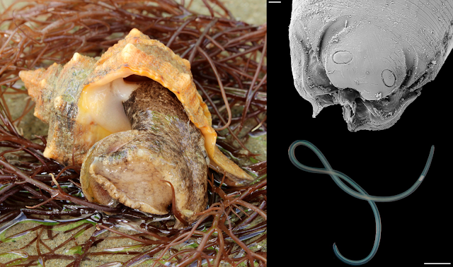
Left: Photo of a Purple-dye Murex by Holger Krisp, used under the Creative Commons (CC BY 3.0) license
Right: (top) SEM close-up photo of Sulcascaris larva's head, (bottom) a fourth-stage Sulcascaris larva  
(Photo of the nematode from Fig. 2 and Fig 4. of the paper)

Larvae of Sulcascaris have recently been reported from scallops and mussels - which raises some concerns since both are popular shellfish that are often eaten only lightly cooked or not at all. A recently published study adds another shellfish to that list - the purple dye murex, Bolinus brandaris. These large predatory snails are so-called because they used to be harvested to obtain a special type of purple dye. But in addition to their historic use in the textile industry, they are also commonly eaten in many parts of the Mediterranean.

A group of researchers in Italy obtained a haul of purple murex from fishermen on the coast of Baia Domizia, Italy, and brought the snails back to their laboratory to dissect them for parasites. Upon detailed examinations of the snails' organs, they found that 9 out of the 56 snails they obtained were infected with Sulcascaris larvae. However, infection intensity was very low, with most of the infected snails being parasitised by just a single nematode larva. These larval worms measured between one to five centimetres long, and were mostly lodged at the base of the snail's proboscis, with a few others found in the mantle cavity - the fleshy bag in a mollusc's body which houses its gills and other organs. 

Because of where those parasites are located in the snails, they can easily get overlooked during routine sanitary inspections, which only involve examining the outer appearance of the snail. The reason why those worms were mostly situated in those parts of the snail's anatomy might be due to their infection pathway. When the eggs of Sulcascaris are released from the turtle host, they settle onto the seafloor where they hatch into larval stages that lie in wait for an encounter with an unlucky murex. As the predatory sea snail moves across the sea floor, searching for prey with its proboscis, those larvae are sucked in via the inhalant current which transport them right into the snail's proboscis and mantle cavity.

Sea turtles with their strong beak and jaws can crack into these tasty snails which are off-limits to other animals, but it also means they end up with Sulcascaris in their gut. While this and previous studies on Sulcasacris have found that most shellfish carried only one or two individual nematodes, a turtle can eat a lot of shellfish, and over time may end up accumulating dozens or even hundreds of those worms in their stomach. When present in large numbers, these nematodes may cause ulcerous gastritis in sea turtles. But aside from that, not as much is known about this worm compared with its more famous, mammal-dwelling relatives, such as Anisakis.

So what does this mean for people who love eating shellfish? Based on prior experiments, it seems that Sulcasacaris can only infect sea turtles, so it is unlikely to become a zoonotic infection if it ends up being ingested by humans. Also, as mentioned above, when they are present, it's only one or two worms in each shellfish, and since purple murex are usually eaten after being cooked, this would kill the worm in the process. So the health risks presented by Sulcasacaris to any seafood consumers are relatively minimal.

However, like other anisakid worms, their tissue and secreted proteins may still potentially cause allergic reactions in some people, even after cooking. But not much is known about that possibility. The researchers suggested that at the very least, commercial fishermen should avoid harvesting snails from areas with sea turtles, since they are likely to be infected with Sulcascaris. This could be a win-win situation for both turtles and people - the turtles get to keep their feeding grounds to themselves, and seafood lovers can safely enjoy some worm-free sea snails. 

As the consumption of fish and other seafood increases around the world, there is a greater need for more studies on the wide variety of parasites that are found in seafood, along with people who have the skills and expertise to identify them - so we can continue to enjoy seafood without unintentionally barging into the life cycle of a parasite (and suffer its associating consequences).

Reference:
Santoro, M., Palomba, M., & Modica, M. V. (2022). Larvae of Sulcascaris sulcata (Nematoda: Anisakidae), a parasite of sea turtles, infect the edible purple dye murex Bolinus brandaris in the Tyrrhenian Sea. Food Control 132: 108547.

May 18, 2021

Anisakis physeteris

Being on top of the food chain sounds like it'd be pretty awesome - all the other animals in the ecosystem are potentially your food and nothing else hunts you. In reality, it also means that there are many parasites out there that see you as prime real estate, a nice place to settle down and start a family. And there's no way for you to avoid them since many of those parasites would be climbing their way up the food chain via the prey animals you have been eating. Nowhere is that more obvious than in the ocean.

The oceans are filled with parasites - not that you'd necessarily know since the vast majority of them are hidden out of plain sight within the body of their hosts. Many of them are parasitic worms that treat the oceanic food web like a transit system, using predator-prey interactions to get from one host to another. This post is about a study on two nematodes that cross path inside some oceanic squids

Left (a, c): Lappetascaris larva (top) embedded in squid mantle muscle, (bottom) viewed under the microscope.
Right (b, d): Anisakis physeteris larva (top) in squid testis, (bottom) viewed under the microscope
Photos from Figure 1 of the paper

A group of researchers from Italy looked at parasitic roundworms that are found in the umbrella squid and the reverse jewel squid. Both of them belong to a group of squid called the "cock-eyed squids", which are commonly found in the mesopelagic zone. The squid that the researchers examined were caught as by-catch from commercial trawling vessels that were operating off the coast of Italy and Naples, and every squid that they looked at were infected with some kind of nematode larvae. 

Most of the nematodes were of a genus called Lappetascaris, along with another species which was identified as Anisakis physeteris. While both of those parasites look superficially similar and sometimes co-infect the same squid, there are some key life history and life cycle differences between them. 

For parasites, a host is not a single homogenous entity, but a collection of different microhabitats, and each parasite species has their own taste when it comes to fine-scale real estate. In this case, the researchers found that A. physeteris mostly settled in the squid's testis whereas Lappetascaris preferred embedding itself in the firm mantle musculature (the part of squid which are sold on the market as "squid tubes").

But these worms don't just differ in the part of the squid they prefer, but also which species of squid they infect. While Lappetascaris was found in both the umbrella squid and the reverse jewel squid, A. physeteris was choosier, and was only found in the umbrella squid. Finally, the two worms complete their life cycles in totally different animals. Lappetascaris reaches maturity in the gut of large teleost fishes such as swordfish and billfish, whereas A. physeteris needs to get into the stomach of a sperm whale - as denoted by its species name (the genus name for sperm whale is Physeter).

This may explain why A. physeteris was only found in the umbrella squid. Compared with the reverse jewel squid, umbrella squid venture into much deeper water which overlaps with the sperm whale's usual hangouts. And this exposes them to infective stages that are being released from sperm whales which have hundreds and thousands of adult Anisakis worms in their gut.

While the popular perception of the sperm whale often depict them as duking it out with the giant squid, the majority of their diet is composed of more modestly sized cephalopods, and the umbrella squid seems to form a major part of their diet. That's not to say umbrella squid is not on the menu of other large oceanic predators like swordfish and billfish too (hence it is also infected by Lappetascaris larvae), but if you are a parasite that is looking for the ideal ride to get you into the belly of a sperm whale, you can't do much better than the umbrella squid. 

What about the Lappetascaris which are sharing that squid with A. physeteris? Well they better hope a swordfish would come along and snatched it up before it ends up in the belly of a marine mammal - an environment that it is ill-equipped to live in.

So while these two worms may sometimes meet in the same squid, they eventually have to go their separate ways - and reaching their respective final hosts would unfortunately spell doom for the other worm in the shared squid. As for the sperm whales, a belly full of yummy squid must inevitably lead to a stomach full of wriggly worms.

Reference:

August 24, 2020

Hexametra angusticaecoides

In 2016, a group of crested geckoes in captivity suddenly got sick soon after they were transferred to a terrarium that previously held some Madagascan mossy geckoes. Within a short period of time, the geckoes started dying of a mysterious illness. Of the ten that were held in that terrarium, only a single gecko survived.

Photos of Hexametra emerging from moribund geckoes from Fig 1, 3, and 4 of the paper

When the dead geckoes were dissected, it was found that they had massive worms that were tearing their ways through the their innards. Some of the worms had even started emerging through the skin.
One gecko was lucky enough to receive treatment in time to save it. It was initially given fenbendazole and pyrantel - two commonly used medications for treating parasitic worm infections - but they had no effects. So a surgical procedure was performed on the lizard to remove the deadly nematodes from under its skin, followed by a dose of levamisole. About two weeks after the surgery, the surviving gecko managed to recover to full health.

Between the nine dead gecko and the sole survivor, over 50 worms were retrieved. The worm in question was Hexametra angusticaecoides - a parasitic nematode which commonly infects reptiles. Some of you who have  been following this blog for a while might recognise the genus Hexametra from a post back in 2016 where another species of that parasite was found in the body of a captive false coral snake.

So how did a bunch of crested geckos ended up with all these worms? Tracking down the original source of infection was a bit tricky, given some were sourced from a breeder in Canada, while other were sourced from a pet shop in Germany. Furthermore, they had been kept separately in different terrariums until they were combined into a single enclosure, soon after which the worms began appearing. However, it could be that particular terrarium which was responsible for the worms. Prior to housing the crested geckoes, that enclosure had been occupied by some wild-caught Madagascan mossy geckoes, Uroplatus sikorae.  

The harm caused by Hexametra to its host is likely due to its relatively large size, and its tendency to move around within the host's body instead of staying in place. Additionally, since the crested gecko is an exotic host for the parasite, this pairing would not have happened naturally. Hosts which have had a history of coevolution with their parasites would have also evolved mechanisms for tolerating or offsetting some of the more harmful effects of the parasite. But hosts which have never encountered those parasites would have no such adaptations and are thus exposed to the full effect of the parasite's presence. On top of that, the stress of captivity might have made the geckoes less able to tolerate any kind of parasitic infection.

The exotic pet trade, in addition to driving the poaching, smuggling and distribution of wildlife worldwide, also bring together animals which would otherwise never come into contact with each other, along with their many parasites. Like the majority of parasites, there is simply insufficient basic information about the ecology, life cycle, and life history for most reptile parasites, let alone what effects they might have if they end up in hosts which they have never encountered before.

Reference:
Barton, D. P., Martelli, P., Luk, W., Zhu, X., & Shamsi, S. (2020). Infection of Hexametra angusticaecoides Chabaud & Brygoo, 1960 (Nematoda: Ascarididae) in a population of captive crested geckoes, Correlophus ciliatus Guichenot (Reptilia: Diplodactylidae). Parasitology, 147: 673-680.

July 16, 2020

Neofoleyellides boerewors

Mosquitoes are mostly known for being blood-suckers. But despite that reputation, they actually spend most of their adult life feeding on nectar - it's only when female mosquitoes are ready to breed that she needs blood to fuel the development of her eggs. Because of this feeding habit, mosquitoes are used by a wide variety of blood-borne parasites to ferry them from one host to another.

In humans, mosquitoes are responsible for transmitting a variety of parasitic infections such as the malaria parasite and filarial worms, as well as a range of different viruses. This also extends to other animals that are fed upon by mosquitoes, which are host to their own array of mosquito-transmitted parasites. There are some species of mosquitoes that specialise in feeding on ectothermic ("cold-blooded") vertebrates such as frogs and toads, and accordingly those mosquitoes are also vectors for a range of parasites that infect those animals.

Left: Microfilaria larva from toad blood, Top left: Late L1 sausage-shaped stage from mosquito thorax, Top right: Adult female worm, Bottom right and left: Infected toad with adult worm in its right eye.
Photos from Figure 3, 5, and 7 of the paper
Neofoleyellides boerewors is a filarial nematode in the Onchoceridae family, a group of parasitic roundworms that includes the parasite that causes river blindness in humans, but the species that infect amphibians are not as well-studied. This paper describes one such species that has been found in the guttural toad, Sclerophrys gutturalis.

The adult worm mostly lives in the toad's body cavity or just under the skin, though some can end up in other parts of the body. For example, in one particularly heavily infected toad, the researchers found 52 adult worms, and one of those worms had even spilled over into the toad's right eye where they caused internal bleeding and blindness.

Inside the toad's body, the adult worm produces larval stages called microfilarials that circulate in the amphibian's blood vessels while waiting for a rendezvous with a hungry mosquito. When the mosquito slurps up a belly full of toad blood, they also end up ingesting a bunch of those baby worms.

Once inside the mosquito, these microfilarial transform into chubby, sausage-shaped worms (indeed, the species name of this parasite, boerewors, is named after a popular type of South African sausage), and proceed to congregate amidst the fat bodies in the thorax, where they can grow by feeding off the mosquito's nutrient reserves. After spending about ten days there, the larvae developed into the infective stage, ready to infect another toad. They migrate to the mosquito's head and move into position at the insect's mouthpart, preparing to disembark into the bloodstreams of another toad the moment that the mosquito begins feeding.

Anurans (frogs and toads) are host to a wide range of parasites, many of which have unique life cycles and life histories which are adaptations to the developmental history of their amphibian hosts. There is still a great deal we don't know about the diverse array of parasites that are found in frogs, toads, and other amphibians.

With many of those amphibians under threat from climate change, habitat destruction, and the dreaded amphibian chytrid fungi, it is highly likely that we may never fully learn about the wonderful adaptations of their associating symbionts - a hidden world of biodiversity that would tragically disappear along with their hosts.

Reference:
Netherlands, E. C., Svitin, R., Cook, C. A., Smit, N. J., Brendonck, L., Vanhove, M. P., & Du Preez, L. H. (2020). Neofoleyellides boerewors n. gen. n. sp.(Nematoda: Onchocercidae) parasitising common toads and mosquito vectors: morphology, life history, experimental transmission and host-vector interaction in situ. International Journal for Parasitology 50: 177-194

May 19, 2020

Anguillicola crassus

Today, we are featuring a guest post by Juliette Villechanoux - an MSc student on the  IMBRSea  programme currently carrying out her professional practice placement (albeit remotely) with Dr. Katie O’Dwyer at the Galway-Mayo Institute of Technology in Ireland. This post is about the “Trojan horse” strategy of Anguillicola crassus nematode in Pomphorhynchus laevis acanthocephalan and its impact on european eels.

The famous “Trojan horse” metaphor referring to a seemingly benign trick but actually hiding sinister intent comes from the Greek mythological Trojan War story. The war began after the Trojan prince stole the Queen of Sparta from her husband. After 10 years of battle, the Greeks finally took down Troy city by the inventive construction of a gigantic hollow wooden horse. They pretended to sail away and offered the horse as a truce. Little did the Trojans know that it was filled with Greek soldiers who by night slaughtered the inhabitants of the city. You will see in this post the “Trojan horse” strategy employed by some cunning parasites, using another parasite feature for their own development and hiding from the host.

(a) Opened  European eel swim bladder showing adult Anguillicola crassus, (b) round goby from the stomach of an eel.
Photos from Figure 1 of Emde et al. (2014)

Anguillicola crassus is a nematode parasite from Japan, introduced to Europe with Japanese eels (Anguilla japonica), their original definitive host. It first infects invertebrates, such as copepods, where it grows to its third larval stage. It can then go on to parasitize several different fish species, which become infected when they ingest the parasitized copepod (this is what’s known as “trophic transmission”). Some of these fish only transport the worm, while others may act as alternative final hosts. Typically, the parasite finally reaches the eel after this final host eats a parasitized fish or crustacean.

The introduction of this invasive nematode species to Europe has had a devastating effect on the overall European eel (Anguilla anguilla) stock leading to a massive decline, and the species becoming classified as critically endangered. The European eel life-cycle is very peculiar: they individually undergo a 5000 km spawning migration from European coasts to the Sargasso Sea at depths fluctuating between 200 and 1000 meters. Anguillicola crassus impacts their survival by infecting their swim bladder and reducing their swimming performance, and possibly leading to the host’s death during their migration journey.

But some fish species have developed an immune response that can cause the nematode’s death. Nonetheless, in the Rhine river, recent studies revealed that invasive A. crassus found an intriguing way to avoid the immune defence of the round goby Neogobius melanostomus by using another European invasive parasite: Pomphorhynchus laevis. This acanthocephalan worm originally invaded the Rhine river from the Ponto-Caspian region using the Danube canal by hiding in the body of its round goby host.

(A) Cysts of encapsulated Pomphorhynchus laevis from the digestive tracts of the round goby, (B) Encapsulated P. laevis illuminate under high light intensity, (C) Digested cysts with A. crassus released (circled in red).
Photos from Figure 1 of Hohenadler et al. (2018)

So how does A. crassus employ a “Trojan horse” strategy to avoid detection by the round goby? When the acanthocephalan infects the round goby, the worm turns into a cocoon-like cyst, and even though the acanthocephalan parasite is encapsulated by the goby’s immune response, its infective power remains. What is even more interesting about this cyst formation is the high intensity of A. crassus nematode larvae within P. laevis cysts in the round goby. Here you have a well packaged trio of non-native European species. This evasion strategy is used by A. crassus to avoid the goby’s immune response and turns the round goby into an unusual second intermediate host due to the distinct geographic origin of the nematode A. crassus, the round goby, and the acanthocephalan, P. laevis.

The cunning nematode uses the cysts as “Trojan horses” facilitating its establishment in the host, like the Greeks in Troy. The relationship between both parasites can be defined as “facultative hyperparasitism” where the cyst gives protection to the nematode, while the acanthocephalan worm continues to develop as normal. This strategy comes to be a considerable problem since it increases the chances of A. crassus infecting European eels as they remain infectious after consumption by the round goby and excystation of the acanthocephalan, along with its nematode passenger. And we know the damaging effects it causes on eel populations, in fact A. crassus has been recognised as one of the 100 “worst” exotic European species because of its impact on the European eel.

This case highlights not only the complexity of the parasite life-cycles involved and the impact of multiple human-driven invasions by invasive species, but also the large impact they can have on native species when combined.

References:
Hohenadler, M.A.A., Honka, K.I., Emde, S. et al. (2018). First evidence for a possible invasional meltdown among invasive fish parasites. Scientific Reports 8, 15085.

Emde, S., Rueckert, S., Kochmann, J., Knopf, K., Sures, B., & Klimpel, S. (2014). Nematode eel parasite found inside acanthocephalan cysts—“Trojan horse” strategy?. Parasites and Vectors, 7, 504.

post written by Juliette Villechanoux

November 8, 2018

Leidynema appendiculatum

A while ago, I wrote a post about a tadpole pinworm, and pinworms are found in the hindgut of a variety of different vertebrate animals. But there are also many pinworms which live in animals without backbone. The reason why pinworms make their home in the gut of those animals is because they are after bacteria which dwell in the the gut of animals which are hindgut fermenters. And there are some insects which provide just the right environment for them to thrive.

Top: Adult female L. appendiculatum (scale bar 500 μm)
Bottom: Adult male L. appendiculatum (scale bar 200 μm)
From Figure 2 of the paper
Pinworms (Oxyurida) are separated into two main groups - the Oxyuroidea which consist of species that infect vertebrate animals (including humans), and the Thelastomatoidea, which consist of species that infect invertebrates such as cockroaches and millipedes. Leidynema appendicaulatum is in the latter group, and it lives in the hindgut of cockroaches. It has been reported from many different species of cockroaches around the world. In Japan, this parasite is most commonly found in the smokybrown cockroach Periplaneta fuliginosa and it is the only parasitic roundworm known to infect that cockroach species.

So is this pinworm also found other cockroach species in Japan? A pair of scientists decided to investigate the presence and development of this parasite in both wild and captive cockroaches. They looked at a mix of cockroaches collected from the wild, from lab-reared colonies, and some that were bought from petshops where they are commonly sold as food for pet reptiles.  They examined 14 species of cockroaches in total, and of those, the smokybrown cockroach was usually found to have L. appendiculatum as expected, but the rest were largely free of the pinworm in question. So what's stopping it from infecting those other cockroaches considering that it has no trouble infecting different cockroach species from around the world?

To find out, the scientist did some infection experiments using colonies of cockroaches which had been raised in the laboratory and never been exposed to any nematode parasites. They fed them food which had pinworm larvae mixed in to see which ones were susceptible. In contrast to what they found for the wild cockroaches, L. appendicaulatum had no trouble making themselves at home in those other cockroach species, going through the same developmental cycle at the usual rate as those living in the smokybrowns. One of the cockroach species which they successfully infected was Blattella nipponica - which was found to be free of the pinworms in the wild.  So why is it that L. appendiculatum can infect captive B. nipponica, but not those out in nature? This might have something to do with those cockroaches' feeding habits.

Pinworms have a relatively simple transmission pathway - the host becomes infected when they ingest fecal matter which are contaminated with the parasite's eggs. So in order for a cockroach to become infected with L. appendiculatum, at some point they have to ingest another cockroach's feces. Unlike the smokybrown cockroach which are common in urban areas and readily feed on detritus, including feces from other cockroaches, B. nipponica mainly live in forests and grasslands, on a more discerning diet of mostly of plant matter, including forest fruits. So out in the wild, B. nipponica would rarely come into contact with the parasite.

Given the right set of circumstances, L. appendiculatum can easily establish itself in many cockroach species, it's just that for some cockroaches, those kind of circumstances just doesn't happened in nature. In the end, you are infected by what you eat.

Reference:
Ozawa, S., & Hasegawa, K. (2018). Broad infectivity of Leidynema appendiculatum (Nematoda: Oxyurida: Thelastomatidae) parasite of the smokybrown cockroach Periplaneta fuliginosa (Blattodea: Blattidae). Ecology and Evolution 8: 3908-3918.

October 8, 2018

Uncinaria sp.

Hookworms are long, skinny gut-dwelling vampires, and there are about 500 million people around the world who are infected with these parasitic roundworms. Hookworm infections cause chronic blood loss and iron deficiencies, which can lead to anemia, lethargy, and hinder childhood development. But it's not just humans who are afflicted by hookworms, there are 68 different described hookworm species and they infect over a hundred different species of mammals from around the world.
Right: Group of South American Fur Seals ( photo by Dick Culbert). Top Left: Intestine of fur seal pup filled with Uncinaria hookworms (from this paper), Bottom Left: The head and mouth of Uncinaria (from this paper)
The life cycle of the typical hookworm is fairly straightforward. Hookworm larvae are hatched from eggs that are deposited in the environment with the host's faeces. When the larval parasite hatches from the egg, it spends about a week developing in the soil and can survive for 3-4 weeks while waiting for a host. If it encounters an appropriate host, the parasite burrow through the host's skin, then take a journey around the body via the blood vessels, through the heart and lungs, and eventually settling down in the intestine where it matures into an adult worm. Much like the human-infecting species, those other wildlife-infecting hookworms can cause chronic illness from their blood-feeding. Additionally, the host is constantly exposed to new infections from other infected hosts in the area. While these parasites can be a debilitating burden on the host, hookworms by themselves aren't usually known to cause host death.

But the fur seal hookworm - Uncinaria - breaks that mould. The adult worm lives in the gut of seal pups, and unlike other hookworms that have a lifespan of at least a year or more, the adult stage of Uncinaria doesn't live that long - most seal pups are free of the hookworms within a month or two after the initial infection. But during that time, Uncinaria takes a massive toll on its host and it is one of the leading causes of pup mortality, responsible for 30-70% of pup deaths during breeding seasons. One of the reason for their lethality is their aggressive feeding habit. Unlike other hookworms which are content to simply hang on to the intestinal wall and steadily sip blood, Uncinaria is a glutton that digs deep into the intestinal wall to get their fill and churn out as many eggs as possible during its short life. Their motto is "live fast, die young, leave a whole lot of eggs".

Along with with its short lifespan and unusually aggressive feeding habit, Uncinaria also differs from other hookworm in having a very convoluted life cycle. Unlike human hookworms, seal pups do not acquire their infection from hookworm larvae in the surrounding environment - instead they get it directly from their mother's milk. Once Uncinaria enters the seal pup's gut, it mature into an adult worm within two weeks and starts producing eggs that are shed from infected pups and get spread all over the rookery grounds. After hatching, the hookworm larvae burrow into any seals that they encounter, and migrate to the belly blubber.
Life cycle of Uncinaria, from Fig. 1 of the paper
Once there, instead of developing any further, the parasite lay dormant until the next breeding season - when that eventually comes around, the Uncinaria larvae in female seals make their way to the mammary glands where they can be on stand-by to infect the next generation of seal pups. As for the adult male seals? Because the transmission cycle relies upon the mother's milk, male seals are effectively a dead end host for this parasite.

So why has Uncinaria evolved to live the way that it does? Well, unlike land-dwelling mammals which can deposit hookworm eggs into the soil for many years and get repeated exposed to new hookworm larvae from their surroundings, Uncinaria does not have those luxuries. Its hosts spend most of their time out at sea and when they do come onto land, they only do so temporarily. The breeding season is the only time when new hosts are around on land for long enough and congregating in sufficient numbers for the parasites to disperse and infect new hosts. Uncinaria only has that brief window of opportunity to complete its life cycle, and to do so successfully means it need to saturate the rookery soil with eggs. And the cost for all those eggs are paid for with the blood of seal pups.

There are a wide range of different factors that determine how harmful a parasite or pathogen is to towards its host. In contrary to popular misconception (or wishful thinking), a well-adapted and successful parasite is not necessarily one that has evolved to live harmoniously with its host, but one that has evolved to get the most out of its host. And under some circumstances, it might mean that the road to successful life cycle completion is one which is paved with dead hosts.

Reference:
Seguel, M., Munoz, F., Perez-Venegas, D., Muller, A., Paves, H., Howerth, E., & Gottdenker, N. (2018). The life history strategy of a fur seal hookworm in relation to pathogenicity and host health status. International Journal for Parasitology: Parasites and Wildlife. 7: 251-260