"So, naturalists observe, a flea has smaller fleas that on him prey; and these have smaller still to bite ’em; and so proceed ad infinitum."
- Jonathan Swift
Showing posts with label fungus. Show all posts

May 14, 2026

Sorochytrium milnesiophthora

Tardigrades (also known as "water bears") have built up a reputation for being pretty much invulnerable (even though Bdelloid rotifers does a lot of tardigrades' tricks and more), but while they can survive being launched into space, they are at the mercy of some fungi that can turn their little bodies into living incubators. One such fungi is a chytrid fungus named Sorochytrium milnesiophthora. And while most people who have heard of chytrid only know about the two species which infects frogs and other amphibians, most chytrids are actually parasites of invertebrates and algae, with some playing important ecosystem roles such as controlling harmful algal blooms and converting otherwise inedible phytoplankton into food for zooplankton, like water fleas

Left: A tardigrade infected with Sorochytrium milnesiophthora, Top Right: S. milnesiophthora zoosporangia (indicated by arrow) in the body of an infected tardigrade, Bottom Right: S. milnesiophthora grown on nutrient agar.
Photos from Figure 1 of the paper

In this study, researchers found S. milnesiophthora growing in tardigrades that were living amongst salted shield lichens collected from Kuljunmaa Island, Finland. Samples of the lichen were dried out for a year, then rehydrated and shaken to isolate the infected tardigrades and extract their fungal cells. When samples of the fungus were grown on a petri dish filled with nutrient agar, it grew into a type of lumpy yellow mould. But S. milnesiophthora takes on a very different form upon encountering a tardigrade.

The story of a water bear's fungal downfall begins with a single zoospore. Unlike most fungi, Sorochytrium and other chytrid-type fungi produce spores which have a flagellum-like tail that allows them to swim through water. Upon contact with the unfortunate tardigrade, the spore fires a tube which punctures the victim's body wall and invades its interior. At this stage, the fungal cells look like round balls which are indistinguishable from regular cells that float around in the tardigrade's body cavity. But over the course of a week, these fungal cells start proliferating throughout the water bear's body, which develops a reddish-brown colour. Once the host dies from being completely taken over by growing fungal cells, S. milnesiophthora squirts newly produced zoospores out of the carcass to begin the infection cycle anew

Aside from its ability to take down the seemingly invulnerable tardigrades, S. milnesiophthora also has something else which makes it stands out - an internal transcribed spacer gene that is 55 thousand megabases long, which is among the longest known for any organisms. This section of DNA, also known as ITS, is commonly used for identifying and classifying different species of organisms, and can serve as a DNA "barcode" to identify different types of fungi. Because it is a relatively short sequence, traces of it can be easily amplified and detected in a sample. Hence they are also often used in metabarcoding, a molecular technique for detecting DNA from a whole bunch of different organisms in a single sample, often used for detecting microorganisms which would be difficult and laborious to find using conventional microscopy. 

But because the ITS gene of S. milnesiophthora is so long, this messes with the primers which are used for metabarcoding, as they were designed for amplifying short sections of DNA. Which means S. milnesiophthora would have been missed by conventional metabarcoding since the primers can't amplify its DNA for detection. So Sorochytrium and other similar fungi might actually be more common than previously thought. Case in point, while examining tardigrades for Sorochytrium, the researchers also found two other species of fungi along with two species of water molds associated with the water bears.

Water bears are charming-looking critters, and it may seem sad to see them die such a brutal death, but all this tardigrade slaying does serve an important ecosystem function. In addition to regulating the tardigrade population to make room for other microscopic animals, S. milnesiophthora may inadvertently be providing food for the ecosystem that exists on lichens. By converting water bears into zoospores, Sorochytrium could be providing other microscopic animals, such as rotifers, with a free meal. While deadly to their hosts, for everything else, chytrid zoospores can be tasty snacks for filter-feeding animals which relish those flagellated spores, as they are more nutritious than those animals' usual fare of algae, bacteria, and organic detritus,

There are vibrant ecosystems everywhere for those with the eyes (and microscope) to see. So next time you see a patch of moss or lichen, take a moment to appreciate the drama of life, death, and rebirth being played out in those microscopic worlds.

Reference:
Dirks, A. C., Vecchi, M., Orozco-Quime, M., Schwarz, E., Calhim, S., & James, T. Y. (2026). Rediscovery of the tardigrade pathogen Sorochytrium milnesiophthora, a blastoclad boasting the longest-known fungal internal transcribed spacer. Mycologia 118: 623-637.

March 13, 2025

Gibellula agroflorestalis

Imagine an infectious disease that turns you into an immobile white statue - while that sounds like something out of a dystopian science fiction story, this is the fate which awaits any spiders that encounter Gibellula, a genus of araneopathogenic (spider-killing) fungi. There are many species of this fungi found all over the world and their spores can infect all kinds of spiders. Gibellula is in the same broad group of fungi as the zombie ant fungi, but instead of producing a single or a few mushroom-like fruiting bodies that emerge from the carcass of their hosts, Gibellula smothers the infected spiders in a dense layer of white, fluffy mould.

Left: Examples of spiders infected with Gibellula agroflorestalis including orb-weavers (a-c) and jumping spider (d), Centre: numerous spore-bearing stalks (conidiophores), their mycelia twisted together, Right: Close-up of the head of a conidiophore.
Photos from Fig 3 and 4 of the paper

This post focuses on Gibellula agroflorestalis, a newly described fungus which was discovered by a group of researchers in the agroforestry systems located at the Abreu e Lima municipality of Brazil. 
Agroforestry is a system of agriculture where instead of cultivating a single type of crop, different crop plants are grown alongside trees and other plants, and these environments can provide habitats for a wider range of organisms. The forests that the researchers studied were composed of fruit plants such bananas, coconuts, limes, and mangoes, growing alongside various trees such as pink trumpet trees, pau brasil, and juazeiro.

So how does one find mouldy spiders amidst this semi-cultivated forest? Well, in short, with a lot of time and effort. The researchers hiked through multiple agroforestry properties while meticulously examining every plant they came across, carefully inspecting the underside of their leaves which is where many fungus-infected spiders end up. On the rare occasion when they come across an infected spider, it was collected intact along with the leaf it was sitting on, placed into a plastic cup, and later stored in silica gel to preserve the specimen. In total, the researchers found 17 infected spiders, ranging from jumping spiders to crab spiders to orb-weavers, alongside other spider hosts which could not be identified, because by that stage they have become more Gibellula than spider, twisted and fungal.

When the researchers examined the fungus under a microscope, they found that the structure and size of its teardrop-shaped spores were different from other known species of Gibellula. Furthermore, DNA analyses showed the fungus to be genetically distinct, and that as a species, G. agroflorestalis is fairly genetically diverse, which is probably why it is able to infect so many different types of spiders.

While there are other species of Gibellula fungi out there which have been recorded from spiders in natural forests, G. agroflorestalis was the first to be recorded in an agroforestry system, showing that these environment may in fact serve as habitats to some unique species. The discovery of G. agroflorestalis reminds us that a lot of unseen biodiversity can be found through careful observations. So it's good to stop and smell the roses, but you should also check underneath the leaves for mouldy spiders.

Reference:
da Rocha Alves, J.E., da Silva Santos, A.C., Pedroso, S.K.B., Melo, R.F.R., & Tiago, P.V. (2025). Untangling a web of spider fungi: Gibellula agroflorestalis (Hypocreales, Ascomycota), a new species of spider parasite from Brazil. Journal of Invertebrate Pathology 209:108278.

April 13, 2023

Rickia wasmannii

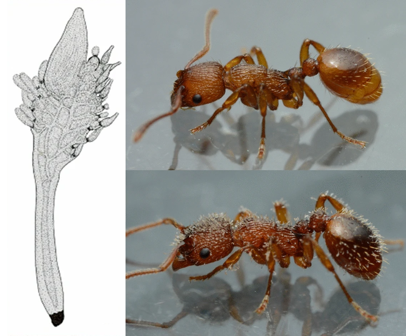
Rickia wasmannii is a fungus that lives on ants, and when it comes to ants and fungi most people usually think of Ophiocordyceps, i.e. the zombie ant fungus - which was the inspiration for The Last of Us series of video games and TV series. But R. wasmannii is not a killer - instead of zombifying its host and digesting the corpse, this fungus seems to reduce animosity and aggression between ants. First of all, let's take a look at what R. wasmannii actually does on ants. 

Left: Illustration of a Rickia wasmannii thallus, Right top: Uninfected ant, Right bottom: ant infected with R. wasmannii
Pictures from Figure 1 of this paper

Rickia wasmannii belongs to a group of fungi called Laboulbeniales, also known more colloquially as "labouls". These fungi have little holdfasts called haustoria that allows them to cling to the ant's cuticle. They are ectoparasites of insects that attach to their host's external surface and suck their hemolymph (insect's equivalent of blood). So in a way they are rather like ticks or lice (and yes, there are labouls that live on ectoparasitic insects, which one might consider as a bit of poetic justice).

But this fungus seems to do more than just suck the ant's blood, as it causes the infected ants and other ants around them to behave differently. Rickia wasmannii changes the host ant's cuticular hydrocarbon or CHC profile. CHC is essentially an ant's ID profile - they use it to recognise nestmates, tell each other apart, and be alerted to strangers from other nests. But R. wasmannii messes with that, scrambling the infected ants' CHC profile, and making them "smell" differently to uninfected ants.

Scientists wanted to find out how the presence of this fungus affects the way ants interact with each other. The challenge with studying ant behaviour is that when you put two ants together, it is difficult to tell apart whether the ant you are observing is responding to the other ant's chemical profile, or if it is responding to the way the other ant is reacting to them. The only way to get a clear observation is to present the ant with something that it would recognise as a fellow ant, but would not muddle the outcome by reacting to the ant that you are trying to observed

The solution turns out to be freeze-killed ants. Ants that are killed in this manner retain their CHC profile, so other ants would treat them just as another live ant, but obviously a dead ant wouldn't react to a live ant's presence and confound the outcome. In addition to those freeze-killed test subjects, scientists also made ant "dummies" which are essentially blank slates in ant forms that they can imbue with whatever chemical signature they were testing. These "dummies" were made by washing ant corpses in hexane to remove their chemical signature. To ants, these specially treated ant corpses are like faceless mannequin, with no identity - until the scientist imbues them with one, by anointing them with a droplet of cuticular extract from another ant.

When ants were presented with dummies that were smeared with the cuticular extract of ants from a different nest, the ants started biting, dragging, or stinging the dummies, much like how they would respond to a live ant from another nest. But when they were presented with either the corpse of a Rickia-infected ant, or dummies that "smell" like a Rickia-infected ant, they were more relaxed and less likely to get aggro. Furthermore, it's not just that the fungus made other ants act differently, the infected ant itself also starts behaving differently. Infected ants are generally less likely to pick a fight with another ant, but especially when facing other infected ants.

As mentioned previously, R. wasmannii seems to change the ant's CHC profile, but one would think scrambling the host ant's profile would make other ants react more aggressively towards them since ants usually have a "stranger danger" response to ants that "smell" different to their nestmates. But the way that R. wasmannii changes how an ant "smell" seems to have a calming effect, and this comes down to a molecule called n-C23 which is present in higher concentration on the cuticle of all infected ants. When the scientist presented ants with dummies that have been smeared with n-C23 and nothing else, almost all hints of aggressive behaviour ceased.

So by increasing n-C23 concentration in its host's cuticle, R. wasmannii has unlocked a life hack that allows it to not just access all areas in an ant colony, but to spread to other nests as well. In the scientists' study population, about half the colonies they studied had the fungus present, and in some nests, all the ants were infected with R. wasmannii. A testament to the fungus' successful manipulation of ant behaviour.

Furthermore the fungus' presence also affects another, very different parasite which also lives with ants - the caterpillar of blue butterflies. These caterpillars are social parasites that convince worker ants into adopting them into their nest. Once they are settled in, they start demanding food from the worker ants and even feed on the ant's developing broods. But the caterpillars don't seem to survive as long in nests which are already hosting R. wasmannii, and in the field, these two parasites co-occur less commonly than expected based on their respective prevalence, which indicates the caterpillar and the fungus are in competition over ant real estate.

By messing with their identity and making them more chilled out, R. wasmannii can turn an ant colony into a fungus party. But the consequences of that ripple out to other ant colonies too, along with the organisms that regularly take up residency in the homes of ants.

Reference:
Csata, E., Casacci, L. P., Ruther, J., Bernadou, A., Heinze, J., & Markó, B. (2023). Non-lethal fungal infection could reduce aggression towards strangers in ants. Communications Biology, 6: 183.

December 17, 2020

Ophiocordyceps sinensis

Ophiocordyceps is a genus of fungi that is probably most well-known for their abilities to usurp and manipulate the behaviour of ants, which gave rise to their more commonly known name - the "zombie ant fungi". But aside from the ant-infecting species, the genus Ophiocordyceps also contains another very well-known insect-zombifying fungus - Ophiocordyceps sinensis, more commonly known as the "caterpillar fungus" - which infects the caterpillars of ghost moths.

Left: O. sinensis fruiting body emerging from a caterpillar, photo by Zhu Liang Yang from here
Right: Ghost moth (top) adult, and (bottom) caterpillar stage, photos from here

While the reputation of the ant-infecting Ophiocordyceps species were built upon their ability to control their host's mind, the roots of O. sinensis' fame is based on the fungus' prized medicinal properties, which has been known and documented for centuries in China where it is known as dōng chóng xià cǎo (冬蟲夏草: which translates into "winter worm, summer grass). It also made an appearance in Moyashimon, a manga (and subsequently, anime) about microbes. Unfortunately, in recent decades, this fungus is currently under threat from a combination of climate change and over-harvesting.

Despite being highly valued and extensively studied for its pharmaceutical potential, the natural ecology of this fungus is not all that well-understood. For example, it is not entirely clear as to how this fungus actually infects its caterpillar host in the first place. Attempts to cultivate the fungus in artificial settings to alleviate harvesting pressure on wild populations have been met with limited success, in terms of producing them on a commercially-viable level.

The host of O. sinensis are ghost moth caterpillars, which live underground munching on the roots of plants. So unlike the ant-infecting zombie fungi that can simply scatter their spores around areas where their ant hosts are likely to walk by, such means of dispersal would be ineffective for reaching caterpillars that spend their entire time underground. Furthermore when scientists examine the soil around fruiting bodies of O. sinensis, the concentration of spores was fairly low, and in any case, they don't seem to disperse very far, with most of the spores found within 20 cm of the fungus fruiting body.

But some of these zombie insect fungi also live a secret double life. When they are not infecting and zombifying or mummifying insects, some of those fungi moonlight as plant symbionts called endophytes. They dwell out of sight within plant tissue, and in some cases providing the plants with various benefits. So perhaps O. sinensis is also leading this double life too? If so, that might be a way through which they are coming into contact with their soil-dwelling caterpillar hosts. 

A group of scientists in China set out to investigate this ecological puzzle at Mount Gongga, in the Sichuan province of China. First of all, they ascertain whether O. sinensis is indeed spending part of its life cycle dwelling as endophytes in the tissue of plants. To do that, they collected plants from areas where the caterpillar fungus was found at the Yanzigous valley, and extracted DNA from the leaves and root tissues of those plants. They then used Quantitative PCR to screen for the presence of O. sinesis. Of the 115 species of plants that were examined, O. sinensis was present in about half of them, across 18 different plant families

Secondly, they also investigated the caterpillars' diet to determine whether they have been eating any of those O.sinesis-positive plants. The scientists collected the caterpillars' gut content, extracted the genetic material they contained, and amplified key sections of DNA that can be used as genetic markers to detect and distinguish different types of plants. From that, they found that those ghost moth caterpillars munched on plants from at least 22 different families, and of the plants that were on the caterpillar's menu, 12 of them had the endophytic stage of O. sinesis in their roots. 

So this might mean that instead of relying upon those spores coming into direct contact with the caterpillars, the way that this fungus completes its life cycle is by using its spores to infect a plant, become established in the plant tissue, then wait for a hungry, hungry caterpillar to come by.

Infecting the host via hiding in their food or prey item (also known as trophic transmission) is a transmission strategy that is usually associated with parasitic worms with complex life-cycles. But here we have a fungus that seem to have convergently evolved this way of reaching its host. While in this case, the hosts (plants and caterpillars) are very different to those that parasitic worms usually infect, functionally it is the same - the hosts become infected through what they eat. Additionally, many of those aforementioned parasitic worms can alter the behaviour and/or appearance of a prey to make it more attractive to a potential host. Can O. sinensis do the same to their host plants to make them more attractive to those soil-dwelling caterpillars?

Given that there are many other fungi which also infect subterraneans insects - this transmission mode might be more common than previously thought, with a wide range of fungi secretly living this double life of being both friends to plants and killers of bugs.

Reference:

April 9, 2018

Massospora cicadina

Periodical cicadas spend most of their lives as juveniles (also known as nymphs), living underground and sucking juices from tree roots. Depending on the species, they keep to this subterranean existence for 13 or 17 years before finally emerging into daylight. And they do so simultaneously in massive numbers. These newly emerged nymphs will climb on to a nearby tree to moult into winged adults. The life of an adult cicada is short and over in about a month. During this period they sing their hearts out and mate until they drop to produce the next generation of cicada nymphs which will return to the soil. But the cicadas aren't the only ones to get busy during this period. Scattered across the landscape are the spores of Massospora cicadina, and for over a decade they have been waiting patiently for the cicadas' return.
Cicadas with and without Masspora infection, note uninfected male cicada which still has the genitalia of an infected female cicada attached. Photos from Figure 1 and 2 of the paper
Massospora cicadina is a parasitic fungus that targets all seven known species of periodical cicadas, and its effects on the host are devastating. Once infected, the cicada is done for - the fungal infection turns the cicada's abdomen into a chalky mass of spores. Surprisingly, despite missing a big chunk of itself, an infected cicada carries on as if it is business as usual - these diseased cicadas keep flying, singing and mating like their uninfected counterparts. But surely there must be more going on beneath that exterior of surprising normality.

A group of researchers investigated if Massospora is doing more to cicadas than just robbing their booties. In particular, they were interested in whether Massospora is altering the cicada's behaviour, as many other insect-infecting fungi are known to do. Since the mid-1990s, they have been spending hundreds of hours documenting the behaviour of both infected and uninfected cicadas. They also collected some of those cicadas and kept them in captivity for closer observations, and played recordings of male cicada songs to them to see how they responded.

There are two ways that cicadas can get infected with Massospora, and how they do so determines what kind of infection they end up with. If a cicada brushed up against some Massospora spores while emerging as a nymph, they end up with what's called a Stage I infection. However, if they picked up the fungus by coming into contact with an infected adult cicada, they would end up with a Stage II infection. Both are equally bad for the cicada, but there are some key differences between them.

Cicadas with Stage I infection tend to crawl around a lot more and leave behind a trail of contagious spores wherever they go. In contrast, those with Stage II infection fly around more often. But aside from that there are also other key behavioural differences, and it relates to what all these cicadas have emerged for - mating. Male cicadas with Stage I infection respond to mating calls the way that female cicadas usually do - with wings flicks that are the cicada's equivalent of "Hey, I'm interested - come and get me!" Any amorous cicadas that respond to this gesture and mate with the infected male also end up contracting the deadly fungus. However those with Stage II infections simply ignored those calls and kept to themselves.

This behavioural change in the infected cicada is more sophisticated that simply turning the male cicada to a "female phenotype". Aside from responding to calls with wing flicks, these male cicadas still behave like other males. The fungus merely added another behavioural response to their repertoire. So what about those with Stage II infection? Why don't they get in on the action?

The spores produced by Stage I infections immediately contagious, so it spreads through the cicada population through physical contact (such as mating). Meanwhile, Stage II infections produce a different type of spores that cannot infect cicadas right away, but can stay dormant and viable in the soil for decades. These spores lie in wait for a future brood of cicadas to emerge, infecting the nymphs as they crawl out of the soil.

In this case, the fungus doesn't need the host to be flirty and rub carapace with other cicadas, they just need it to be a diligent little crop-duster that sprinkle fungal spores all over the landscape. By doing so, Massospora is well-prepared for the next emergence event, when the festival of frantic cicadas and fungal booty-snatchers can start all over again.

Cooley, J. R., Marshall, D. C., & Hill, K. B. (2018). A specialized fungal parasite (Massospora cicadina) hijacks the sexual signals of periodical cicadas (Hemiptera: Cicadidae: Magicicada). Scientific Reports 8(1), 1432.

October 6, 2017

Arthrophaga myriapodina

The forests around Ithaca, New York is the scene of an arthropod murder mystery. The killer seems to cover their track well and leave no obvious clues behind - aside from the dried, empty husk of dead millipedes clinging to the top of fence posts, branches, and fallen logs. So who or what is the macabre killer leaving the desiccated corpses of millipedes in prominent places? There are pathogens with similar modus operandi that infect and mummify insects; most of them are fungi, and a few of them have been previously featured on this blog, the most well-known example being the "zombie ant fungus". So what is the identity of this millipede killer?

(A) Typical posture of zombified millipedes infected with Arthrophaga myriapodina, (B, C) fungal structures erupting from between the segments of zombified millipedes. Photos from Fig. 3 of the paper

To find out, a group of scientists collected zombified millipedes and examined their fungal infection in detail using microscopes and by sequencing specific sections of their DNA which are used to identify and distinguish different fungi species. With this, they were able to identify and describe the zombie millipede fungus - they named it Arthrophaga myriapodina. This fungus that belongs to a group called the Entomophorales - a group of fungi consisting mostly of insect killers. For example a few months ago, I wrote about another entomophorale fungus that zombifies soldier beetles.

But A. myriapodina is the first species of that group documented to target millipedes. And while this study is the first time that this fungus has been formally described in detail and given a scientific name, such "zombie millipedes" have been known from as long ago as 1886, with some specimens stored in herbarium collections dating back from the early 20th century.

Given this millipede-infecting fungus has had such a long, but under-studied history, these scientists compared their freshly collected zombie millipedes with similar specimens held in museum collections, along with photographs of similar zombified millipedes hosted on sites such as Flickr, BugGuide, iNaturalist and other online photo-sharing sites. Through the combination of collecting fresh specimens, examining museum collections, and searching for online photos, they were able to establish that this fungus is found throughout Northeastern North America, with a few sighting from Texas and California.

As mentioned above, A. myriapodina has a modus operandi similar to many fungi that infect insects. The fungal spores find their way into the host's body and proliferate, eventually taking over the host entirely. When the fungus is ready to reproduce, it changes the host's behaviour so that it would carry it to a position that maximise spore dispersal. For A. myriapodina, this means anywhere elevated, whether it is the top of a fallen log, tree branches, or bridge abutments. Once in position, the fungus  emerge from the zombified millipedes in the form of powdery masses that seep out from between the segments. After they have dispersed their spores, the remaining fungal mass withers away, leaving an empty corpse and a fairy ring of infective spores.

The climbing behaviour that A. myriapodina induces in millipedes is comparable to those caused by zombie ant fungi. It is also a remarkable example of convergent evolution with a group of viruses known as baculoviruses which infect caterpillars and cause them to climb to their deaths. Those viruses induces a syndrome called Wipfelkrankheit or "treetop disease" that makes infected caterpillar climb to a high place before melting their bodies and raining droplets of virus-laden caterpillar goo into the forest canopy.

The emergence of zombie millipedes also seems to be weather dependent, because they are typically sighted a day or two after a bout of heavy rain. Perhaps heavy inundation acts as a trigger for the fungus to produce its spores. More research is needed to understand how rainfall and other seasonal pattern affects the life-cycle and outbreak of this fungal killer.

Reference:
Hodge, K. T., Hajek, A. E., & Gryganskyi, A. (2017). The first entomophthoralean killing millipedes, Arthrophaga myriapodina n. gen. n. sp., causes climbing before host death. Journal of Invertebrate Pathology 149: 135-140.

P.S. Some of you might know through my activities on Twitter (@The_Episiarch) that when I'm not writing these posts on new scientific papers about parasites, I also do illustrations, many of which are inspired by parasites and for the last two years I have been doing a series of illustrations known as "Parasite Monster Girls". So in keeping with the theme of this post, my most recent piece is Cordelia - a Parasite Monster Girl version of Cordyceps-infected zombie ants.

June 16, 2017

Eryniopsis lampyridarum

Mind-controlling fungi that manipulate ants have become quite well-know among the general public due to their ability to induce a "zombie-like" state in their host, but ants are not the only insects that can get infected by fungi, nor are they the only insects to get mind controlled by them. The study featured in this post is about a zombie beetle fungus call Eryniopsis lampyridarum which infects the goldenrod soldier beetle. Despite its name, the goldenrod soldier beetle is not as formidable as its name might indicate. The name is actually based on the first described soldier beetle species which has a colour pattern that resembles the coat of 17th-19th century British soldiers.

From Fig. 2 of the paper
The presence of E. lampyridarum in these beetles has been known for over a century, but relatively little research has been conducted on this pairing aside from some basic ecological research conducted in the 1970s and 1980s. It was not until now that someone has investigated this parasite-host interaction in close details, and provide descriptions of the fungal structure

From Fig. 4 & 5 of the paper
When the fungal infection in a beetle ripens, the infected insect will seek out a flower and clamp their mandibles around it in a vice-like grip. This is rather reminiscent of some zombie ant fungi which cause their hosts to position themselves on the underside of leaves where they can sprinkle spores into the path of uninfected ants. But the zombie beetles don't clamp themselves to leaves, nor do they bite down on just any old flowers, they only chose those from the Asteraceae - better known as daisies. After biting down on a daisy, the infected beetle succumbs to the infection. But the fungus is not done with its host quite yet.

Slowly, the dead beetle's wing covers and wings unfurl throughout the night, revealing a bloated abdomen brimming with fungal growth. By dawn the wings and their covers are full extended. So why have daisies as the final resting place for these zombie beetles? Also why unfold the wings and their covers at night just before daybreak?

For soldier beetles daisies, are like pubs or cafe - that's where they congregate to feed and possibly socialise with other beetles. So by placing itself on a flower, the zombie beetle is in prime position to meet its uninfected cousins. Unlike the zombie ant fungus which sprinkle its spores onto the ground to infect foraging worker ants, the spores of E. lampyridarum stays on the zombie beetle because that's where uninfected beetles are likely to come into contact with them.

With the fungal bodies sprouting from the abdomen, it seems that unfolding the wings would help expose the infective spores to potential host. However, there might be another reason for the wings to be unfolded. The researchers of this study suggested it actually serves the function of making the fungus-ridden corpse more attractive to uninfected beetles. Having the zombie beetle's wings open just before daybreak is also tailored to suit the daily routine of these beetles which are more likely to visit daisies in the morning. You can imagine that an unsuspecting goldenrod soldier beetle would visit a flower for a drink in the morning, meet some attractive looking beetles while it is there, only to end up with a fungal infection that will eventually take over them in body and mind

While some degree of mind-control is involved in getting the beetles to bite down on flowers, unfolding the wings seems to be a purely mechanical process. The wing unfolds long after the host has died, but the fungal growth propagate in such a way that it pushes the connective tissue at base of the beetle's wings and forces them to unfold. The fungus acts like the hand in a puppet, animating the beetle's dead body as if it is some kind of chitinous marionette.

But not all the infected beetles eventually become flower-clampers, some infected beetles simply die without ever climbing onto or clamping onto a daisy. In that case, the beetle are filled with thousands of resting spores, which unlike the ones on the zombie beetles, are not immediately infective. But those spores can last for a long time in the environment. For those beetles, when their bodies hit the ground and are broken apart by scavengers and microbes, they end up seeding the soil with a bank of viable spores.

So whereas the purpose of the infective spores on those flower-clamping zombie beetle is to spread the infection far and wide in the moment, those resting spores are an investment for the future - they are hardy and resistant, and their purpose is to wait in the soil for the next season, when they will unleash a brand new wave of zombifying plague.

Reference:
Steinkraus, D. C., Hajek, A. E., & Liebherr, J. K. (2017). Zombie soldier beetles: Epizootics in the goldenrod soldier beetle, Chauliognathus pensylvanicus (Coleoptera: Cantharidae) caused by Eryniopsis lampyridarum (Entomophthoromycotina: Entomophthoraceae). Journal of Invertebrate Pathology 148: 51–59

April 28, 2017

Arthrorhynchus nycteribiae

Bat flies are ectoparasites that cling to bats and suck their blood. As their name indicates, they are actually flies, but their bodies have been so heavily modified for their parasitic life style that they are barely recognisable as such. Many of them look like spiders with their long crawling legs which allow them to climb all over a bat's furry coat, and some species have even lost their wings. They can be very picky about what species of bat they parasitise, and most bat flies are specialists that are only found on one or two bat species. While they are a pest to bats, these bat flies also have their own ectoparasites to deal with, in the form of a group of fungi, and this post is on a study which examined some of them.
Bat fly Penicillidia conspicua with Arthrorhynchus nycteribiae attached
from Fig. 3. of the paper

These fungi belong to a group call Laboulbeniales, and are more commonly known as the "labouls". The live on the cuticle of their hosts and are not as invasive as other insect-infecting fungi. Labouls are found on a variety of different terrestrial arthropods including mites, millipedes and insects, but most species of labouls are found on beetles - which is to be expected somewhat since most species of terrestrial arthropods are beetles.

Labouls that infect bat flies have been found all over the world, but they in the environment where they do occur, they are relatively rare. In one study, scientists screened over 2500 bat flies and found only 56 laboul-infected flies. In Europe, there are four species of labouls that live on bat flies, all of them belong to the genus Arthrorhynchus. The fungi described in this study came from bat flies which lived on bats in the mountainous region of Hungary and parts of Romania. The samples were collected as a part of a long term bat surveys which took place between 1998 to 2015.

During the course of the survey, researchers caught bats with mist nets which were placed close to roosting sites. The bats that they caught were inspected for bat flies, and then released right after the researchers finished picking off their bat flies. They end up screening 1594 bats and collected a total of 1494 bat flies. Most of the bat flies the researchers collected were free from labouls, and of the eleven bat fly species they came across, only three were hosting labouls from two species - Arthrorhynchus eucampsipodae and Arthrorhynchus nycteribiae. The most commonly infected bat fly was the spider-look-alike bat fly Penicillidia conspicua - about a quarter of all the P. conspicua they found were infected with A. nycteribiae, and they seem to be the preferred host for that fungus.

Regardless of host fly species, the laboul fungi have an overwhelming preference for infecting female flies. This might be due to female flies simply being better hosts for the fungi - they live for longer than male flies (which gives them more opportunity to pick up laboul infections), they grow bigger, and have higher fat reserves (especially during pregnancy - yes, bat flies get pregnant), all of which makes them better hosts for the labouls than male bat flies.

There is still much that we do not known about these ectoparasites of ectoparasites - do all the bat fly labouls have a single common ancestor that initially jumped onto bat flies from some other insect host, then diversified into different species? Or did the different laboul species independently colonised bat flies on their own? Given mixed species roosts are pretty common among bats, how does this affect the transmission and evolution of these fungi on the bat flies? Additional do the labouls affect the interactions between the bat flies and their hosts?

Parasites can themselves become parasitised. Even on the backs of flies that live on the backs of bats, there is an undiscovered world of biological diversity - and we have barely scratched its surface.

Reference:
Haelewaters, D. et al. (2017). Parasites of parasites of bats: Laboulbeniales (Fungi: Ascomycota) on bat flies (Diptera: Nycteribiidae) in central Europe. Parasites & Vectors 10(1): 96.

January 29, 2017

Ophiocordyceps pseudolloydii

The Cordyceps fungus has become a fixture in popular media, at least as the go-to comparison/cause for fictional human zombies. The nominal Cordyceps that most people think of is probably Ophiocordyceps unilateralis - the infamous "zombie ant fungus". But what most people don't realise is that there isn't just "the Cordyceps fungus" - that is just a single species out of many ant-infecting fungi in the Ophiocordyceps genus. That's right - there are multiple species of zombie ant fungi and they are all different. Each of them have evolved their own ways of getting the most out of their ant hosts.

Photo of infected ants from Fig. 1 and Fig. 2 of this paper
The species featured in today's blog post is Ophiocrodyceps pseudolloydii, and it is found in central Taiwan. This fungus specifically targets a tiny ant called Dolichoderus thoracicus. In the forest of central Taiwan are so-called "ant graveyards" - areas with high density of Cordyceps-infected zombie ants. Such sights are familiar to scientists who study these ant-fungi relationships, indeed, such "ant graveyards" have been found in other parts of the world where ants and Cordyceps fungi co-occur.

A group of scientists set out to document the behaviour and position of ants which have been mummified by O. pseudolloydii. One key thing they observed was that no matter where the zombie ants were found in the forest, the head of the dead ant tends to be pointed towards the direction of openings in the forest canopy. This indicates that the fungus might be using sunlight that comes through the canopy as a cue to steer the host ants into position.

Like other ant-infected Cordyceps fungi, O. pseudolloydii places the host ant in a position which is ideal for spreading its spores, without being dried out in open air. This usually means placing the ant underneath a leaf. But the fungus needs some way of anchoring the ant to the leaf before it can mummify the host and start sprouting into a fruiting body. Ophiocordyceps unilateralis induces a "death grip" in the zombified ants, whereby the ant locks its mandible around the vein of a leaf to secure it in place.

But O. pseudolloydii does not do that - instead of using the ant's mandible, O. pseudolloydii simply sprout a dense mass of fungal tissue which binds the ant to the underside of a leaf. So why doesn't it simply do what its more famous cousin does and make the ant bite down on a leaf vein? Possibly because the ant which O. pseudolloydii infects is much smaller than the carpenter ant which O. unilateralis parasitises. Compared with the carpenter ant workers which can grow up to 25 millimetres (about an inch) in length, the workers of D. thoracicus are merely 4 millimetres long. With such a tiny host a dense mat of fungal tissue is enough to anchor the ant in place.

By doing so, this might allow the fungus to save on making the mind-altering chemical to induce the leaf-vein biting behaviour, which can possibly allow it to produce more spores instead. All Ophiocordyceps pseudolloydii needs to do is make sure the ant is intoxicated enough to crawl to the right spot, and once that is done, the fungus will take care of the rest.

Reference:
Chung, T. Y., Sun, P. F., Kuo, J. I., Lee, Y. I., Lin, C. C., & Chou, J. Y. (2017). Zombie ant heads are oriented relative to solar cues. Fungal Ecology 25: 22-28.

September 22, 2016

Plectocarpon lichenum

Lichens can be found all over the world, even in the most barren and inhospitable environments (even near active volcanoes). They grow on exposed surface like moss, but they are very different to those plants. Lichens are the outcome of a highly successful conglomerate resulting from the fusion of a pair of very different lineages of fungi combined with a photosynthetic alga. Together they form a beneficial tripartite that has allowed lichen to colonise environments all over the globe

Photo of parasitised lichen from Fig 2. of this paper
However, lichen are also involved in other forms of symbiosis which are more deleterious - to the lichen anyway. There are parasitic fungi call lichenicolous fungi that have evolved to parasitised lichens. There are about 1500 known species of such fungi and they vary in their specificity and their harmfulness, and some of them form galls on their host. In addition to such parasites, lichen also has to contend with a range of animals that feed on them, including (but not limited to) reindeerscaterpillars, and snails.

Since lichens are constantly being attacked from multiple fronts, some species have evolved various forms of chemical defences that make themselves less appetising to animals that try to eat them. But no counter-measures ever survive intact in evolution's battlefield, and lichen-infecting fungi like Plectocarpon lichenum can mess with their host's attempt at avoiding being eaten.

The scientists in this study looked at how these parasitic fungi affect their lichen host, specifically how tasty they might be to other animals. Plectocarpon lichenum infects a species of lichen call Lobaria pulmonaria. Generally, snails prefer eating those parasitised lichens over unparasitised lichens, but they avoid eating the parasitic galls themselves (see the photo above where the snail has neatly grazed the lichen around the parasitic galls).

So on top of already drawing nutrient away from its host, this parasitic fungus also make the lichen more delicious to the lichen's predators - a double whammy. Now this isn't like other cases featured on this blog where the parasite alters the host to make it more edible because it would help get the parasite transmitted. It doesn't benefit P. lichenum to have the snail munching on its host, but this is simply a side effect (a tasty one for the snail) of the infection. But how is how is P. lichenum causing this?

The scientists measured the level of carbon and nitrogen in both parasitised and unparasitised lichen, and found those with the parasitic galls had lower concentration of carbon. In addition to altering the nutritional content of L. pulmonaria, parasitised lichen also had lower level of defensive chemicals. So is this reduction in defensive chemicals the reason why the snails preferred parasitised lichen? While it seems to make intuitive sense, that turned out not to be the case. When they remove the influence of those defensive compounds in both parasitised and unparasitised lichen by rinsing them with acetone, the snails still preferred the parasitised lichen.

So lichen-infecting fungi makes their lichen host more tasty to snails - but that's not the full story. When they investigated a related host-parasite pairing - in this case Lobaria scrobiculata infected by Plectocarpon scrobiculatae, they found that the presence of L. scrobiculata did not make any difference to their palatability to snails

Despite being in the same genus, these two parasitic fungi affected their lichen host differently. In any case, the effects that P. lichenum have on its lichen host was what the scientists had predicted, but not for the reasons they had thought. This shows that ecological interactions are often messy and complicated, and the dynamics found in one particular relationship or species may not be applicable to another - even if they are closely related.

Reference:
Asplund, J., Gauslaa, Y., & Merinero, S. (2016). The role of fungal parasites in tri‐trophic interactions involving lichens and lichen‐feeding snails. New Phytologist 211: 1352–1357

August 6, 2014

Ballocephala sphaerospora

This is the second post in a series of blog posts written by students from my third year Evolutionary Parasitology unit (ZOOL329/529) class of 2014. This particular post was written by Danielle Mills Waterfield on a paper published all the way back in 1951 on a fungus that infects everyone's favourite cuddly extremophile - the tardigrade (you can read the previous post about bizarre copepods that infect sea slugs here). 
Photo by Bob Goldstein & Vicky Madden
What can survive extreme pressures greater than the bottom of the Mariana Trench, withstand very high levels of radiation, and even survive in the vacuum of space? Is it a bird, is it a plane? No, it is a kind of microscopic organism call the tardigrade.

It is also called the water bear as resembles a kind of adorable multi-legged. furless teddy bear that you can’t really hug because it is too small. The tardigrade has a strong defence against most life threatening circumstances it encounters (dehydrating and dying for a while until conditions improve), but this almost invincible micro-beast is not so resistant to certain types of threats. Like every other living thing, this little creature can fall victim to merciless parasites.

In 1950 Charles Dreshler was given a leaf mold gathered form a roadside near Oxford. He observed the mold under a microscope and grew the sample in a Petri dish. To his surprise he found tardigrades being killed by a parasitic fungus. The fungus he discovered was Ballocephala sphaerospora, a member of the order Entomophthorales which has a name that literally means ‘insect destroyer’. But this fungus infects more than just insects - it also infects worms, mites and even tardigrades.

Firstly the fungal spores can and do stick to anywhere on the cuticle of the tardigrade, this is strange though because there is no evidence of an adhesive surface or other structure on the spores that allow them to stick to a tardigrade, yet they are still able to attach themselves. A while after establishing contact with the water bear, the fungus begins its takeover. The tardigrade's cuticle is still a barrier, but the fungus has a way of bypassing that. The spore develops what is called a ‘germ tube’, a long outgrowth that penetrates into the tardigrade's body.

After the germ tube is inserted into the body, the fungus starts to grow what looks like branches all through the inside of the tardigrade. It continues to spread as it feeds; the branches taking up all the room inside, squishing and crushing the animalcule's organs, and it eventually kills the tardigrade. However, some the branches becomes abjointed and will drift within the body of the host becoming much like a harmless floating husk. This continues until the water bear's organs fail, but now that the host is dead, asexual reproduction can take place!

The branches of the fungus' hyphae grow outwards from within, pushing back out through the tardigrade’s cuticle like a sowing needle and face upwards. From here, the hyphae start growing little bud like structures that fill with more of the tardigrades fleshy fluids for energy until finally, once full, the bud is walled off becoming its own little spore calla a conidia. That little conidia, can start another fungal infection elsewhere by either falling off and lying in wait for an unsuspecting tardigrade to walk into it and stick on, or wait until there are tardigrades nearby and conditions are favourable before falling off. Entomophthorales also have another trick up their sleeve - the little conidia spores have the ability to shoot off into the air via a rupture at their base. This allows the fungi to spread further and find neighbouring tardigrades, restarting the cycle as the fungus continues its reign of takeover.

Drechsler, C. (1951). An entomophthoraceous tardigrade parasite producing small conidia on propulsive cells in spicate heads. Bulletin of the Torrey Botanical Club 78: 183-200.

This post was written by Danielle Mills Waterfield

P.S. For a superb illustration of Ballocephala sphaerospora by Lizzie Harper, click here.

November 12, 2013

Ophiocordyceps sessilis

There are many species of fungi that infect insects and some of the most well-known species are the ones that infect ants, better known to most as the "zombie ant fungus". We have previously featured one such fungus and its ant-jacking antics on this blog. But while most people might think that there's just a single zombie ant fungus out there which is responsible for creating this intriguing wonder (or nightmare) of nature, there are actually many different species of such fungi and they are found all over the world infecting various different insects. In the Ophiocordyceps genus alone there are over a hundred species and there might be some undescribed fungi that are hiding in plain sight because they have been misidentified and misclassified as a previously known species.
Photo of Ophiocordyceps sessilis from
Fig. 1 of the paper

Today, we are going to be featuring one such fungus and it hails from Japan where they are called Kobugata-aritake which means the "bump-neck ant fungus". The fungi specimen described in the paper we are discussing today were originally collected in 2006 from a forest near the village of Iitate, Fukushima. They were initially thought to be specimen of a fairly commonly found species call Ophiocordyceps pulvinata, but upon reexamination, researchers noticed a number of key differences which separated O. sessilis from O. pulvinata.

Both fungi were found sprouting from dead ants which had their mandibles clamped tightly around a branch in the typical "zombie ant" pose, but whereas O. pulvinata produce a bulbous fruiting body that sprouts from the back of the ant's head (see photo on lower left), ants infected with O sessilis are covered in spiny fruiting bodies jutting out all over the ant's body (see photo on upper right).

Further difference between the two fungi can be seen under the microscope; O. pulvinata produce discrete spores that are long and slim, but the spores of O. sessilis look like beads on a necklace which readily breaks apart into small "part-spores". These part-spores of O. sessilis can also germinate on malt-extract agar plates within two days, growing into soft, velvety colonies of fungal mass, whereas O. pulvinata spores failed to grow on such artificial medium. Finally, comparisons of sequences from selected genetic markers revealed that O. sessilis is clearly a very different species to O. pulvinata.

Photo of Ophiocordyceps pulvinata from
Fig. 1 of the paper
A peculiar thing the researchers noticed is that O. sessilis is only ever found in ants that are also infected with O. pulvinata. They suggested that O. sessilis is actually a parasite of O. pulvinata itself and noted other Ophiocordyceps species are often found in pairs, so what had previously be considered as coinfections may in fact be a case of hyperparasitism (whereby a parasite is itself infected by a parasite).

However, there is another possibility that the researchers did not mention in their paper, which was that O. sessilis needs O. pulvinata to pave the way in order for them to colonise the ant's body. An example of this is can be found among fluke-snail host-parasite systems. Like most digenean trematodes, the blood fluke Austrobilharzia terrigalensis they needs to infect a snail for the asexual part of its life cycle, but unlike those other species, A. terrigalensis cannot infect a snail on its own and is always found in snails that are already infected with another species of fluke. The coinfecting species always appear shriveled and emaciated in the presence of A. terrigalensis and it has been suggested that while A. terrigalensis lacks the ability to subvert or suppress the immune defences of snails, they are capable of colonising a snail once its defences have been knocked out by another species, at which point they barge in, overpower the resident parasite and take over the host.

So either O. sessillis is a hyperparasite (or a "mycoparasite" - a parasite of a fungus) of O. pulvinata, or it cannot colonise a host on its own and instead piggybacks on O. pulvinata, eventually usurping it and taking over the ant for its own. Either way, it appears that O. sessilis is a fungus that can hijack a fungus which is used to hijacking ants.

Reference:
Kaitsu, Y., Shimizu, K., Tanaka, E., Shimano, S., Uchiyama, S., Tanaka, C., & Kinjo, N. (2013). Ophiocordyceps sessilis sp. nov., a new species of Ophiocordyceps on Camponotus ants in Japan. Mycological Progress 12: 755-761.

P.S. I recently wrote an article for The Conversation about parasites that can survive freezing - including the hairworm (otherwise known as the parasite that gives crickets nightmares). To read it, just follow this link here.

August 9, 2013

Ascosphaera apis

This is the second post in a series of blog posts written by students from my third year Evolutionary Parasitology unit (ZOOL329/529) class of 2013. This particular post was written by Karen McDonald on a paper published in 2008 on how bees use resin to protect their hive against fungal parasites (you can read the previous post about toxic birds and their lice here).

Animals have evolved many different strategies to fight parasite infections; from eating tough or poisonous leaves (which would normally never be chosen as part of their diet), dirt bathing, grooming themselves with plants that contain chemicals that kill parasites, living in hostile environments that parasites can't tolerate, to drinking toxic substances like alcohol to kill internal parasites. Animals in general are individuals and care only for their own personal well-being and so the parasite-ridding strategies animals use really only affect their own health and well-being. But bees, on the other hand, are different.

SEM photo of Ascosphaera apis sporeball from here
Bees are communal animals and each bee is an important part of the hive community. The article I am going to talk about today shows that bees don't act on a self-motivated level where they are only concerned with their own well-being, instead bees work only to improve and support the whole hive community. Wild bees always smother the inside of their nests with sticky plant resin and the reason for this was never really understood. Domesticated bees don't use much, if any resin at all. They have been selectively bred to not use it because the sticky resin makes opening the hive and removing the honey and combs very difficult. But domestic bees are also plagued by many, often destructive, parasites.

In 2008 researchers decided to document whether the amount of plant resin that domestic bees use in their hives has an effect on fungal parasite levels in that hive. Two groups of hives were set up; the first group of 12 had the inside of each box painted with thick resin to replicate the nests of wild bees, the second group of 11 boxes were only painted with the type and quantity of resin used by commercial apiaries. Bees from both groups were fed with pollen infected with Chalkbrood, which they ate and/or carried back to their hives.

Photo of chalkbrood-infect larvae from here
Chalkbrood (Ascosphaera apis) is a fungal infection of bee larvae, causing them to die and mummify in the nest (see photo on the right). Adult bees are not affected by the parasite but they do carry it in their bodies and drop spores throughout the nest infecting young bees. Normally, as mentioned above, infected animals are usually only concerned with their own well-being and so the researchers were interested in seeing whether the adults would react to the threat to the larvae or ignore the parasite menace because it did not affect them personally.

Within days, the bees immediately began collecting more resin for their nests. Normally, there are only a few bees in each hive that forage for resin, the majority forage for pollen or nectar. Bees do not eat resin; its only function is to line the nest, so not much energy is used by the hive community to collect it. But when the hive is under threat from a parasite like Chalkbrood, more bees begin to forage for resin and a lot of energy is used to find it.  The nests painted with resin, although infected at the same level, also had a reduced level of infection compared to the commercial standard nests, but the level of infection in all nests dropped as the amount of resin in the nest increased. The bees were using the resin as a form of  social immunity rather than self-immunity.

References:
Simone-Finstrom M.D., Spivak M., (2012) Increased Resin Collection after Parasite Challenge: A Case of Self-Medication in Honey Bees? PloS One, 7(3): e34601. Doi: 10.1371/journal.pone.0034601

This post was written by Karen McDonald

December 18, 2012

Metarhizium anisopliae

Today, we are featuring the insect-killing fungus Metarhizium anisopliae. I have previously written about a related species that specialises on orthopterans (grasshoppers, locusts) and all species in the Metarhizium genus are dyed-in-the-wool insect killers - some of them are used as biological insecticides. There is even ongoing research looking into ways of loading them with scorpion venom to fight mosquitoes which spread malaria

Metarhizium anisopilae growth from termite cadaver
Image from Fig. 1 of the paper
Metarhizium anisopliae infects a variety of insects and in the study we are featuring today, the host they were presented with were termites. But M. anisopliae is not alone in their taste for these blind social insects. Termites can also fall victim to Aspergillus nomius - a fungus that usually lives as a saprophyte (feeding off dead things), but can sometimes be a parasite when the opportunity arises. Aspergillus nomius can grow very well by feasting on dead termites, but it has one problem; being an opportunistic "sometime" parasite, it is not very good at actually killing termites - in fact it is very bad at it.

When healthy termites are exposed to the spores of A. nomius, they are unaffected. Termites only succumb when exposed to an extremely high dose of spores (five million spores per gram of sand in the enclosure the termites were housed in) and even then, after more than 10 days, only a tenth of the exposed population died. However, when exposed to M. anisopliae at a much lower dose (five hundred thousand spores per gram of sand), the termites died in droves, as expected. When the termite population was exposed to a fifth of the dose of M. anisophliae as had been tested with A. nomius (one million spores per gram of sand), the entire experimental population was wiped out after a week
Aspergillus nomius growth from termite cadaver
Image from Fig. 1 of the paper

In additional experiments where termites were exposed simultaneously to equal doses of spores from both fungi, they died at the same rate as those exposed to the equivalent dose of M. anisopilae sans A. nomius, showing that the M. anisopliae was the true killer and A. nomius did not contribute to bringing down the termites. But despite its role in mixed infection, the dedicated parasite M. anisopliae did not get to reap all the reward for its work in mixed company. Instead, it is out-competed by the opportunistic A. nomius, with termites cadaver killed by mixed infections sprouting more A. nomius.

This study illustrates the context-dependency nature of harm and competition. Ecological competition between parasites often involves trade-offs in a number of traits, and traits that allow a parasite to successfully overcome a host's defences do not necessarily makes it a good competitor when confronted with other parasites. In this particular case, the usually saprophytic A. nomius can't take down a healthy termites on its own, but given the chance through a true killer M. anisopliae, it'll step in and take over completely.

Reference:
Chouvenc, T., Efstathion, C.A., Elliott, M.L., Su, NY. (2012) Resource competition between two fungal parasites in subterranean termites. Naturwissenschaften 99: 949-958

August 27, 2012

Metschnikowia bicuspidata

If you are a regular reader of this blog, at some point you would have read about the concept of coevolutionary arms races between hosts and parasites (see this for example). Previously, we have featured Pasteuria ramosa - a bacterial parasite of the waterflea Daphnia. Pasteuria ramosa is very picky about its host - specific strains are compatible only with specific host genetic lines, and as we have talked about in that previous post, this parasite is very harmful. Because of how virulent P. ramosa is to waterfleas and because the resistance by the host is dependent upon being the lucky genotype that is not compatible with whatever strain of the parasite which is most common at the time, this sets up an ideal situation for a Red Queen-style evolutionary arms race (and it is one that has been going on for long time).

Uninfected (top right) and
infected waterflea (lower left)
Photo by Meghan Duffy
But in some areas where P. ramosa is found, it also co-occurs with a different parasite - the one that we are featuring today: Metschnikowia bicuspidata. It is a yeast that also infects Daphnia (other fungal parasites also named Metschnikowia biscuspidata have been reported to cause disease in shrimps, crabs, even fish - but it is more likely that they are similar-looking fungi that have been lumped together). The study we are looking at today was conducted by a collaborative group of three researchers who wanted to find out what happens when waterfleas are confronted by both parasites.

Under such circumstances, will the presence of M. bicuspidata exacerbate the existing arms race between Daphnia and P. ramosa, or will it simply get in the way? If resistance for P. ramosa is also associated with resistance to M. bicuspidata, then it means Daphnia has a general mechanism for resisting both parasites. This scenario will simply select for general parasite resistance in the Daphnia population, reducing the level of genetic variation in the population (the raw material for ongoing Red Queen-style evolutionary arms race). On another hand, if Daphnia resistant to P. ramosa are negatively associated with resistance to M. bicuspidata, then it means resistance for one parasite will come at the cost to another - this trade-off in defending against two different parasites sets up an additional selective pressure that can potentially accelerate the arms race.

There are a number of key differences between the two parasites. While P. ramosa reduces the reproductive capacity of the host more than M. bicuspidata, the latter kills the host quicker. Metschnikowia bicuspidata is extremely lethal, killing infected waterfleas within 2-3 weeks of infection (whereas waterfleas can live up to 5-7 weeks after being infected by P. ramosa). The fungus releases its spores after the waterflea dies, and those infective spores can even survive passage through a fish's gut if their host Daphnia is eaten.

And unlike P. ramosa, infection success of M. bicuspidata depends not so much on encountering a host with the right genes, but through sheer persistence - the more often a waterflea is exposed to M. bicuspidata spores, the more likely that they become infected. This difference also manifests in the nature of outbreaks caused by the two parasites. Outbreaks of P. ramosa tend to be rarer and more limited, especially in genetically diverse populations, whereas M. biscuspidata is more prone to massive outbreaks that spread widely across the whole population. Even though it is not as discriminate about host genotype as P. ramosa, it is not as if M. bicuspidata does not influence the evolution of its host. But the way it affects host evolution is different to that of P. ramosa - instead of selecting for specific genotypes, it influences how much the waterfleas allocate their resources into either reproduction or parasite resistance.

In this study, the researchers found that different genetic lines of waterfleas varied considerably in their resistance to M. bicuspidata, but a waterflea's resistance to the fungal parasite did not in turn predict how well it also resisted P. ramosa. Instead, as found in previous studies, infection success of different P. ramosa strains depended upon the specific combination of host genotype and parasite genotype. This indicates that waterfleas have very different ways of resisting the two parasites, and that resistance to one does not lend protection to the other, but at the same time, nor does protection against one parasite increases a waterflea's vulnerability to the other.

Therefore, as far as the Red Queen arms race between waterfleas and P.ramosa is concerned, even though M. bicuspidata looms as a significant threat to the waterflea population, it is unlikely to significantly alter the coevolutionary dynamics between Daphnia and P. ramosa.

Reference:
Auld SKJR, Hall SR, Duffy MA (2012) Epidemiology of a Daphnia-Multiparasite System and Its Implications for the Red Queen. PLoS ONE 7(6): e39564. doi:10.1371/journal.pone.0039564