"So, naturalists observe, a flea has smaller fleas that on him prey; and these have smaller still to bite ’em; and so proceed ad infinitum."
- Jonathan Swift
Showing posts with label arthropod. Show all posts

November 12, 2025

Cymbasoma sp.

Floating amidst the ocean's plankton is a tiny monster, it has no mouth and it must mate, after which it will give birth to a new generation of little monsters that will grow within the bodies of worms. Everything about this tiny crustacean sounds like a science fiction monster, starting with the group's name - Monstrilloida, meaning "tiny monster" - coined by a scientist who found their life cycle and appearance to be delightfully bizarre.

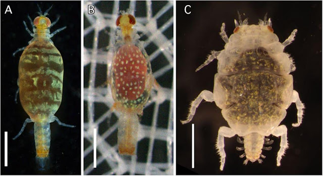
Left: Copepodid stage of a female Cymbasoma dissected from a Haplosyllis worm, Right: Adult stage of a female Cymbasoma 
Photos from Fig. 2 and Fig. 4 of the paper

Adult monstrilloid are free-swimming and they don't feed, but as juveniles, they live as parasites that can grow inside various marine invertebrates including snails, mussels, and polychaete worms. In polychaete worms, they can grow pretty large in relation to their host, and when they reach adulthood, they bust out of the host like it's a novelty birthday cake. In that way, their life cycles are comparable to the hairworms that parasitise crickets and mantids. 

Unlike other planktonic copepods that often swim by flicking their long antennae, the antennae of adult monstrilloids are fixed, so instead they have powerful swimming legs that allow them to kick their way through the water.  And while the adult stage of these weird little crustaceans are sometimes found in plankton trawl samples, their juvenile stage are much more elusive. Out of the 195 known species of monstrilloids the parasitic juvenile stage has only been identified for seven species, since they are hidden away in the bodies of their hosts. As a result, it has been over a century since anyone has investigated those parasitic juveniles in detail.

In this study, scientists in Japan were examining pieces of sponge that had been washed up on Tancha Beach at Okinawa Island. Those sponges turned out to be home for hundreds of Haplosyllis polychaete worms, but the worms themselves were also occupied by monstrilloids. This was also the case for sponge worms from Diamond Beach on another part of the island, which turned out to be an absolute haven for the little monsters, with over half of the worms hosting monstrilloids. This abundance of monstrilloids at Okinawa Island presented an amazing opportunity for scientists to get a better look at the parasitic stage of these copepods. 

In order to find out more about these enigmatic crustaceans, scientists first had to coax the host worms out of their spongey home, and they did that by taking chunks of the sponges and kept them in water without aeration. As oxygen level dropped, the worms were forced to abandon their sponge to seek more oxygenated water, at which point they could be collected and examined under the microscope. Monstrilloids are relatively large and highly visible as the bulk of the copepod stretches out the worm's body wall to transparency. 

Among these sponge-dwelling polychaete worms, the scientists found the larvae of two monstrilloid genera - Cymbasoma and Monstrilla, the former is coloured pale pink while the latter is teal green, but only the female copepods are so eye-catching due to their ovaries. The males are colourless and transparent. These larvae also live up to the monstrilloid name - they are banana-shaped, with a single eye, enclosed in a translucent sheath, and have a pair of long feeding tubes which it uses to slurp up nutrients from the host's body. When they reach maturity, the copepod uses those same tubes to make its exit by tearing a hole through the worm's body wall. Once free of the host's body, the monstrilloid shrugs off its juvenile exoskeleton to transform into an adult and takes its place among the zooplankton. 

In order to complete its life cycle, monstrilloids have to survive in three very different environments - the open ocean as adults, the sea floor (briefly) as nauplii, and inside the body of animals as juveniles. In the words of one of the scientists who study these little monsters, they are simply an awesome group of crustaceans.

Reference:

October 8, 2025

Dolops discoidalis

The electric eel, also known as poraquê, is a formidable animal. Not only is it capable of stunning its prey with an electrifying shock, it can leap out of the water to deliver a powerful jolt to any larger animals (including humans) unwise enough to approach it. Anyone would think twice about laying a finger on one of those living tasers. And yet, there's Dolops discoidalis, a humble little fish louse which makes its living by clinging to and sucking blood from these slippery shockers.

Top left: an adult Dolops discoirdalis, Top right: A D. discoidalis on the skin of an electric eel. Bottom: A electric eel in an observation tank, with a hand wearing protective glove reaching towards it with a pair of tweezers.
Photos from Fig. 1 of the paper

Dolops discoidalis is a branchiuran - a group of ectoparasitic crustaceans which are related to the endoparasitic tongue worms. Branchiurans are commonly called "fish lice" and they cling onto the skin of their slippery hosts with all kinds of gnarly equipment. Some of them such as Argulus, have a pair of massive suckers which have been modified from the crustacean's mouthpart. But in the case of Dolops, they have a pair of stout, sickle-like hooks - and they'll need those to cling to a smooth-skinned fish like the electric eel.

A group of researchers were collecting electric eels at the Xingu River to study their physiology and behaviour when they noticed that the eels were not alone. The six electric eels they collected were all covered in little fish lice, which the researchers identified as belonging to a species called Dolops discoidalis, and it is the first ectoparasite to be reported from the electric eel. Each of those fish lice was about the size of your fingernails, and they scuttled freely over the eel's skin.

Electric eels are usually solitary animals, but sometimes they get together to hunt in packs, which may provide opportunities for these parasitic crustaceans to jump onto new hosts. Dolops discoidalis is widely distributed across the Amazon Basin, living on many different types of fishes, but it seem to have a preference for smooth scaleless fish, such as the spotted tiger shovelnose catfish.

It is unclear why D. discoidalis isn't affected by their host's powerful electrical discharges, but then again the electric eel is not the only electrical fish to host external parasites. Torpedo rays are known to harbour tiny blood-sucking isopods which are kind of like "ticks of the sea", and they also sometimes fall victim to parasitic snails that slowly creep up on those electrifying sea pancakes to suck their blood. So there might be other species of ectoparasites living on the electric eel which we don't know about yet. It's just that handling a slippery fish that can shock you with electricity can be extremely tricky, let alone trying to study the tiny things living on it.

So while being a living taser is certainly useful for getting a feed or scaring off any would-be predators, it is no deterrent to much tinier creatures which might see you as just another big, moveable feast.

Reference:
Silva, J. O. S., Sousa, L., de Paula, E. A., Takemoto, R. M., & Carvalho, L. N. (2025). First documented case of ectoparasitism in electric eel: Dolops discoidalis (Branchiura: Argulidae) infesting Electrophorus voltai (Teleostei: Gymnotidae). Parasitology International 109:103114.

June 10, 2025

Sacculina pugettiae

Sacculina pilosella is a parasite of spider crabs (Scyra ferox), and while it is technically a barnacle, you're going to have to abandon all your preconceived notion of what a barnacle, or for that matter, an animal looks like in order to understand these parasites. These parasitic barnacles are called rhizocephalans and they are sometimes visible as a blob poking out of a crab's belly. While that is already far from what a conventional barnacle looks like, that's just the parasite's reproductive organ - the rest of its body is composed of an extensive network of roots that spread deep into the crab's body.

Spider crabs infected with Sacculina pugettiae (left) and Parasacculina pilosella (right).
Photos from Figure 2 of the paper

It was previously thought that those spider crabs only have a single species of rhizocephalan barnacle parasitising them, the aforementioned Sacculina pilosella, but DNA analyses of rhizocephalan specimens revealed that the spider crab is actually being tag-teamed by TWO parasitic barnacles hiding in plain sight. Turns out that what scientists have been calling "Sacculina pilosella" is actually two entirely different rhizocephalan species - Sacculina pugettiae and Parasacculina pilosella. It shouldn't be a surprise that their differences have gone unnoticed considering the body of these barnacles is just a blob with a mass of fine roots. Both species share the same breeding season between June to September, during the summer months, and sometimes they even infect the same crab simultaneously.

They do have some minor anatomical differences on the blob-like reproductive organ, but even when compared side-by-side, they can be tricky to tell apart. There is another anatomical feature which might provide a more reliable clue to the parasite's true identity, but that is only visible on a microscopic level. As previously mentioned, the body of a rhizocephalan is a massive network of rootlets, but not all those roots are made the same. Some of the roots, called trophic roots, absorb nutrients and are situated in the host's body cavity.

But the barnacle also grows a different type of roots that invade the host's brain. And it is those brain-invading roots that offer a way of distinguishing those two different species. Sacculina pugettiae has roots that end in microscopic goblets whereas P. pilosella has regularly shaped tapered ends to their brain-invading roots. While the significance of those microscope goblets is not clear, their presence is a reliable way to tell those barnacle species apart.

Often, when two different species of parasites are sharing the same host, this can result in a turf war, especially if they are body-snatchers that take up much of the host's body. Some parasites have even evolved specialised stages to fight off competitors. Since rhizocephalans have extensive roots that proliferate throughout the host's body, you would think that two such parasites living in the same crabs would inevitably end up butting heads (well, roots) with each other. But that's not what the scientists found. Somehow, these barnacles were able to share the same crab without conflict, both being able to successfully grow and reproduce, their rootlets intertwined with each other as they tickled the host's brain stem and absorb the crab's lifeblood in harmony.

Reference:
Lianguzova, A. D., Poliushkevich, L. O., Laskova, E. P., Golubinskaya, D. D., Arbuzova, N. A., Petruniak, A. M., & Miroliubov, A. M. (2025). Two in one: A case study of two rhizocephalan species invading the nervous tissue of one host. Journal of Zoology 325: 185-195.

March 13, 2025

Gibellula agroflorestalis

Imagine an infectious disease that turns you into an immobile white statue - while that sounds like something out of a dystopian science fiction story, this is the fate which awaits any spiders that encounter Gibellula, a genus of araneopathogenic (spider-killing) fungi. There are many species of this fungi found all over the world and their spores can infect all kinds of spiders. Gibellula is in the same broad group of fungi as the zombie ant fungi, but instead of producing a single or a few mushroom-like fruiting bodies that emerge from the carcass of their hosts, Gibellula smothers the infected spiders in a dense layer of white, fluffy mould.

Left: Examples of spiders infected with Gibellula agroflorestalis including orb-weavers (a-c) and jumping spider (d), Centre: numerous spore-bearing stalks (conidiophores), their mycelia twisted together, Right: Close-up of the head of a conidiophore.
Photos from Fig 3 and 4 of the paper

This post focuses on Gibellula agroflorestalis, a newly described fungus which was discovered by a group of researchers in the agroforestry systems located at the Abreu e Lima municipality of Brazil. 
Agroforestry is a system of agriculture where instead of cultivating a single type of crop, different crop plants are grown alongside trees and other plants, and these environments can provide habitats for a wider range of organisms. The forests that the researchers studied were composed of fruit plants such bananas, coconuts, limes, and mangoes, growing alongside various trees such as pink trumpet trees, pau brasil, and juazeiro.

So how does one find mouldy spiders amidst this semi-cultivated forest? Well, in short, with a lot of time and effort. The researchers hiked through multiple agroforestry properties while meticulously examining every plant they came across, carefully inspecting the underside of their leaves which is where many fungus-infected spiders end up. On the rare occasion when they come across an infected spider, it was collected intact along with the leaf it was sitting on, placed into a plastic cup, and later stored in silica gel to preserve the specimen. In total, the researchers found 17 infected spiders, ranging from jumping spiders to crab spiders to orb-weavers, alongside other spider hosts which could not be identified, because by that stage they have become more Gibellula than spider, twisted and fungal.

When the researchers examined the fungus under a microscope, they found that the structure and size of its teardrop-shaped spores were different from other known species of Gibellula. Furthermore, DNA analyses showed the fungus to be genetically distinct, and that as a species, G. agroflorestalis is fairly genetically diverse, which is probably why it is able to infect so many different types of spiders.

While there are other species of Gibellula fungi out there which have been recorded from spiders in natural forests, G. agroflorestalis was the first to be recorded in an agroforestry system, showing that these environment may in fact serve as habitats to some unique species. The discovery of G. agroflorestalis reminds us that a lot of unseen biodiversity can be found through careful observations. So it's good to stop and smell the roses, but you should also check underneath the leaves for mouldy spiders.

Reference:
da Rocha Alves, J.E., da Silva Santos, A.C., Pedroso, S.K.B., Melo, R.F.R., & Tiago, P.V. (2025). Untangling a web of spider fungi: Gibellula agroflorestalis (Hypocreales, Ascomycota), a new species of spider parasite from Brazil. Journal of Invertebrate Pathology 209:108278.

January 14, 2025

Cymothoa indica (et al.)

Tongue-biters are among the most (in)famous parasites found in fish, but they aren't the only type of isopods that parasitise fish, nor is the mouth the only spot ripe for parasitism - there are many other parts of a fish's body where an isopod can make itself at home. Why, right behind the fish's mouth are its gills, and this cosy, well-aerated and blood-rich location is where some isopods reside. There are also others that cling to the fish's skin where they gnaw and suck on host tissue, and even some that just burrow into the fish's body cavity for extra coziness.

Photo collage showing a range of cymothoid isopods on various fishes: (a) Cymothoa indica male (smaller one in the photo) and female attaching to the buccal chamber of Datnoides polota; (b) Cymothoa indica attaching to the mouth of Jonhius sp.; (c) Nerocila loveni attaching to the skin in the ventrolateral region of Deveximentum Interruptum; (d) Nerocila orbignyi attaching to the tail skin of Mugil cephalus; (e) Agarna malayi attaching to the gill cavity of Nematolosus nasus; (f) Joryma sawayah male (smaller one in the photo) and female attaching to the gill cavity of Nematolosus nasus.
From Figure 1 of the paper

So there are many different ways to parasitise a fish and cymothoid isopods are particularly adept at doing so. But some isopods are pickier than others when it comes to which fish they parasitise, and it seems to have something to do with where they live on a fish. The study featured in this post looked at factors that may have driven the preference of these parasites. To do this, the researchers studied fish collected from commercial trawlers at harbours and fish landing centres along the north-eastern coast of India, from Petuaghat down to Gopalpur.

The researchers examined a total of 5798 fish, of which 923 (from 59 fish species) were parasitised by 21 different species of cymothoid isopods. With this massive dataset, they were able to compare the host preference of tongue-biters, gill-biters, and the skin-biters, noting how many different species of fish each of them parasitise, and the characteristics of the fish they infect. From their analyses, it seems that generally speaking gill-biters tend to be most specific - they stick to a single fish species and are mostly found in pelagic schooling fish. In contrast, tongue-biters tend to infect fish that hang out near the seafloor, and are less selective about their host species. And the skin-biters are happy to just go after whatever fish they come across.

This trend might have something to do with the life histories of those isopods. The gill-biters have free-swimming larvae that reach their host by getting sucked into the respiratory current of fish swimming through the water column, and if those fish are in a school, there would be plenty of hosts available nearby for the next generation of gill-biters. On another hand, tongue biters have larvae that hang out on the seafloor, waiting to ambush any foraging fish that come near, so they are more likely to encounter a wider range of fish. But even though tongue-biters can infect more fish species than the gill-biters, each species of tongue-biter definitely has a "type".

For example, take Cymothoa indica, a tongue-biter which is found in a wide range of fish species across seven different families - while that seems like it has pretty broad taste, its hosts all tend to be shallow water fish that live and feed near the seafloor. Similarly, another tongue-biter - Catoessa boscii - infect seven different fish species, but all those fishes are similarly shaped, as they are mostly deep-bodied fishes such as jacks and scads. Meanwhile, the skin-biters have larvae that roam freely around the water, and can launch its attack from the seafloor or while rapidly looping in the water column. Essentially if it runs into a fish, it just latches on and starts gnawing.

Parasitic isopods are found in/on fish all over the world, and they have significant impact on fisheries and aquaculture. But despite their ubiquity, they are relatively under-studied, with most of the published research on their taxonomy, biogeography and patterns of host associations coming from only a handful of specialist researchers across the globe. Studies like the one featured in this post can provide us with some much needed insight into the secret lives of these widely found parasites.

Reference:


November 14, 2023

Stylops ater

Strepisptera is an order of parasitic insects with some very unique characteristics.They are also known as twisted wing parasites, based on the twisted hindwings on the male parasite. They infect many different orders of insects, but mostly target wasps and bees where they up take up residency in the host's abdomen. If you know what to look for, you can immediately spot their presence. In fact, there's even a special term for describing bees and wasps that are parasitised - they get "stylopized".

Top: A male Stylops ater (indicated by red arrow) attempting to mate with a female in a bee's abdomen.
Bottom left: Female Stylops ater adult (indicated by red arrow) in a bee's abdomen,
Bottom right: Male Stylops ater pupa casing (indicated by red arrow) in a bee's abdomen. 
From Fig. 1 of the paper

And it's not just the hindwings of stresipterans that are a bit twisted, these insects have extreme sexual dimorphism, so much so that if you didn't know any better, you'd think the females and males are completely different types of animals. The female stresipteran looks like a grub, and she spends her entire life inside the abdomen of the host, with just her head partially poking out from between the segments of the host's abdomen.

In contrast, the males have a pair of giant compound eyes, prominent branched antennae and the "twisted wings" that give this group of insects its name. They have a short and frantic adulthood - after emerging from the host, he only lives for a few hours and his sole mission in life is to find and mate with an elusive female strepisteran, hidden away in the abdomen of a host insect. And he does the deed with an appendage that entomologist Tom Houslay once vividly called a "stabby cock dagger". The technical term for this form of mating is "hypodermic insemination" - where the male basically stabs and inject his sperm into the female, and the sperm somehow find their way to the eggs. Strespiterans are not alone in having this type of appendage, male bed bugs also have a stabby cock dagger - but that's another story.

The study being featured in this post focuses on Stylops ater, a species which parasitises Andrena vaga, the grey-backed mining bee. Unlike the honeybees that most people are familiar with, these are solitary bees, with no castes. And while they do gather into an aggregation to nest, each bee just builds and looks after their own nest. The researchers examined a population of these bees in Lower Saxony, Germany. They sampled over two periods, during late winter, when all 508 bees they looked at were stylopized, and late spring, when they only managed to find two stylopized bees out of a total of 150.

Almost two-third of the stylopized bees were female, but these parasites seem to prefer hosts that are of the same sex as themselves. Since female bees live longer and can provide more nutrients than male bees, this works out well for the life history of female Stylops as it gives them more time and nutrients to grow her brood. After mating, the female Stylops can release up to 7000 offspring, which crawl off to find other bees. While each larva is merely 0.2 millimetre long, they can traverse long distances by hitching a ride on the hair, pollen sacks, or even the crop of bees, to end up in a new bee nest, filled with fresh hosts.

While most bees only hosted a single parasite, some had two or three, and the researchers did find one very unlucky bee that was harbouring four Stylops in its abdomen. But even a single Stylops can take a severe toll on its host. In fact, this parasite is so demanding that it wouldn't grow as big if it had to share its host with another Stylops. As a result, bees infected with Stylops are unable to develop eggs or only produce poorly developed eggs.

But aside from effectively sterilising the bee, Stylops also tinkers its host's biological clock, making it emerge out of hibernation a few weeks earlier than uninfected bees - hence why the researchers found so many stylopized bees in late winter. Making the bees such early risers ensures that there will be plenty of female Stylops around for the male Stylops to find, which will be emerging at that time to live out their extremely short lives. It also gives the female Stylops' larvae more time to develop, so they will be able to crawl off in time to find new hosts in the bee's brood cells. This type of behaviour manipulation is comparable to what's found in Sphaerularia vespae, a nematode that alters the seasonal biological clock of hornet queens.

In order to make these changes to the bee's internal clock, Stylops would have to manipulate the host's hormones, but this also results in some side effects on the bee's body. Female bees that get stylopized tend to have a hairier back, skinnier legs, and the hairs on said legs become shorter and more sparse. In short, they take on characteristics that are more similar to that of regular male bees.

So next time you are out and about, keep an eye out for a bee that looks a bit different from the rest. It might be flying under the influence of a parasite tucked away in its abdomen, looking to make a rendezvous with her short-lived partner.

Reference:

July 11, 2023

Diexanthema hakuhomaruae

The study in this post takes us to one of the darkest corners of the deep sea, over 7000 m below sea level in the Kuril-Kamchatka Trench, located in the northwestern Pacific. Living in this dark and oppressive environment are isopods called Eugerdella kurabyssalis. And despite the crushing pressure, these crustaceans like it just fine, in fact they are the most abundant isopod down in those depths. But such success and abundance can also attract the attention of parasites, and this post is about a newly described parasitic copepod called Diexanthema hakuhomaruae.

Left: Diexanthema hakuhomaruae (indicated by white arrow) attached to the leg of its Eugerdella kurabyssalis isopod host. Right: Close-up of D. hakuhomaruae, the arrow indicating the copepod's ovaries. Photos from Figure 1 of the paper

Those who are familiar with this blog would know that parasitic copepods come in all kinds of shapes  that would defy most people's idea of what a crustacean is "supposed" to look like. And D. hakuhomaruae is no different - its tiny body is ROUND and if anything, it looks almost like a legless tick. And much like a tick, D. hakuhomaruae attaches itself stubbornly to the leg of its host.

Diexanthema hakuhomaruae belongs to the Nicothoidae family, a group of parasitic copepods that contains about 140 known species. They live on a variety of crustacean hosts, including tanaidaceans, ostracods, amphipods, cumaceans, mysid shrimps, and lobsters. Most of them have a rotund, almost spherical body, greatly reduced or no legs at all, and a specialised mouthpart that ends in a sucker with syringe-like mandibles. And much like the ticks that they resemble, these copepods feed by stabbing their mouth syringe into their host's body and sucking up that crustacean blood (hemolymph) on tap. Some species such as Choniomyzon infaltus are specialised egg parasites - their balloon-shaped bodies allow them to hide amidst broods of their hosts and feed on their eggs without being discovered.

There are currently six other known species of Diexanthema, all of them are parasites of deep sea isopods. And Diexanthema is not alone in its preference - there are other nicothoid copepods that have also been found parasitising deep sea isopods. What makes D. hakuhomaruae special is that it is the first to be found from the Hadal Zone. All other Diexanthema species have been reported from depths of 1300 to 3500 metres below sea level, but none of them had gone down as deep as D. hakuhomaruae.

It is unknown whether D. hakuhomaruae feeds on the host's fluid or if it is an egg parasite, or how it even completes itself life cycle in the hadal zone - as you can imagine, discovering and describing such parasites in an environment like the deep sea is challenging enough as it is. Studying the life style and ecology of these deep sea parasites with current technology is next to impossible. Even so, this description shows that parasitism is indeed ubiquitous on this planet, and wherever you find life, you can be sure that some of them will be parasites

Reference:
Kakui, K., Fukuchi, J., & Ohta, M. (2023). Diexanthema hakuhomaruae sp. nov.(Copepoda: Siphonostomatoida: Nicothoidae) from the Hadal Zone in the Northwestern Pacific, with an 18S Molecular Phylogeny. Acta Parasitologica 68: 413-419.

May 13, 2023

Dendrogaster nike

Parasitic crustaceans can evolve into some pretty funky forms and they have been featured multiple times on this blog. These crustaceans don't so much flaunt, but completely toss out all your expectations of what a crustacean or even an arthropod is "supposed" to look like. And among the best examples of that is Dendrogaster.

Left: Female Dendrogaster nike side view, with attached male (bracketed in the square), Top right: Female D. nike frontal view, Bottom right: Male D. nike. Photos from Fig. 2. of the paper

Dendrogaster is a genus of crustaceans that live as internal parasites of sea stars, nestled snugly within the body cavity of its host. So much so that their body shape seems to have evolved into somewhat resembling that of its host. In contrast to other crustaceans, Instead of having hard carapaces, segments, or jointed legs, Dendrogaster has multiple branching lobes, like some kind of fleshy, parasitic antler. They belong to a group of crustaceans called Ascothoracida - a sister group to the barnacles, who themselves are no strangers to the way that evolving towards parasitism can warp their body.

Dendrogaster rivals those parasitic barnacles in the "WTF Evolution?" department, and despite how bizarre they may look to us, they are not some rare oddity lurking in an obscure corner of the world. There are 35 known species of Dendrogaster and they have been found parasitising eighteen different families of sea stars all over the world, ranging from those dwelling in the shallows, to those inhabiting the deep sea over 2500 m below sea level. It seems that wherever sea stars went, Dendrogaster followed.

The paper featured in this blog post adds another species to this roster of evolutionary weirdos. This newly described species was found from sea stars living 1970 m below sea level, collected during a biodiversity survey at the An'ei Seamount, an offshore marine protected area off the eastern coast of Japan. The host was Asthenactis agni - a sea star which itself was newly discovered and described just late last year. This parasitic crustacean has multiple, wing-like branches protruding from its body, and it is this appearance which inspired its scientific name, Dendrogaster nike, named after Nike, the Greek winged goddess of victory.

But that's only how the female of the species looks like. The male is less than a quarter the size of its partner, and unlike the female Dendrogaster with its multiple protruding branches, the male is comparatively unremarkable, with a simple ovoid-shaped body and a pair of long thin testes dangling from it. It is usually found attached to its much larger and more flamboyant partner, floating inside the body cavity of a sea star.

Dendrogaster nike is just one of many new species of Dendrogaster that have been described over the last few years. In 2020, there were three other species of Dendrogaster which had been discovered from sea stars collected from the depths of the bathyal zone. It seems that sea stars from the deep sea are particularly favoured by this parasitic crustacean, and there are probably many other species of Dendrogaster yet to be discovered which are lurking in the abyss.

When scientists compared the DNA sequences of different Dendrogaster species, they found that the genus seems to be divided into two main sub-groups - those who stuck to the shallows, and those who ended up partying in the deep. While the evolutionary pathways of many parasites somewhat parallel that of their hosts, for Dendrogaster, it followed the hosts' habitats instead. This may provide some insight into the evolutionary origin of this bizarre, but widely found group of parasitic crustaceans.

When life hands you a sea star, sometimes it comes with a free Dendrogaster.

Reference:
Jimi, N., Kobayashi, I., Moritaki, T., Woo, S. P., Tsuchida, S., & Fujiwara, Y. (2023). Insights into the diversification of deep-sea endoparasites: Phylogenetic relationships within Dendrogaster (Crustacea: Ascothoracida) and a new species description from a western Pacific seamount. Deep Sea Research Part I: Oceanographic Research Papers 196: 104025.

April 13, 2023

Rickia wasmannii

Rickia wasmannii is a fungus that lives on ants, and when it comes to ants and fungi most people usually think of Ophiocordyceps, i.e. the zombie ant fungus - which was the inspiration for The Last of Us series of video games and TV series. But R. wasmannii is not a killer - instead of zombifying its host and digesting the corpse, this fungus seems to reduce animosity and aggression between ants. First of all, let's take a look at what R. wasmannii actually does on ants. 

Left: Illustration of a Rickia wasmannii thallus, Right top: Uninfected ant, Right bottom: ant infected with R. wasmannii
Pictures from Figure 1 of this paper

Rickia wasmannii belongs to a group of fungi called Laboulbeniales, also known more colloquially as "labouls". These fungi have little holdfasts called haustoria that allows them to cling to the ant's cuticle. They are ectoparasites of insects that attach to their host's external surface and suck their hemolymph (insect's equivalent of blood). So in a way they are rather like ticks or lice (and yes, there are labouls that live on ectoparasitic insects, which one might consider as a bit of poetic justice).

But this fungus seems to do more than just suck the ant's blood, as it causes the infected ants and other ants around them to behave differently. Rickia wasmannii changes the host ant's cuticular hydrocarbon or CHC profile. CHC is essentially an ant's ID profile - they use it to recognise nestmates, tell each other apart, and be alerted to strangers from other nests. But R. wasmannii messes with that, scrambling the infected ants' CHC profile, and making them "smell" differently to uninfected ants.

Scientists wanted to find out how the presence of this fungus affects the way ants interact with each other. The challenge with studying ant behaviour is that when you put two ants together, it is difficult to tell apart whether the ant you are observing is responding to the other ant's chemical profile, or if it is responding to the way the other ant is reacting to them. The only way to get a clear observation is to present the ant with something that it would recognise as a fellow ant, but would not muddle the outcome by reacting to the ant that you are trying to observed

The solution turns out to be freeze-killed ants. Ants that are killed in this manner retain their CHC profile, so other ants would treat them just as another live ant, but obviously a dead ant wouldn't react to a live ant's presence and confound the outcome. In addition to those freeze-killed test subjects, scientists also made ant "dummies" which are essentially blank slates in ant forms that they can imbue with whatever chemical signature they were testing. These "dummies" were made by washing ant corpses in hexane to remove their chemical signature. To ants, these specially treated ant corpses are like faceless mannequin, with no identity - until the scientist imbues them with one, by anointing them with a droplet of cuticular extract from another ant.

When ants were presented with dummies that were smeared with the cuticular extract of ants from a different nest, the ants started biting, dragging, or stinging the dummies, much like how they would respond to a live ant from another nest. But when they were presented with either the corpse of a Rickia-infected ant, or dummies that "smell" like a Rickia-infected ant, they were more relaxed and less likely to get aggro. Furthermore, it's not just that the fungus made other ants act differently, the infected ant itself also starts behaving differently. Infected ants are generally less likely to pick a fight with another ant, but especially when facing other infected ants.

As mentioned previously, R. wasmannii seems to change the ant's CHC profile, but one would think scrambling the host ant's profile would make other ants react more aggressively towards them since ants usually have a "stranger danger" response to ants that "smell" different to their nestmates. But the way that R. wasmannii changes how an ant "smell" seems to have a calming effect, and this comes down to a molecule called n-C23 which is present in higher concentration on the cuticle of all infected ants. When the scientist presented ants with dummies that have been smeared with n-C23 and nothing else, almost all hints of aggressive behaviour ceased.

So by increasing n-C23 concentration in its host's cuticle, R. wasmannii has unlocked a life hack that allows it to not just access all areas in an ant colony, but to spread to other nests as well. In the scientists' study population, about half the colonies they studied had the fungus present, and in some nests, all the ants were infected with R. wasmannii. A testament to the fungus' successful manipulation of ant behaviour.

Furthermore the fungus' presence also affects another, very different parasite which also lives with ants - the caterpillar of blue butterflies. These caterpillars are social parasites that convince worker ants into adopting them into their nest. Once they are settled in, they start demanding food from the worker ants and even feed on the ant's developing broods. But the caterpillars don't seem to survive as long in nests which are already hosting R. wasmannii, and in the field, these two parasites co-occur less commonly than expected based on their respective prevalence, which indicates the caterpillar and the fungus are in competition over ant real estate.

By messing with their identity and making them more chilled out, R. wasmannii can turn an ant colony into a fungus party. But the consequences of that ripple out to other ant colonies too, along with the organisms that regularly take up residency in the homes of ants.

Reference:
Csata, E., Casacci, L. P., Ruther, J., Bernadou, A., Heinze, J., & Markó, B. (2023). Non-lethal fungal infection could reduce aggression towards strangers in ants. Communications Biology, 6: 183.

September 20, 2022

Hymenoepimecis bicolor

Over the course of human history, numerous species of plants, animals, and other organisms have been taken from their original habitats and introduced (either intentionally or accidentally) to other parts of the world. Some of those introduced species become "invasives" in their new home, partly due to the lack of natural enemies. But while many invasive species get a brief moment of respite from their old adversaries, the local parasite and predators quickly catch on that the new arrival could be added to their menus too. This was the case for a species of spider that has been introduced to Brazil, where it ended up attracting the attention of a mind-controlling wasp.

Left top: Hymenoepimecis bicolor larva on a spider host, Left bottom: Developing H. bicolor cocoon in a cocoon web,
Right: an adult H. bicolor wasp.
Photo of H. bicolor larva from Fig. 2 of this paper, Photos of cocoon and adult H. bicolor from Fig. 7 and 9 of this paper

Hymenoepimecis bicolor is a species of parasitoid wasp that belongs to a subfamily of wasps called Pimplinae. All the members of that subfamily are a spider's worst nightmares. These wasps specialise in attacking spiders at every stage of their lives - some species go after the unhatched eggs in silk sacs, while others tackle fully-grown adult spiders. Not only that, some of them are also masterful mind manipulators that induce their host spider into spinning a special web called a "cocoon web" that secures the wasp's developing cocoon. And Hymenoepimecis bicolor just happens to be one such manipulator.

Among the pimpline wasps, each species have their own host preferences and in the case of H. bicolor, one of its usual hosts is the golden silk orb-weaver (Trichonephila clavipes), a spider which is native to Brazil. When a female H. bicolor spots a potential host, she flies in and grapples with it, immobilising the spider by stabbing it in the mouth with her ovipositor, before checking it for other wasp eggs, and then planting one of her own. The thing about the golden silk orb-weaver is that while the juvenile spiders are relatively easy for H. bicolor to handle, once the spiders reach adulthood, they become more dangerous for the wasp to tackle. 

Nevertheless, limited host availability means that the wasp sometimes need to go after the bigger spiders anyway, with demand being so high that some spiders end up being parasitised by two wasp larvae at the same time. But the arrival of the tropical tent-web spider (Cyrtophora citricola) has provided H. bicolor with some new options.

The tropical tent-web spider has spread to many parts of the world by hitchhiking in shipments of fruit, potted plants, or packing material, and it has made its way to South America about three decades ago. And it just so happens that their size and habits place them firmly in the sight of H. bicolor. Not only is the tent-web spider in the preferred size range for H. bicolor to parasitise, much like the native orb-weavers, this introduced spider constructs open webs that leave them exposed to attacks. Researchers found that the H. bicolor larvae are able to successfully parasitise the tent-web spider just as well as the native spiders.

While H. bicolor larvae grow well and pupate as usual on the tent-web spiders, it seems that they haven't yet achieved complete mastery over this new-fangled host. As mentioned earlier, when these wasps are ready to pupate, they commandeer their spider hosts to weave a special "cocoon web" that suspends the developing wasp cocoon in mid-air. This makes the cocoon less accessible to any would-be predators or hyperparasitoids. Hymenoepimecis  bicolor embellish that with an added layer of security, by inducing the spider to also build a series "barrier threads" around the cocoon that further bar entry, as well as making the web more stable

This is where the introduced spider host falls short. While the parasitised tent-web spider is able to produce the usual cocoon web with the necessary structure to support and suspend the developing cocoon, it lacks the finishing touches of those additional barrier threads. Ironically, compared with the spiders that H. bicolor usually targets, the regular webs made by the tent-web spider actually needs less modification to make it suitable for the wasp's cocoon.

Based on what's known about these wasps, when it is ready to pupate, the wasp larva produces a cocktail of chemicals that place the spider under its spell. But in this case, it looks like that cocktail formula needs a bit of tweaking to work its full magic on the introduced tent-web spider. While not perfect, it serves its purpose well enough, and the introduction of this spider has allowed a parasitoid wasp to expand its host horizons.

Reference:

June 18, 2022

Sarcotaces izawai

Parasitic copepods are a weird bunch, and many of them look nothing like what most people would recognise as a crustacean. But even among those weirdos, Sarcotaces stands out, because during the course of its evolution, it has turned into a big teardrop-shaped blob living inside a fish's body.

Top left: Staining of the fish body due to Sarcotaces, Bottom left: Sarcotaces extracted from fish flesh
Right: A Female Sarcotaces specimen (about 4 cm in length)
Photos from Fig. 1 and Graphical Abstract of the paper.

There are seven known species of Sarcotaces, all of which are parasites that dwell in fleshy galls embedded in the muscles of fish. The female of the species can grow up to about 5 cm long. They belong to a family of copepods called Philichthyidae which all specialise in living within nooks and crannies of a fish's body, including their skull, sensory canals, or inside galls just beneath the fish's skin. The study featured in this blog post described a newly discovered species of Sarcotaces - Sarcotaces izawai.

Specimens of S. izawai were retrieved from a consignment of frozen fish which were originally destined for the fish market, but were redirected to researchers when the County Veterinary Inspector of Szczecin noticed signs of infection in some of the fish. In total, 29 fish were taken to the University of Szczecin for further examination. Nine of those fish were found to harbour the gall of Sarcotaces - where there was once fish muscle had been turned into a black void, a dark fleshy cavern where the female Sarcotaces resided alongside her tiny males and microscopic larvae.

The black liquid associated with this copepod is what gave Sarcotaces its German name - "Tintenbeutel" which means "ink bag", and why in parts of Australia, they're called "Iodine Worms". Even in the other fish where no Sarcotaces were found, the fish's flesh were tainted with an ink-stained void, which most likely meant a Sarcotaces had once lived there, but was inadvertently removed when the fish were being processed. While the presence of this parasite does not pose any health hazards to any would-be consumers, the inky stain in the fish's flesh do render them off-putting to any would-be buyers on the market. But, because of this, the researchers were given an opportunity to conduct detailed scanning electron microscopy on the copepod, and provided the first DNA barcode for this unique genus of parasite based on its COI gene.

While the female S. izawai is very distinct and noticeable, the male is rather inconspicuous - they grow to about 3 mm in length, and are comparatively tiny and fairly nondescript. In comparison, the female is shaped like a knobbly radish, and grows to 2.5 to 5 cm in length or 10-20 times the length of the male. This size difference is comparable to that of a human and a sperm whale. It also means that a single female could be accompanied by multiple males. Indeed, the researchers found one female who was accompanied by 18 suitors in her flesh gall.

While very little is known about how the microscopic, free-swimming larvae of Sarcotaces gets into a fish in the first place,  it seems that the growth and development of the female Sarcotaces takes place entirely within the sac-like gall. This flesh bag has a tiny opening to the outside world that the copepod usually keeps plugged using the pointy tip of her body, and unplugs to release larvae into the surrounding waters. Because of this, the researchers consider Sarcotaces as a "mesoparasite", because while they largely live within the fish's body, they still maintain some contact with the outside world with the tip of the body plugging up that hole.

As an added layer to that study, the consignment of frozen fish that the researchers examined have been been frozen and thawed multiple times, and were "pan-dressed" - in that their head, fins, and the guts have been taken out - this might be why some of the fish had the characteristic inky stain of Sarcotaces even though the parasite was absent. This made the identification of those fish rather difficult simply through visual inspection. While the consignment of fish were labelled as Pseudophycis bachus - red codling - from "The Falklands", the researchers found this to be a case of seafood identity fraud.

When they did some DNA analyses they found that the fish were actually Mora moro - a species of deep sea cod which is found in temperate seas across many parts of the world, but has not been recorded from the Falklands. It is likely that the fish wholesalers were trying to use the mislabelling to bypass regional quotas or conceal catches from restricted waters.

This type of seafood mislabelling is very common around the world, and presents problems for consumer protection, food safety and supply, fisheries regulations, and conservation. In this case, not only did the ink-stained fillets of these Sarcotaces-infected fish provide scientists with an opportunity to examine a poorly-understood parasite, the presence of this tubby copepod also helped draw attention to a case of seafood identity fraud.

Reference:

May 20, 2022

Guimaraesiella sp.

Quite a few years ago I wrote a blog post about a study on some bird lice that hitch-hike on louse flies as a way of reaching new hosts - this type of interaction whereby an organism attach itself to the body of another as a way of getting around is called "phoresy". And while it is a fascinating interaction with important ecological implications, this phenomenon is not particularly well-studied. Well, the paper that is being featured in this blog post revisited that field of research, and used multiple approaches to investigate this type of interaction. And the researchers behind it did so by combining literature review, traditional parasitology, DNA barcoding, and citizen science.

Left: Guimaraesiella lice found on from louse flies. Right: Louse fly with lice attached (indicated by red arrows). 
From Figure 3 of the paper.

The researchers of this study were trying to figure out how common phoresy is among bird lice, and who exactly is hitch-hiking on what. They conducted a review of the existing scientific literature on phoretic relationships between lice and louse flies, and found that many of the older records were unusable because they lack sufficient details regarding species identity of the lice involved. Furthermore, while phoretic behaviour in lice is most well-documented in North America and Europe, there are other parts of the world with much richer avian fauna (and thus more bird lice species), but phoretic behaviour of bird lice in those regions are not as well-studied.

To address this, the researchers came up with a way of collecting lice and louse flies from a large number of birds, and did so with some help from members of the public. As a part of long-term project to monitor bird mortality from vehicle and building collisions, ordinary citizens in Singapore were encouraged to report any dead birds that they come across. Through this, the researchers were able to track down and collect over a hundred recently deceased birds for this study. They then screened the dead birds for lice and louse flies, which were identified based on their morphology and their DNA.

In total, they screened 131 birds composed of 54 different species, and collected 603 lice and 32 louse flies. Of those, 22 birds had louse flies on them, but only three of the louse flies also happened to be carrying hitch-hiking lice, which were identified as belonging to the genus Guimaraesiella. Amidst all that, they found something unexpected - one of the birds, a Blue-winged pitta (Pitta moluccensis) was infected with louse flies carrying Guimaraesiella lice. This is the first time that Guimaraesiella lice has been found on pittas, as those birds are usually infected with lice in the Picicola genus.

It is likely that riding on louse flies is how Guimaraesiella ended up on the pitta. Indeed, lice in that genus appear to live on a wider range of birds compared with most bird lice, which are often confined to a single or handful of closely related host species, and its hitch-hiking habit may be the key to their success. While bird lice are very adept at climbing around and between their host's feathers, they are completely helpless off the host's body. This doesn't give them much opportunity to branch out and onto other bird species as they can only climb onto a new host through direct contact.

But since louse flies feed on a variety of different bird hosts, travelling on one of those flying blood-suckers can open up a whole new world of possibilities for lice that engage in phoresy. The species of Guimaraesiella lice they found on the pitta has also been found on at least 24 other species of birds, possibly more. Considering that the louse fly that Guimaraesiella rides on - Ornithophila metallica - feeds from over a hundred different bird genera, perhaps it is surprising that Guimaraesiella hasn't been found from even more bird species. So while the louse fly presents its hitch-hiker lice with many different species of birds, those well-travelled lice still stay fairly selective when it comes to where they settle on. These lice are like Goldilocks when it comes to picking a new feathery home - it needs to be just the right fit.

The approach taken by the researchers in this study to recover and screen large numbers of birds for louse flies and lice can also be applied to other parts of the world. This would help us obtain a more complete understanding of how widespread hitch-hiking lice actually are, and the role this behaviour has played in the evolution of these ectoparasitic insects.

Reference:
Lee, L., Tan, D. J., Oboňa, J., Gustafsson, D. R., Ang, Y., & Meier, R. (2022). Hitchhiking into the future on a fly: Toward a better understanding of phoresy and avian louse evolution (Phthiraptera) by screening bird carcasses for phoretic lice on hippoboscid flies (Diptera). Systematic Entomology 47: 420-429.

March 17, 2022

Thaumastognathia bicorniger

Gnathiidae is a family of parasitic isopods that can be considered as ticks of the sea. I make that comparison not only because gnathiids are blood-feeding arthropods, but like ticks, their life cycle involves going through a series of feeding and non-feeding stages. The blood-hungry fish-seeking stage is called a zuphea that, much like how a tick would on land, attaches itself onto passing fish and starts feeding to its heart's content. Once it is fully engorged with a belly full of blood, it becomes what's called a praniza, which drops off the fish to grow and moult into its next stage. Gnathiid isopods need to go through alternating between the zuphea and the praniza stage at least three consecutive times before they can reach full maturity.

Thaumastognathia bicorniger stripe (left) and spots (centre) pigemented third stage praniza, and adult male (right)
From Fig. 2. of the paper

The paper featured today is about Thaumastognathia bicorniger, a gnathiid isopod which has recently been described from the waters of Japan. The researchers who described this species found the isopod on various chimaera and sharks that were caught by fishing vessels operating in the waters of Suruga Bay and around Kumejima Island. Additionally, they were also able to obtain previously collected specimens of this isopod that had been stored at the laboratory of fish pathology at Nihon University. Those specimens were originally collected from various different cartilaginous fishes that were caught by fishing vessels off the southern coast of central Japan.

Based on their samples, this isopod has been recorded to feast on the blood of at least ten different species of cartilaginous fishes including nine species of sharks from six different families, along with one species of chimaera (also known as ratfish, in this case the Silver Chimaera). Thaumastognathia bicorniger larvae were always found in the gill chamber of their hosts, where they attached themselves to the blood-rich gill filaments. These isopods are tiny, with the third stage praniza larva measuring about 3.7-4.8 mm long, so having one or two of them would merely pose a minor inconvenience to the host. 

However, some sharks were found to be infected with dozens or even hundreds of those tiny blood-suckers. Of those, the Blotchy Swellshark (Cephaloscyllium umbratile), the Shortspine Spurdog (Squalus mitsukurii), and the Starspotted smooth-hound (Mustelus manazo) appeared to be among this gnathiid's favourite hosts, as they were commonly found to be infected with at least 50 T. bicorniger larvae and some even harboured hundreds of those blood-sucking isopods in their gill chambers. Additionally, much like how ticks are known to carry various pathogens, gnathiid isopods have also been implicated in the transmission of blood-borne parasites in coral reef fishes.

The juvenile stages of T. bicorniger seem to come in two different colour patterns - spotty and stripey. This was only visible in the live or freshly caught specimens as the colour faded rapidly when they are preserved in ethanol. Genetic analysis revealed that despite their superficial differences, those two colour morphs belong to the same species, and it is unclear whether the different colour patterns signify anything, as they're not associated with a particular haplotype, sex, nor host species.

The researchers kept some of the gnathiid larvae alive in captivity to see if any of them would metamorphose into an adult stage - but only one successfully moulted into an adult male. Among gnathiid isopods, there is a high degree of sexual dimorphism - the male gnathiids have squat body with big mandibles, while in contrast, female gnathiid have a larger rotund body for brooding eggs into larvae. Neither of which look anything like a "typical" isopod like a woodlouse or even the infamous tongue-biter parasite and its cymothoid relatives.

For other species of gnathiid isopods, metamorphosing from the third-stage praniza into a mature adult is a relatively brief process. After their last feeding session, some species would take just a week or two to mature into a reproductive adult, while others may take up to two months at most. However, T. bicorniger took a whooping 204 days to moult from a third stage praniza into an adult. So why does T. bicorniger take so long to mature compared with other species of gnathiid isopods?

Gnathiid metabolism and growth is greatly affected by water temperature, and many of the gnathiids that have very short development time are found in warmer, tropical waters. In this study researchers kept their T. bicorniger at 10-20°C in their lab, which is slightly cooler than the water temperature that those other known gnathiids are regularly exposed to. However, there is a species of Antarctic gnathiid - Gnathiia calva - which only took 6 weeks to transform into an adult despite living in waters that were kept at 0 to -1°C.

Alternatively it might have something to do with the fishes that they were feeding on. Many sharks have high levels of urea in their blood, which may make their blood more difficult to digest for any would-be blood-suckers. Lamprey that feed on basking sharks are specially adapted to excrete large volumes of urea which is found in their host's blood. The need to detoxify your food would most likely complicate the digestion process, decrease the blood's nutritional value, which would result in cost to development time. But then again there is another gnathiid species - Gnathia trimaculata - which infects Blacktip reef shark (Carcharinus melanopterus) and it only takes 6 (for males) or 24 days (for female) to moult into an adult.

So for now, the reason(s) why T. bicorniger seems to take such a long time to grow into an adult compared with other species of gnathiid isopods, remains a unsolved mystery.

Reference:
Ota, Y., Kurashima, A., & Horie, T. (2022). First Record of Elasmobranch Hosts for the Gnathiid Isopod Crustacean Thaumastognathia: Description of Thaumastognathia bicorniger sp. nov. Zoological Science, 39: 124-139

July 14, 2021

Echinophthirius horridus

Lice are common parasites of mammals. Humans alone are host to three different species of lice, and it's not just humans or land mammals that can get infected with lice. Pinnipeds - seals and sea lions - also have to contend with being covered in those ectoparasites. Unlike many other ectoparasites in the sea which have been bestowed with the name of "lice" such as salmon lice, tongue-biter lice, or whale lice (all of which are crustaceans), seal lice are true lice, in that they are parasitic insects belonging to the order called Phthiraptera.

Left: An adult seal louse, Right: two opened seal lice eggs (nits) glued to a strand of seal fur hair
From Fig. 1 of the paper

When the ancestors of modern pinnipeds took to the sea some time in the Oligocene about 30 million years ago, the lice followed them into the water, and in the process, they have to deal with all the challenges associated with living on an aquatic host. Seal lice belong to a family of lice called the Echinophthiridae and they have some specialised adaptations for living on hosts that spend most of their time immersed in sea water. This include elongated spiracles (the opening insects use to breathe) with mechanism for closing, a dense covering of spines and scales, and stout clamp-like claws that allow them to grip tightly onto their hosts' fur.

Blood-sucking arthropods such as ticks, fleas, and lice are often responsible for transmitting a wide variety of parasites and pathogens. And it seems that seal lice can also play a similar role in the sea. While performing routine diagnostics on 54 harbour seals and a very heavily-infected grey seal pup that were hospitalised at the Sealcentre Piteterburn (a seal rehabilitation and research centre in Netherlands), a group of scientists were able to use that opportunity to collect a massive number of seal lice from those marine mammals. They ended up collecting 200 lice from the harbour seals, and another 1000 from that one very heavily infested seal pup.

Those researchers divided the lice into batches of 1-20 lice, based on the individual host that they came from (the lice from the heavily infected seal pups were divided into multiple batches of 15 lice), then grind them up, and examined the lice slurry by subjecting it to polymerase chain reactions that amplified the DNA of known seal parasites and pathogens.

The DNA analyses showed that the seal heartworm (Acanthocheilonema spirocauda) was the most commonly found parasite, with it being detected in about one-third of the lice samples. While most people would associate "heartworm" with the dog heartworm (Dirofilaria immitis), that species is just one out of many different filarial roundworms that live in the heart of mammals. The findings of this study corroborates with previously published research which have found heartworm larvae dwelling in the gut of seal lice, demonstrating that these ectoparasitic insects play a key role in the transmission and life cycle of these nematodes.

Alongside the heartworm, there were also some bacterial pathogens lurking in those lice. Some of the lice from the grey seal pup were also carrying Anaplasma phagocytophilum, the bacteria which causes tick-borne fever and as their name indicates, are usually carried by ticks. Additionally, a few of the lice from that seal pup and some of the harbour seals were also carrying a species of Mycoplasma bacteria. This microbe is commonly found in seals and other marine mammals, but when it gets transmitted to humans, it is also associated with a disease known as "seal fingers". However, unlike the heartworm, it is unclear if the lice actually play a role in the transmission of these bacterial pathogens, or if they were incidental infections that simply came with living on a seal host.

It is worth noting that while pinnipeds had retained an heirloom of their terrestrial ancestry in the form of lice, another group of marine mammals - the whales - have acquired their own unique suite of ectoparasites which are unlike that of any other mammals. They have "whale lice" which are actually crustaceans in the same group as sandhoppers, along with pennellid copepods - a family of parasitic copepods that usually infect fish, with the exception of one species which has evolved to parasitise whales.

So why are there no "true lice" on whales? Well, for all their adeptness at clinging to their host, lice ultimately depend on the presence of hair or similar structures to hang onto their host. When a seal dives underwater, the layer of fur forms a covering that the lice can shelter underneath. But no such shelter exists on the smooth, hair-free surface of a whale. As a result, while whales have escaped the lice (and have picked up other parasites in the process), pinnipeds have kept their fur, and along with it, their lice and the worms that they carry.

Reference:

June 16, 2021

Allokepon hendersoni

Crabs have some pretty scary parasites infecting them. They range from worms that use them as vehicles to complete their complex life cycles, to parasitic dinoflagellates that turn their muscles into bitter slurry, and on top of those, there are also other crustaceans that can take over their body, and in some cases, castrate them in the process. These body-snatching crustaceans come in two main types - bopyrids and rhizocephalans. 

Top: Bopyrid isopod and an infected crab, Bottom: Rhizocephalan barnacle with infected crab.
Photos modified from the graphical abstract of the paper

Bopyrids are parasitic isopods in the same suborder as the infamous tongue biter parasite, but instead of going into a fish's mouth, they go inside the body of crabs and make themselves at home, often causing a characteristic bulge on the infected crab's carapace. And then, you have the rhizocephalans, which are freaky barnacles that have a body composed of a network of roots which wrap themselves around the crab's internal organs.

Each of them inflict their own respective flavour of pain on their crab hosts.

This study looked at the effects that these parasitic crustaceans have on the two-spotted swimming crab (Charybdis bimaculata), which is host to both parasitic isopods and barnacles. Here representing the bopyrid isopods, we have a species named Allokepon hendersoni. And fronting for the barnacles, is an as yet undescribed species of rhizocephalan. As hinted at earlier, these two body-snatcher parasites seem to have different effects on the crabs - but what exactly are they?

When scientists compared infected crabs with uninfected crabs, they found the effects to be most pronounced in male crabs, with both species of parasites causing a reduction in weight and claw size of their hosts. This is most likely due to the energetic drain associated with hosting these crustaceans, since they can grow to alarmingly large sizes when compared with their hosts. 

While reducing the claw size may leave the host crab less able to compete with uninfected males, on another hand (or claw as the case may be), it would not be in the interest of the parasite for its host to be getting into too many fights and risk injuries anyway, so it can be a beneficial side-effect from the parasite's perspective

But there were some changes which were more specific to particular parasite species. Male crabs infected with Allokepon had a narrower abdominal flap (the triangular flap on the "belly" of a crab). In male crabs, this flap would usually serve to protect the gonopods - which are specialised appendages that arthropods use in reproduction - but given the crab is already hosting such a demanding resident in its body, it wouldn't be getting up to any of that any time soon.

In contrast, the rhizocephalan barnacle had the opposite effect on the crab and widened that flap - this is part of a whole suite of changes that these parasites induce in male hosts. Male crabs that are infected by rhizocephalans develop characteristics which are associated with female crabs in both appearance and behaviour. In female crabs, the wider abdominal flap serves to cradle and brood the eggs before they hatch. So the barnacle essentially "feminise" the male crabs so that they can become better babysitters for the barnacle's offspring.

Fortunately for this crab population, infection rate was very low. Of the 2601 crabs the scientists examined, only 14 were infected with the isopod, and 21 infected with the rhizocephalan barnacle, though the isopod seems to have a preference for infecting male crabs, whereas the barnacle was less discriminate.

But if you are the unfortunate crab that gets infected, you are in for a bad time either way.

Reference:
Corral, J. M., Henmi, Y., & Itani, G. (2021). Differences in the parasitic effects of a bopyrid isopod and rhizocephalan barnacle on the portunid crab, Charybdis bimaculata. Parasitology International, 81: 102283.